. 2006 Sep 20;25(18):4187-94.
doi: 10.1038/sj.emboj.7601299. Epub 2006 Aug 31.

High-resolution cryo-EM maps show the nucleotide binding pocket of KIF1A in open and closed conformations

Affiliations

High-resolution cryo-EM maps show the nucleotide binding pocket of KIF1A in open and closed conformations

Masahide Kikkawa et al. EMBO J. .

Abstract

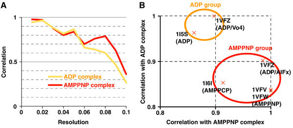
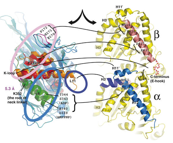
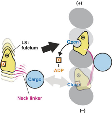
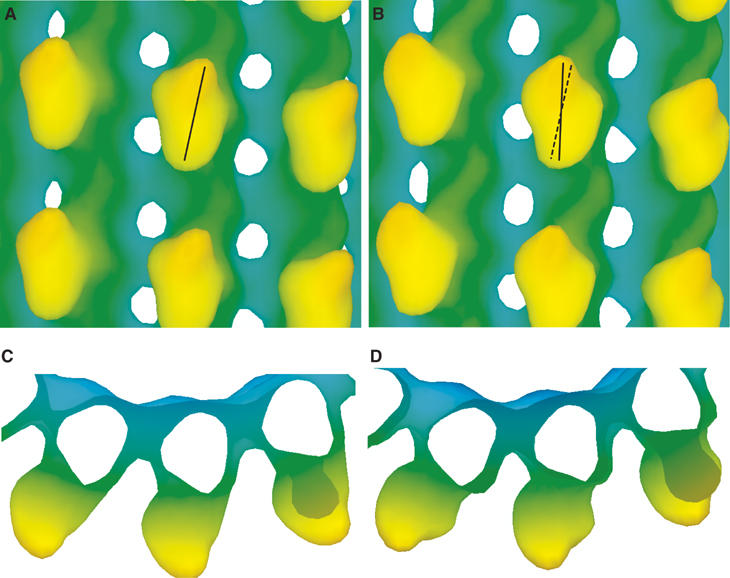
Kinesin is an ATP-driven microtubule (MT)-based motor fundamental to organelle transport. Although a number of kinesin crystal structures have been solved, the structural evidence for coupling between the bound nucleotide and the conformation of kinesin is elusive. In addition, the structural basis of the MT-induced ATPase activity of kinesin is not clear because of the absence of the MT in the structure. Here, we report cryo-electron microscopy structures of the monomeric kinesin KIF1A-MT complex in two nucleotide states at about 10 A resolution, sufficient to reveal the secondary structure. These high-resolution maps visualized clear structural changes that suggest a mechanical pathway from the nucleotide to the neck linker via the motor core rotation. In addition, new nucleotide binding pocket conformations are observed that are different from X-ray crystallographic structures; it is closed in the 5'-adenylyl-imidodiphosphate state, but open in the ADP state. These results suggest a structural model of biased diffusion movement of monomeric kinesin motor.


Figures

Figure 1
Figure 1
Evaluation of resolution of maps and docking of atomic models. (A) Fourier shell correlations between the KIF1A–MT complex density maps and atomic models generated from the crystal structures of KIF1A and tubulin. (B) Correlation between the KIF1A–MT complex density maps and KIF1A atomic models. Each cross represents correlation values of the atomic model specified by PDB ID. Correlation values were normalized by the maximum value among the atomic models.
Figure 2
Figure 2
Cryo-EM maps of KIF1A–MT complexes in the ADP (A, C, E) and AMPPNP states (B, D, F). (A, B) Isosurface representation of KIF1A–MT complexes. (C–F) Fitting of the X-ray crystal structures into cryo-EM maps. MTs are shown with their plus-end up and assignment of α- and β-tubulin is based on that of Krebs et al (2004). The blue chickenwires are contoured at 0.7σ of the density map, with a mesh size of 1 Å. A 20.6° rotation around the axis shown in (D) and (F) explains conformational changes of the kinesin core from the ADP state to the AMPPNP state.
Figure 3
Figure 3
Detailed views of the 3D reconstructions of the KIF1A–MT complex. (A) Right and (B) left sides of the MT protofilament outer surface in the AMPPNP complex showing the similarity between the tubulin atomic model and map. Blue and pink chickenwires represent 0.7σ and 1.5σ of the density map, respectively. Solvent-exposed helices (H3–5, H11 and H12) are well resolved. A small shift of α-tubulin helix H3 is indicated with an open arrowhead, possibly as a result of interactions with loop L11 of KIF1A. (C, D) Top view of the switch II helix from the plus-end of the MT (C) in the ADP and (D) AMPPNP states. Both ADP (orange) and AMPPNP (red) KIF1A switch II helices are fitted into the complexes. The interface between KIF1A and MT is depicted with dashed lines. Asterisks indicate the possible points of steric clashes between KIF1A and tubulin atomic models.
Figure 4
Figure 4
Experimental and simulated density maps surrounding NBP. (A, B) Slab view around the KIF1A NBP. (A) ADP is mostly exposed to solvent (open arrowhead), while (B) the phosphate portion of AMPPNP is enclosed by density (filled arrowhead). Nucleotides are surrounded by switch I and switch II loops. In the AMPPNP state, a well-conserved E253 (illustrated with a spacefill model) is in direct contact with α-tubulin helix H11′. The displacement of E253 is explained by rotation of the core. The rotation axis of the core is shown in (B). (C) Simulated 10 Å density map of KCBP (PDB ID:1SDM) displaying a closed conformation. Blue and pink chickenwires represent 0.7σ and 1.5σ, respectively.
Figure 5
Figure 5
MT-centric superposition of the ADP and AMPPNP KIF1A crystal structures from KIF1A–MT complexes, presenting interactions between KIF1A residues and tubulin. Interacting residues observed in ADP (orange), AMPPNP (red) or both complexes (black) are depicted with lines connecting to the corresponding tubulin-binding site.
Figure 6
Figure 6
‘Biased-capturing' model of monomeric kinesins. The model explains the structural basis of plus-end biased binding of monomeric kinesin motors. The monomeric kinesin motor domain is linked to a bead, and the linker determines the direction of rotation of the motor core. The core rotate clockwise only when the motor moves to the forward binding site, which allows ADP release and strong binding of kinesin to the MT.
Figure 7
Figure 7
Simulated 25 Å resolution density maps of the human kinesin–microtubule complexes, (A, C) in the ADP state and (B, D) in the AMPPNP complex. The atomic models of the complexes were constructed based on our docking results derived from the KIF1A–microtubule complex.

Similar articles

Cited by

References

    1. Al-Bassam J, Cui Y, Klopfenstein D, Carragher BO, Vale RD, Milligan RA (2003) Distinct conformations of the kinesin Unc104 neck regulate a monomer to dimer motor transition. J Cell Biol 163: 743–753 - PMC - PubMed
    1. Carter NJ, Cross RA (2005) Mechanics of the kinesin step. Nature 435: 308–312 - PubMed
    1. Chacon P, Wriggers W (2002) Multi-resolution contour-based fitting of macromolecular structures. J Mol Biol 317: 375–384 - PubMed
    1. Garcia-Saez I, Yen T, Wade RH, Kozielski F (2004) Crystal structure of the motor domain of the human kinetochore protein CENP-E. J Mol Biol 340: 1107–1116 - PubMed
    1. Hackney DD (1988) Kinesin ATPase: rate-limiting ADP release. Proc Natl Acad Sci USA 85: 6314–6318 - PMC - PubMed

Publication types

MeSH terms