. 2006 Sep 20;25(18):4293-304.
doi: 10.1038/sj.emboj.7601260. Epub 2006 Aug 31.

Mechanism of RecA-mediated homologous recombination revisited by single molecule nanomanipulation

Affiliations

Mechanism of RecA-mediated homologous recombination revisited by single molecule nanomanipulation

Renaud Fulconis et al. EMBO J. .

Abstract

The mechanisms of RecA-mediated three-strand homologous recombination are investigated at the single-molecule level, using magnetic tweezers. Probing the mechanical response of DNA molecules and nucleoprotein filaments in tension and in torsion allows a monitoring of the progression of the exchange in real time, both from the point of view of the RecA-bound single-stranded DNA and from that of the naked double-stranded DNA (dsDNA). We show that strand exchange is able to generate torsion even along a molecule with freely rotating ends. RecA readily depolymerizes during the reaction, a process presenting numerous advantages for the cell's 'protein economy' and for the management of topological constraints. Invasion of an untwisted dsDNA by a nucleoprotein filament leads to an exchanged duplex that remains topologically linked to the exchanged single strand, suggesting multiple initiations of strand exchange on the same molecule. Overall, our results seem to support several important assumptions of the monomer redistribution model.


Figures

Figure 1
Figure 1
Experimental design: schematic view. (A) ssDNA-RecA+dsDNA assay. An ssDNA molecule (14 kb) is held in the magnetic tweezers, then covered by RecA and incubated with homologous dsDNA of various sizes. (B) dsDNA+ssDNA-RecA assay. A dsDNA molecule (14 kb) is held in the magnetic tweezers, partially untwisted, and incubated with preassembled RecA-ssDNA fibers, the ssDNA being homologous to the central part of the dsDNA. Please note that the number of molecules involved in the exchange reaction, and the topology of the formed complex are just provided as examples of what could happen, and do not imply any a priori assumption of the real mechanism of strand exchange, or of the actual final product of the exchange.
Figure 2
Figure 2
ssDNA-RecA+dsDNA assay: force versus extension curves. (A) Symbols represent the experimental data, and the full lines the best-fit using the extended analytical interpolation of the worm like chain model (Bouchiat et al, 1999). This approximation yields an analytical expression consistent with the exact numerical solution within better than 2%. Since we used forces at most 10 pN, corrections for elastic deformation of the DNA backbone are below 5% (Cizeau and Viovy, 1997) and were thus neglected; +, black: ssDNA before recombination (recorded in Binding Buffer); The best-fit parameters obtained using the worm like chain model are: l=5.4 μm and ξ=6.2 nm; × , blue: same molecule after extensive RecA polymerization (recorded in Binding Buffer+2 mM Mg2+ to ensure good nucleoprotein filament stability). Best-fit parameters: l=6.9 μm and ξ=449.2 nm; ○, red: same molecule after recombination with 14 kb homologous dsDNA and rinsing to get rid of RecA (in Binding Buffer); □, green: 14 kb dsDNA used as a reference (in Binding buffer); best-fit parameters: l=4.7 μm and ξ=56.7 nm. In this example, the reaction product curve (○) was fitted with an ‘hybrid' curve consisting of 30% single-stranded DNA and 70% double-stranded DNA. Both increasing and decreasing force scans data are plotted, and were fitted independently with the model, leading to the two plain lines. They are almost superposed, showing the very weak hysteresis and good reproducibility of the experiments. 71±1%. (B) Histogram of the fraction of dsDNA in the hybrid molecule formed in presence of ATP, relative to the homology between the probe molecule and the substrate. Blue: experiments with 14 kb homologous DNA; red: experiments with 3.5 kb homologous DNA; green: experiments with sonicated 14 kb homologous DNA. (C) Histogram of the fraction of dsDNA in the hybrid molecule formed in the presence of ATPγS, relative to the homology between the probe molecule and the substrate. Same color meaning as in (B).
Figure 3
Figure 3
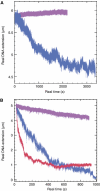
Kinetics of the ssDNA-RecA+dsDNA in solution assay. The filament length was recorded immediately after injection of Binding Buffer+12 mM Mg2++14 kb homologous dsDNA, except otherwise mentioned. (A) Blue line: Nucleoprotein filaments held in the tweezer at 10 pN; purple line: same with heterologous dsDNA. (B) Same under a stretching force of 0.1 pN; blue line: length recorded immediately after injecting buffer+homologous 14 kb dsDNA; red line: length recorded after having maintained the same molecule for 10 min at 10 pN and then lowered the force back to 0.1 pN (see text for details); purple line: control performed in the same buffer, at 0.1 pN in the absence of homologous dsDNA.
Figure 4
Figure 4
Kinetics of the dsDNA+ssDNA-RecA in solution assay. A 14 kb dsDNA is held at a stretching force of 2.3 pN, and 3.5 kb preformed homologous nucleoprotein filaments are injected at the beginning of recording. N is the number of turns applied on the dsDNA using the tweezers. Purple: applying negative supercoiling progressively by 100 turns steps every 600 s, in order to compensate for the untwisting action of the double strand invasion by the nucleofilament (see text). The degree of supercoiling corresponding to different sections of the curve are indicated in the figure; green: same conditions, except for imposing 400 turns negative supercoiling in one step at the beginning of incubation; red: same as green, except that the nucleoprotein filaments were assembled in the presence of ATP-γ-S instead of ATP; blue: control: same conditions as for the green curve, in the absence of nucleoprotein filament. Gray: control: same conditions as for the green curve, in the presence of heterologous nucleoprotein filament.
Figure 5
Figure 5
dsDNA+ssDNA-RecA assay. Extension versus supercoiling curves. The measures are carried out in Binding Buffer, under 0.3 pN force. (A) Blue, dsDNA profile before incubation with nucleoprotein filaments; red, same molecule after 30 min incubation with 3.5 kb homologous preformed nucleoprotein filaments. The red plot is followed reversibly upon series of increasing and decreasing scans (increasing and decreasing σ, respectively). (B) Plot obtained after incubation with the same ssDNA, in the presence of SSB and in the absence of RecA. Black: naked DNA; red: first increasing scan from −420 to +100 turns. Green: ‘return' scan from +100 to −420 turns; After the first return scan, the green curve, identical to that of naked dsDNA, is reversibly followed during both increasing and decreasing scans. (C) Plots obtained after incubation with nucleoprotein filament assembled in the presence of ATPγS. Each color represents a different sequence of one increasing scan (plots with symbols) and one decreasing (full line without symbols) scan. This series of curves displays a stable hysteresis loop.
Figure 6
Figure 6
Proposed mechanism for depolymerization-induced strand exchange. dsDNA is invaded by spooling (black arrow) on a synapsis progressing 5′–3′ on the ssDNA nucleoprotein filament. Depolymerization of RecA at the rear of the synapsis releases naked dsDNA and an exchanged ssDNA, which can, depending on the environment, develop secondary structures or be covered by SSB or RecA (not represented here). The red arrow represent a behavior suggested by Bianco et al (1998), for the passage of heterologies: Upon arrest of synapsis progression by the heterology, the synapsis rotation should be reversed by the release of torsional stress stored on DNA, induced by continuing RecA depolymerization no more compensated by strand invasion. This results in an untwisting of the heterologous zone if the invading and invaded DNAs are topologically linked downstream (not represented).

Similar articles

Cited by

References

    1. Adzuma K (1992) Stable synapsis of homologous DNA molecules mediated by the Escherichia coli RecA protein involves local exchange of DNA strands. Gene Dev 6: 1679–1694 - PubMed
    1. Bennink ML, Scharer OD, Kanaar R, Sakata-Sogawa K, Schins JM, Kanger JS, de Grooth BG, Greve J (1999) Single-molecule manipulation of double-stranded DNA using optical tweezers: interaction studies of DNA with RecA and YOYO-1. Cytometry 36: 200–208 - PubMed
    1. Bianco PR, Tracy RB, Kowalczykowski SC (1998) DNA strand exchange proteins: a biochemical and physical comparison. Front Biosci 3: D570–D603 - PubMed
    1. Bouchiat C, Wang MD, Allemand JF, Strick T, Block SM, Croquette V (1999) Estimating the persistence length of a worm-like chain molecule from force-extension measurements. Biophys J 76: 409–413 - PMC - PubMed
    1. Bugreeva I, Bugreev D, Nevinsky G (2005) Formation of nucleoprotein RecA filament on single-stranded DNA. FEBS J 272: 2734–2745 - PubMed

Publication types

LinkOut - more resources