

Rapamycin reduces disease activity and normalizes T cell activation-induced calcium fluxing in patients with systemic lupus erythematosus
- PMID: 16947529
- PMCID: PMC4034146
- DOI: 10.1002/art.22085
Rapamycin reduces disease activity and normalizes T cell activation-induced calcium fluxing in patients with systemic lupus erythematosus
Abstract
Objective: Systemic lupus erythematosus (SLE) is an autoimmune disease of unknown origin. Current treatment options are often ineffective or poorly tolerated. Recent observations have revealed mitochondrial hyperpolarization and enhanced Ca2+ fluxing in T cells from SLE patients. Rapamycin, a lipophilic macrolide antibiotic that regulates mitochondrial transmembrane potential and Ca2+ fluxing, has been used safely and effectively to treat renal transplant rejection since 1999. In addition, rapamycin has been shown to ameliorate T cell function and to prolong survival in lupus-prone MRL/lpr mice. We therefore undertook the present study to investigate whether rapamycin is beneficial in patients with SLE.
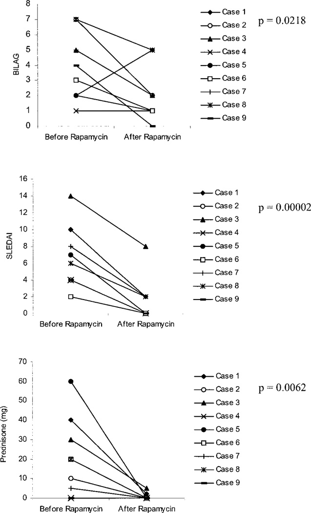
Methods: Nine patients with clinically active SLE that had been treated unsuccessfully with other immunosuppressive medications began therapy with rapamycin, 2 mg/day orally. Disease activity was assessed with the British Isles Lupus Assessment Group (BILAG) score, SLE Disease Activity Index (SLEDAI), and requirement for prednisone therapy. Mitochondrial transmembrane potential and Ca2+ fluxing were assessed by flow cytometry.
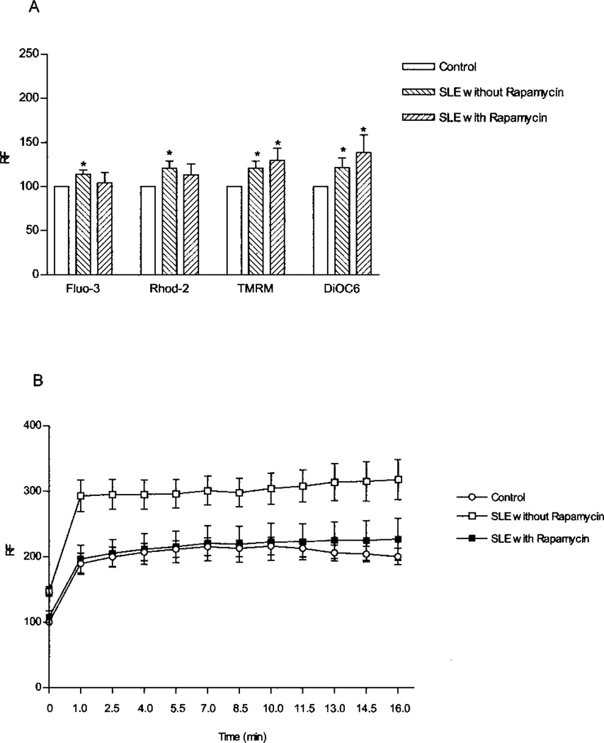
Results: In patients treated with rapamycin, the BILAG score was reduced by a mean +/- SEM of 1.93 +/- 0.9 (P = 0.0218), the SLEDAI by 5.3 +/- 0.8 (P = 0.00002), and concurrent prednisone use by 26.4 +/- 6.7 mg/day (P = 0.0062) compared with pre-rapamycin treatment. While mitochondrial hyperpolarization persisted, pretreatment cytosolic and mitochondrial Ca2+ levels and T cell activation-induced rapid Ca2+ fluxing were normalized in rapamycin-treated patients.
Conclusion: Rapamycin appears to be a safe and effective therapy for SLE that has been refractory to traditional medications. Mitochondrial dysfunction and Ca2+ fluxing could serve as biomarkers to guide decisions regarding future therapeutic interventions in SLE.
Figures
References
-
- Vassilopoulos D, Kovacs B, Tsokos GC. TCR/CD3 complex-mediated signal transduction pathway in T cells and T cell lines from patients with systemic lupus erythematosus. J Immunol. 1995;155:2269–2281. - PubMed
-
- Kyttaris VC, Tsokos GC. T lymphocytes in systemic lupus erythematosus: an update [review] Curr Opin Rheumatol. 2004;16:548–552. - PubMed
Publication types
MeSH terms
Substances
Grants and funding
LinkOut - more resources
Full Text Sources
Other Literature Sources
Medical
Miscellaneous
