. 2006 Sep;2(9):e86.
doi: 10.1371/journal.ppat.0020086.

Yersinia has a tropism for B and T cell zones of lymph nodes that is independent of the type III secretion system

Affiliations

Yersinia has a tropism for B and T cell zones of lymph nodes that is independent of the type III secretion system

Joan-Miquel Balada-Llasat et al. PLoS Pathog. 2006 Sep.

Abstract

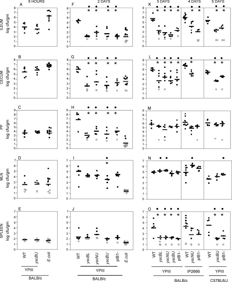
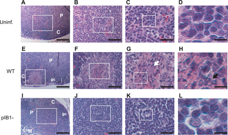
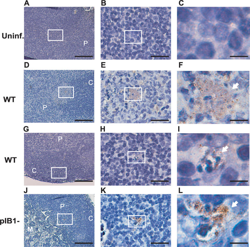
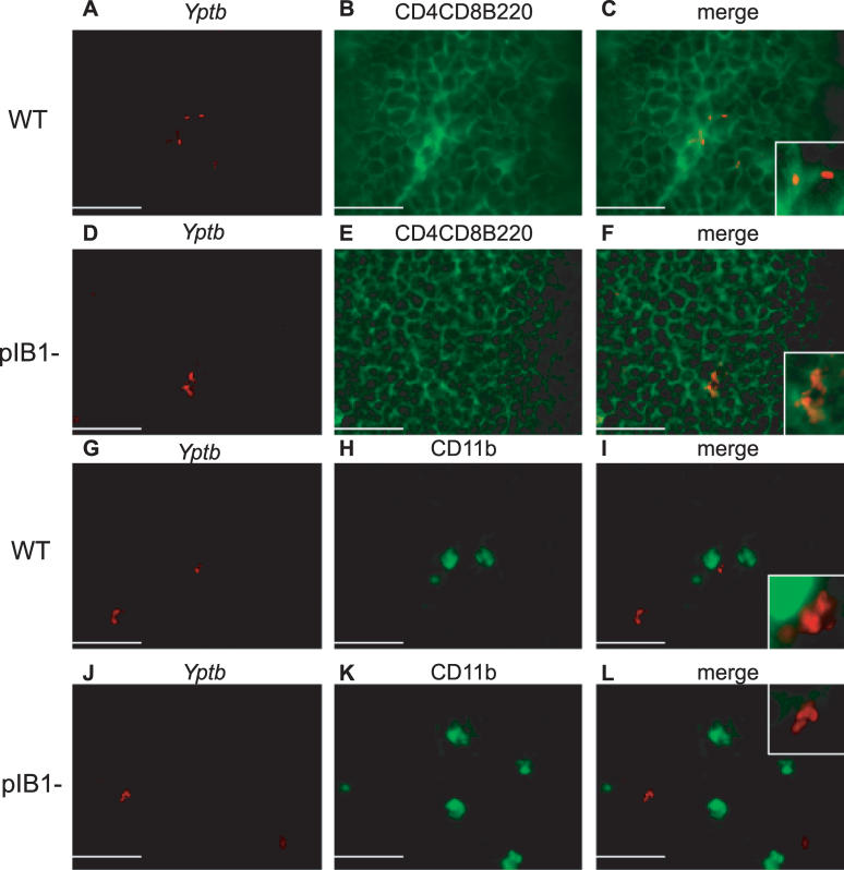
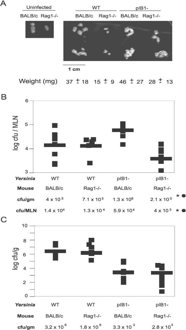
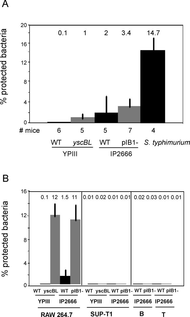
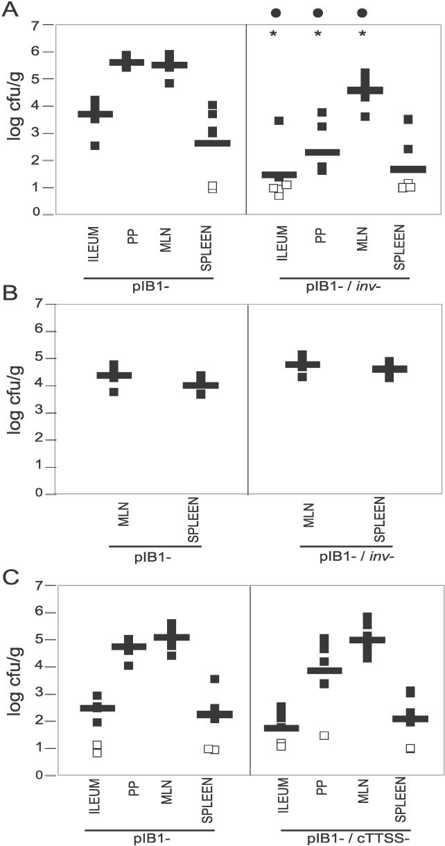
Pathogenic Yersinia have a pronounced tropism for lymphatic tissues and harbor a virulence plasmid that encodes a type III secretion system, pTTSS, that transports Yops into host cells. Yops are critical virulence factors that prevent phagocytosis by macrophages and neutrophils and Yersinia mutants lacking one or more Yops are defective for survival in lymphatic tissues, liver, and gastrointestinal tract. However, here we demonstrate that Y. pseudotuberculosis (Yptb) mutants lacking the pTTSS survived as well as or better than wild-type (WT) Yptb in the mesenteric lymph nodes (MLN). Infection with pTTSS mutants caused lymphadenitis with little necrosis, whereas infection with WT Yptb provoked lymphadenitis with multiple necrotic suppurative foci. Gentamicin protection assays and microscopic examination of the MLN revealed that pTTSS mutants resided extracellularly adjacent to B and T lymphocytes in the cortex and paracortex. WT Yptb was found extracellularly adjacent to neutrophils and macrophages in necrotic areas and adjacent to B and T lymphocytes in less-inflamed areas. To determine whether lymphocytes protected pTTSS mutants from phagocytic cells, Rag1(-/-) mice were infected with pTTSS mutants or WT Yptb. pTTSS mutants but not WT, were impaired for survival in MLN of Rag1(-/-) mice, suggesting that lymphocyte-rich regions constitute a protective niche for pTTSS mutants. Finally, we show that invasin and the chromosomally encoded TTSS were not required for Yptb survival in MLN. In summary, chromosomally encoded factors are sufficient for Yptb replication in the cortex and paracortex of MLN; the pTTSS enables Yersinia to survive within phagocyte-rich areas of lymph nodes, and spread to other tissues.


Conflict of interest statement

Competing interests. The authors have declared that no competing interests exist.

Figures

Figure 1
Figure 1. pTTSS Is Not Required for MLN Colonization
BALB/c or C57BL6/J mice were intragastrically inoculated with 2 × 10⁸ WT YPIII, YPIII yscBL, YPIII yscNU, YPIII yscBU, or YPIII pIB1, and colonization levels of the ileum (A), (F), and (K), cecum (B), (G), and (L), PP (C), (H), and (M), MLN (D), (I), and (N), and spleen (E), (J), and (O) were determined at 6 h (A–E), 2 d (F–J), and 5 d (K–O) post-inoculation. BALB/c mice were intragastrically inoculated with 2 × 10⁸ mouse commensal E. coli, and colonization levels of tissues were studied at 6 h (A–E) and 2 d post-inoculation (F–J). BALB/c mice were intragastrically inoculated with 2 × 10⁹ of WT IP2666, IP2666 yscNU, or IP2666 pIB1−, and colonization levels were determined at 4 d post-inoculation ( [K–O], middle section). Data are from 4–12 mice from at least two different experiments. All data was combined for each strain, tissue, and time point. Each square represents the log cfu/g tissue from one mouse; open squares indicate that less than 10 cfu were recovered per tissue, and bars represent the geometric mean. Asterisks (*) and black circles (•) indicate statistically significant differences between the WT and the mutants, calculated by the t test (*p < 0.01) or by Mann-Whitney (•U < 0.05), respectively.
Figure 2
Figure 2. MLN Histopathology during WT or pIB1 Infection
BALB/c mice were intragastrically inoculated with 2 × 10⁹ WT or IP2666 pIB1, and MLN were processed for H&E staining 4 d later. MLN sections from uninfected (Uninf.) (A–D), WT infected (E–H), and pIB1 infected (I–L) are shown at 15×, 40×, 90×, and 450× magnification. White boxes indicate magnified areas in the next slide. White arrow points to the bacterial foci, black arrow to neutrophils. Scale bars correspond to 133 μm for 15× magnification, 50 μm for 40× magnification, 22 μm for 90× magnification, and 4.4 μm for 450× magnification. Pictures shown are representative of multiple fields and samples from 16 MLN infected with WT or 14 MLN infected with pIB1. C, cortex; gc, germinal center; M, medulla; P, paracortex.
Figure 3
Figure 3. Yptb Localizes in the Cortex and Paracortex of the MLN
BALB/c mice were intragastrically inoculated with 2 × 10⁹ WT or IP2666 pIB1. At 4 d post-inoculation, MLN were harvested and stained for Yptb followed by hematoxylin staining. Picture sections of uninfected (A–C), WT infected (D–I), and pIB1 infected (J–L) MLN were taken at 150×, 900×, and 4,500× magnification. White boxes indicate magnified areas in the next slide. Arrows point to Yptb microcolonies. Scale bars correspond to 133 μm for 15× magnification, 22 μm for 90× magnification, and 4.4 μm for 450× magnification. Pictures shown are representative of multiple fields and samples from MLN infected with WT or pIB1. C, cortex; gc, germinal center; M, medulla; P, paracortex.
Figure 4
Figure 4. pTTSS Mutants Are Adjacent to B and T Lymphocytes Whereas WT Is Adjacent to B and T Lymphocytes and CD11b+ Cells
BALB/c mice were intragastrically inoculated with 2 × 10⁹ WT IP2666 or IP2666 pIB1. At 4 d post-inoculation, MLN were harvested, sectioned, and examined by fluorescence microscopy. Staining with antibodies to Yptb (red) (A), (D), (G), and (J), to CD4-CD8-B220 (green) (B) and (E), or to CD11b (green) (H) and (K) was performed, and images merged (C), (F), (I), and (L). Pictures show the cortex–paracortex and are representative of multiple fields and samples for WT-infected mice and pIB1 mice stained with CD4-CD8-B220. Fewer fields had pIB1 bacteria and CD11b+ cells. Scale bars correspond to 22 μm for 900× magnification.
Figure 5
Figure 5. B and T Lymphocytes Are Important for pIB1 Colonization in the MLN
BALB/c or Rag1−/− mice were intragastrically inoculated with 2 × 10⁹ WT IP2666 or IP2666 pIB1. (A) Size and weight of MLN from BALB/c or Rag1−/− mice that were either uninfected or infected with WT IP2666 or IP2666 pIB1 were measured. (B) and (C) Colonization of the MLN or luminal content of the ileum 4 d post-intragastric inoculation of BALB/c or isogenic Rag1−/− mice was determined. Each square indicates the cfu from one mouse calculated as log cfu/MLN (B) or the log cfu/g of luminal content of the ileum (C); bars represent the geometric mean. Each experiment was performed with two to three mice and repeated three times. Asterisks (*) and black circles (•) indicate statistically significant differences between the number of pIB1 recovered from BALB/c mice versus Rag1−/−, calculated by the t test (*p < 0.05) or by Mann-Whitney (•U < 0.05), respectively.
Figure 6
Figure 6. Yptb pTTSS Mutants Remain Extracellular in the MLN and Are Not Efficiently Internalized by B or T Lymphocytes in Culture
(A) Single-cell suspensions of Yptb-infected MLN were assayed for the presence of intracellular bacteria using a gentamicin protection assay. BALB/c mice were intragastrically inoculated with WT YPIII (n = 6 mice), YPIII yscBL (n = 5 mice), WT IP2666 (n = 5 mice), IP2666 pIB1 (n = 7 mice), or S. typhimurium (n = 4 mice). Four days post-inoculation with IP2666 strains or 5 d post-inoculation with YPIII strains, the percentage of intracellular bacteria was assessed by generating a single-cell suspension of the MLN and treating half the suspension with gentamicin and half without gentamicin (see Materials and Methods). (B) Murine macrophage RAW264.7 cells, human T cells SUP-T1, and B and T lymphocytes isolated from MLN (see Materials and Methods) were infected with WT YPIII, YPIII yscBL, WT IP2666, or IP2666 pIB1 strains at MOI of 10:1 for 30 min, and then treated with gentamicin for 90 min. The data are presented as 100 times the number of gentamicin-resistant bacteria divided by the number of input bacteria. Data from one representative experiment done in triplicate is shown. All experiments were performed at least three times.
Figure 7
Figure 7. Invasin and cTTSS Are Not Essential for Yptb Survival in the MLN
(A) BALB/c mice were intragastrically inoculated with 2 × 10⁸ YPIII pIB1 or YPIII pIB1inv−, and colonization levels of the ileum, PP, MLN, and spleen were determined 5 d post-inoculation. (B) BALB/c mice were i.p. inoculated with 2 × 10⁶ YPIII pIB1 or YPIII pIB1 inv, and their colonization levels in the MLN and spleen were determined 3 d post-inoculation. (C) BALB/c mice were inoculated intragastrically with 2 × 10⁸ YPIII pIB1 or YPIII pIB1cTTSS, and colonization levels of the ileum, PP, MLN, and spleen were determined. Data are from 6–11 mice from at least two different experiments. Each square represents log 10 cfu/g tissue recovered from one mouse. Bars represent the geometric mean. Open squares indicate than less than 10 cfu were recovered per tissue. Asterisks (*) and black circles (•) indicate statistically significant differences between the pIB1 and either pIB1 inv or pIB1cTTSS strains, calculated by the t test (*p < 0.01) or by Mann-Whitney (•U < 0.05), respectively.

References

    1. Putzker M, Sauer H, Sobe D. Plague and other human infections caused by Yersinia species. Clin Lab. 2001;47:453–466. - PubMed
    1. Achtman M, Zurth K, Morelli G, Torrea G, Guiyoule A, et al. Yersinia pestis, the cause of plague, is a recently emerged clone of Yersinia pseudotuberculosis . Proc Natl Acad Sci U S A. 1999;96:14043–14048. - PMC - PubMed
    1. Brubaker RR. Factors promoting acute and chronic diseases caused by yersiniae. Clin Microbiol Rev. 1991;4:309–324. - PMC - PubMed
    1. Smego RA, Frean J, Koornhof HJ. Yersiniosis I: Microbiological and clinicoepidemiological aspects of plague and non-plague Yersinia infections. Eur J Clin Microbiol Infect Dis. 1999;18:1–15. - PubMed
    1. Ibrahim A, Goebel BM, Liesack W, Griffiths M, Stackebrandt E. The phylogeny of the genus Yersinia based on 16S rDNA sequences. FEMS Microbiol Lett. 1993;114:173–177. - PubMed

Publication types

MeSH terms