Review
. 2006 Sep;68(3):193-209.
doi: 10.1111/j.1399-0039.2006.00649.x.

Mannose-binding lectin in innate immunity: past, present and future

Affiliations
Review

Mannose-binding lectin in innate immunity: past, present and future

R M Dommett et al. Tissue Antigens. 2006 Sep.

Abstract

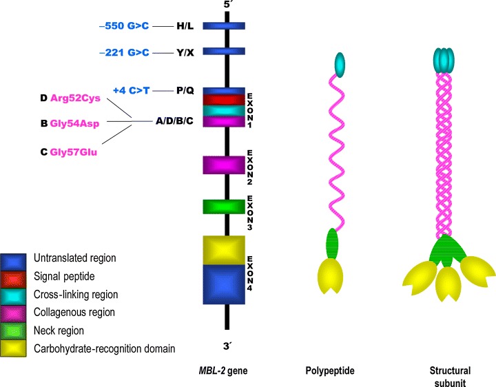
The human collectin, mannose-binding lectin (MBL), is an important protein of the humoral innate immune system. With multiple carbohydrate-recognition domains, it is able to bind to sugar groups displayed on the surfaces of a wide range of microorganisms and thereby provide first-line defence. Importantly, it also activates the complement system through a distinctive third pathway, independent of both antibody and the C1 complex. Three single point mutations in exon 1 of the expressed human MBL-2 gene appear to impair the generation of functional oligomers. Such deficiencies of functional protein are common in certain populations, e.g. in sub-Saharan Africa, but virtually absent in others, e.g. indigenous Australians. MBL disease association studies have been a fruitful area of research and implicate a role for MBL in infective, inflammatory and autoimmune disease processes. Overall, there appears to be a genetic balance in which individuals generally benefit from high levels of the protein. However, in certain situations, reduced levels of circulating MBL may be beneficial to the host and this may explain the persistence of the deleterious gene polymorphisms in many population groups.


Figures

Figure 1
Figure 1
Structure of the human MBL‐2 gene and the encoded protein product. Positions of the exon 1 and promoter polymorphisms are shown. Different regions of the polypeptide are encoded by different exons of the MBL gene. Three identical 32‐kDa polypeptides form a structural subunit, based on formation of a collagenous triple helix. Oligomerization of the structural subunit results in MBL molecules of differing size, but the tetrameric form shown in Figure 2 is probably the most common. MBL, mannose‐binding lectin.
Figure 2
Figure 2
Tetramer of human MBL structural subunits (Figure 1). The subunits are cross‐linked by disulphide bonds in the N‐terminal regions. Each MASP‐2 molecule is believed to bind close to the hinge point of the collagenous region. The number of MASPs able to bind to a given MBL tetramer is not definitively known, but the arrangement shown would be consistent with the model for rat MBL proposed by Feinberg et al. (24). The details of MASP‐1, MASP‐3 and MAp19 binding to MBL remain unclear. MBL, mannose‐binding lectin; MASP, MBL‐associated serine proteases; MASP‐1, MBL‐associated serine protease‐2; MASP‐2, MBL‐associated serine protease‐2; MASP‐3, MBL‐associated serine protease‐3.
Figure 3
Figure 3
Complement activation pathway. The lectin pathway of complement is activated by MBL and ficolins. On binding to appropriate targets, MBL–MASP‐2 complexes cleave C4 and C2 to form C3 convertase (C4bC2a). MBL–MASP‐1 complexes may activate C3 directly. Ficolins also work in combination with the MASPs. The classical and alternative pathways also generate C3 convertase enzymes, which cleave C3. The lytic pathway (C5–C9) is common to all three routes of C3 cleavage. MBL, mannose‐binding lectin; MASP, MBL‐associated serine proteases; MASP‐1, MBL‐associated serine protease‐2; MASP‐2, MBL‐associated serine protease‐2.
Figure 4
Figure 4
MBL haplotype frequencies have been shown to differ in various populations. Variant A (wild type) is found in association with four different promoter haplotypes, HYPA, LYQA, LYPA and LXPA. The B, C and D variant exon 1 alleles are in linkage disequilibrium with three different promoter haplotypes, LYPB, LYQC and HYPD. Haplotype frequency data are taken from published population studies. 1, Chiriguanos, Argentina (56); 2, Mapuche, Argentina (56); 3, Eskimos, Greenland (56); 4, Caucasians, Spain (151); 5, Caucasians, Denmark (56); 6, Mozambique (56); 7, Kenya (56); 8, Korea (152); 9, Japan (153); 10, Warlpiri, Australia (53). MBL, mannose‐binding lectin.
Figure 5
Figure 5
Serum MBL levels, MBL haplotype and development of SIRS. Serum MBL level is plotted against MBL haplotype (exon 1 and X/Y promoter polymorphisms, where O indicates the presence of B, C or D variants). Haplotypes are grouped from those associated with the highest serum levels (YA/YA), to those associated with the lowest levels (YO/YO). Red circles show cases that developed SIRS; open circles, cases that did not develop SIRS [Adapted from Fidler et al. (103)]. MBL, mannose‐binding lectin; SIRS, systemic inflammatory response syndrome.
Figure 6
Figure 6
Schematic representation illustrating how both high and low serum MBL levels may impact the health of a given host. IL, interleukin; MBL, mannose‐binding lectin.

Similar articles

Cited by

References

    1. Burnet FM, McCrea JF. Inhibitory and inactivating action of normal ferret sera against an influenza virus strain. Aust J Exp Biol Med Sci 1946: 24: 277–82. - PubMed
    1. Anders EM, Hartley CA, Jackson DC. Bovine and mouse serum beta inhibitors of influenza A viruses are mannose‐binding lectins. Proc Natl Acad Sci U S A 1990: 87: 4485–9. - PMC - PubMed
    1. Kawakami M, Ihara I, Suzuki A, Harada Y. Properties of a new complement‐dependent bactericidal factor specific for Ra chemotype salmonella in sera of conventional and germ‐free mice. J Immunol 1982: 129: 2198–201. - PubMed
    1. Kawakami M, Ihara I, Ihara S, Suzuki A, Fukui K. A group of bactericidal factors conserved by vertebrates for more than 300 million years. J Immunol 1984: 132: 2578–81. - PubMed
    1. Robinson D, Phillips NC, Winchester B. Affinity chromatography of human liver alpha‐D‐mannosidase. FEBS Lett 1975: 53: 110–2. - PubMed

MeSH terms