. 2006;7(9):R80.
doi: 10.1186/gb-2006-7-9-R80.

The human urinary proteome contains more than 1500 proteins, including a large proportion of membrane proteins

Affiliations

The human urinary proteome contains more than 1500 proteins, including a large proportion of membrane proteins

Jun Adachi et al. Genome Biol. 2006.

Abstract

Background: Urine is a desirable material for the diagnosis and classification of diseases because of the convenience of its collection in large amounts; however, all of the urinary proteome catalogs currently being generated have limitations in their depth and confidence of identification. Our laboratory has developed methods for the in-depth characterization of body fluids; these involve a linear ion trap-Fourier transform (LTQ-FT) and a linear ion trap-orbitrap (LTQ-Orbitrap) mass spectrometer. Here we applied these methods to the analysis of the human urinary proteome.

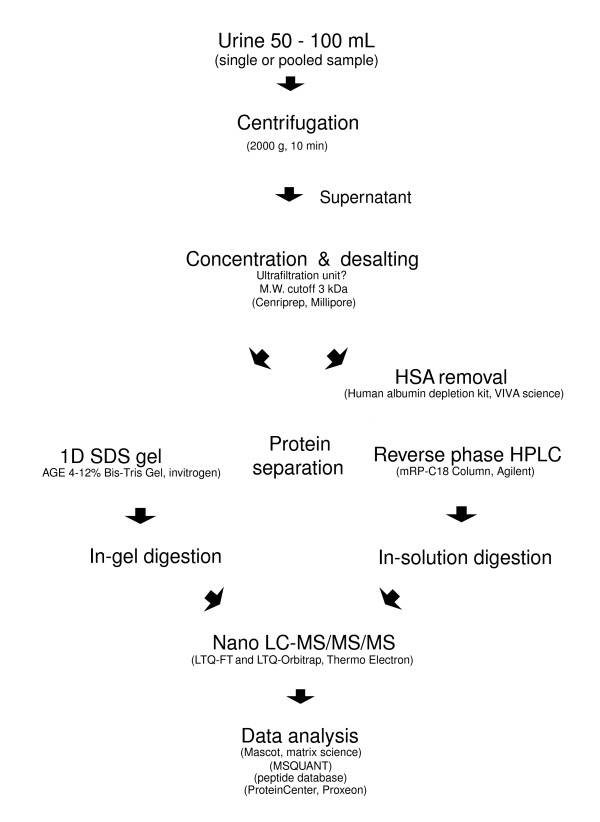
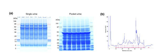
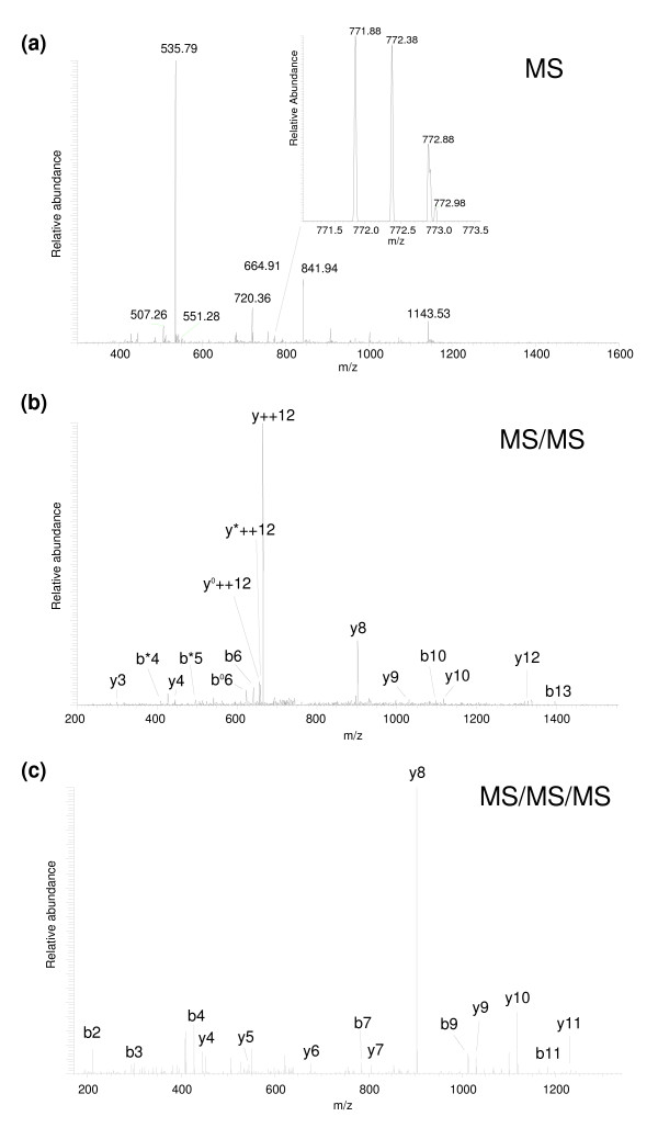
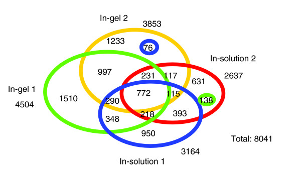
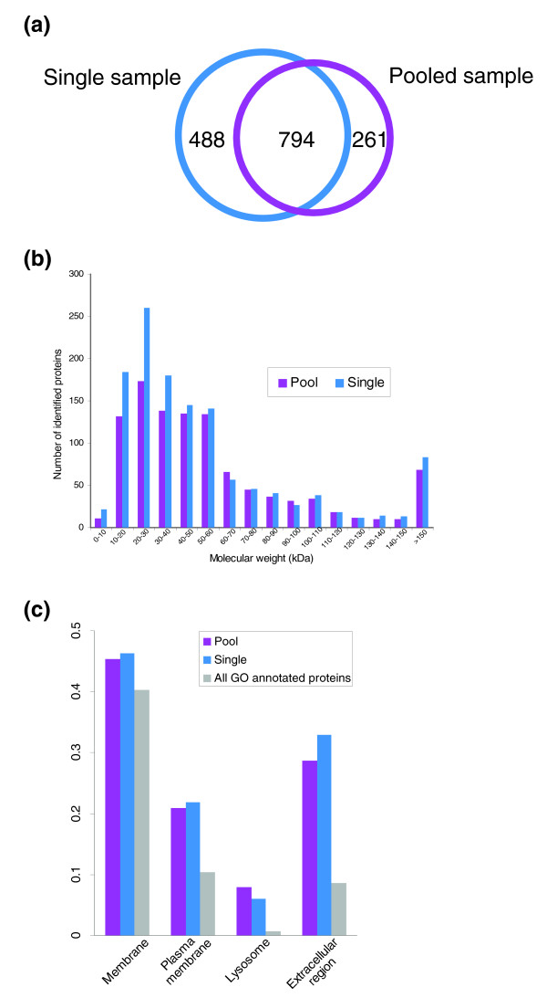
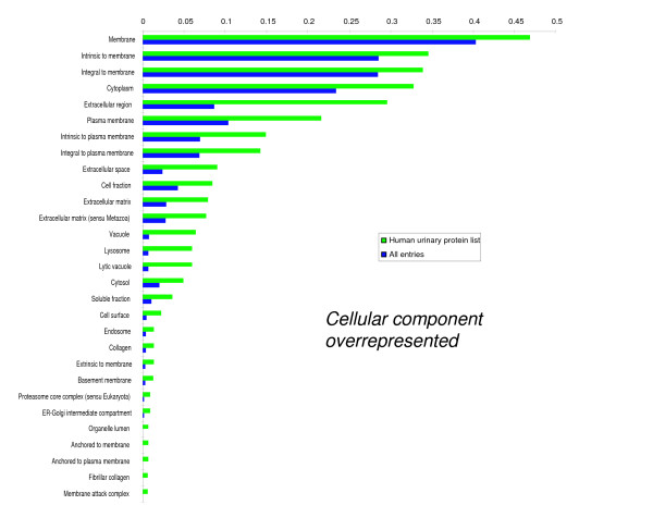
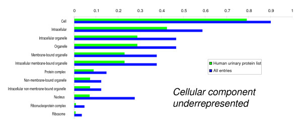
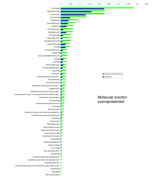
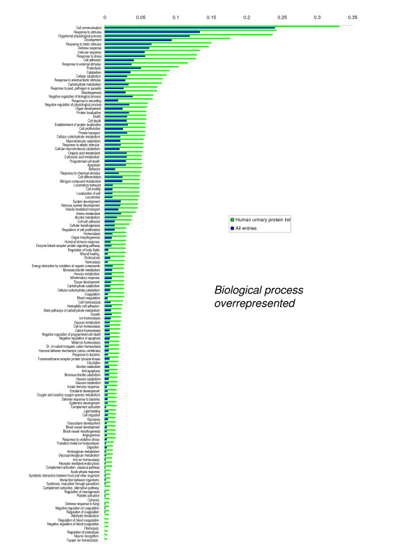
Results: We employed one-dimensional sodium dodecyl sulfate polyacrylamide gel electrophoresis and reverse phase high-performance liquid chromatography for protein separation and fractionation. Fractionated proteins were digested in-gel or in-solution, and digests were analyzed with the LTQ-FT and LTQ-Orbitrap at parts per million accuracy and with two consecutive stages of mass spectrometric fragmentation. We identified 1543 proteins in urine obtained from ten healthy donors, while essentially eliminating false-positive identifications. Surprisingly, nearly half of the annotated proteins were membrane proteins according to Gene Ontology (GO) analysis. Furthermore, extracellular, lysosomal, and plasma membrane proteins were enriched in the urine compared with all GO entries. Plasma membrane proteins are probably present in urine by secretion in exosomes.

Conclusion: Our analysis provides a high-confidence set of proteins present in human urinary proteome and provides a useful reference for comparing datasets obtained using different methodologies. The urinary proteome is unexpectedly complex and may prove useful in biomarker discovery in the future.


Figures

Figure 1
Figure 1
An overview of the procedure used for analysis of the urinary proteome. 1D, one-dimensional; HPLC, high-performance liquid chromatography; HSA, human serum albumin; MW, molecular weight; LC, liquid chromatography; MS, mass spectrometry; SDS, sodium dodecyl sulfate.
Figure 2
Figure 2
Urinary protein separation by one-dimensional SDS gel and reverse-phase HPLC. (a) 150 μg urinary protein (25 μg/lane) from single sample and pooled sample were applied on a 4-12% Bis-Tris gel. Gel was stained by colloidal Coomassie and cut into 14 pieces (in-gel 1 set) or 10 pieces (in-gel 2 set) for single urine sample, and cut into 10 pieces for pooled urine sample. (b) 250 μg of urinary protein was applied to Vivapure Anti-HSA Kit to deplete serum albumin. The albumin-depleted protein mixture was dissolved in 6 mol/l urea and 1.0% acetic acid solution, and separated on mRP-C18 High-Recovery protein column at 80°C using linear multi-segment gradient, as described in the Materials and Methods section. HPLC, high-performance liquid chromatography; SDS, sodium dodecyl sulfate.
Figure 3
Figure 3
Two consecutive stages of mass spectrometric fragmentation (MS3). The precursor of peptide DVPNSQPEMVEAVK (a; see insert) was selected for fragmentation from a full scan of mass to charge ratio range. The doubly charged y12 fragment ion (b) was subsequently fragmented. Characteristic pattern for charged directed fragmentation is observed in MS3 spectra (c) and confirms the identification of the above peptide. See Steen and Mann [65] for an introduction to peptide sequencing and confidence of peptide identification. MS, mass spectrometry.
Figure 4
Figure 4
Diagram of peptides found in multiple datasets. All overlaps of peptides are shown (two way, three way, and four way) for all four input datasets: in-gel 1 (green), in-gel 2 (yellow), in-solution 1 (blue), and in-solution 2 (red). Numbers represent the number of shared peptides in the respective overlapping areas.
Figure 5
Figure 5
Comparison of identified proteins in urine of a single person and pooled urine from nine persons. (a) Overlapping proteins, (b) molecular weight distribution, and (c) cellular localization were compared. The ratio of membrane, plasma membrane, lysosome, and extracellular region proteins in each dataset were calculated using BiNGO, as described in the Materials and Methods section. GO, Gene Ontology.
Figure 6
Figure 6
Significantly over-represented GO cellular component terms for the set of identified urinary proteins. The set of identified urinary proteins was compared with the entire list of IPI entries (IPI_Human, version 3.13, 57050 protein sequences), and significantly over-represented and underrepresented GO terms (P < 0.001) are shown. The ratio shown is the number of urinary and entire IPI proteins annotated to each GO term divided by the number of urinary and entire IPI proteins linked to at least one annotation term within the indicated GO cellular component, molecular function, and biological process categories. GO, Gene Ontology; IPI, International Protein Index.
Figure 7
Figure 7
Significantly under-represented GO cellular component, molecular function and biological process terms for the set of identified urinary proteins. Each term was selected as described in the legend to Figure 6. GO, Gene Ontology.
Figure 8
Figure 8
Significantly over-represented GO molecular function terms for the set of identified urinary proteins. Each term was selected as described in the legend for Figure 6. GO, Gene Ontology.
Figure 9
Figure 9
Significantly under-represented GO molecular function terms for the set of identified urinary proteins. Each term was selected as described in the legend of Figure 6. GO, Gene Ontology.
Figure 10
Figure 10
Significantly over-represented GO biological process terms for the set of identified urinary proteins. Each term was selected as described in the legend of Figure 6. GO, Gene Ontology.
Figure 11
Figure 11
Significantly under-represented GO biological process terms for the set of identified urinary proteins. Each term was selected as described in the legend of Figure 6. GO, Gene Ontology.
Figure 12
Figure 12
Comparison between proteins identified in the present study and five recently published proteomic datasets.

References

    1. Brenner B, (editor) The Kidney. Philadelphia, PA: WB Saunders; 2000.
    1. Brunzel NA. Fundamentals of Urine & Body Fluid Analysis. Philadelphia, PA: Saunders; 2004.
    1. Haraldsson B, Sorensson J. Why do we not all have proteinuria? An update of our current understanding of the glomerular barrier. News Physiol Sci. 2004;19:7–10. doi: 10.1152/nips.01461.2003. - DOI - PubMed
    1. Maunsbach AB. Absorption of I125-labeled homologous albumin by rat kidney proximal tubule cells. A study of microperfused single proximal tubules by electron microscopic autoradiography and histochemistry. 1966. J Am Soc Nephrol. 1997;8:323–351. discussion 327-331. - PubMed
    1. Burne MJ, Osicka TM, Comper WD. Fractional clearance of high molecular weight proteins in conscious rats using a continuous infusion method. Kidney Int. 1999;55:261–270. doi: 10.1046/j.1523-1755.1999.00234.x. - DOI - PubMed

Publication types