. 2006 Nov 15;299(2):345-55.
doi: 10.1016/j.ydbio.2006.07.039. Epub 2006 Aug 5.

Meiotic resumption in response to luteinizing hormone is independent of a Gi family G protein or calcium in the mouse oocyte

Affiliations

Meiotic resumption in response to luteinizing hormone is independent of a Gi family G protein or calcium in the mouse oocyte

Lisa M Mehlmann et al. Dev Biol. .

Abstract

The signaling pathway by which luteinizing hormone (LH) acts on the somatic cells of vertebrate ovarian follicles to stimulate meiotic resumption in the oocyte requires a decrease in cAMP in the oocyte, but how cAMP is decreased is unknown. Activation of Gi family G proteins can lower cAMP by inhibiting adenylate cyclase or stimulating a cyclic nucleotide phosphodiesterase, but we show here that inhibition of this class of G proteins by injection of pertussis toxin into follicle-enclosed mouse oocytes does not prevent meiotic resumption in response to LH. Likewise, elevation of Ca2+ can lower cAMP through its action on Ca2+-sensitive adenylate cyclases or phosphodiesterases, but inhibition of a Ca2+ rise by injection of EGTA into follicle-enclosed mouse oocytes does not inhibit the LH response. Thus, neither of these well-known mechanisms of cAMP regulation can account for LH signaling to the oocyte in the mouse ovary.


Figures

Figure One
Figure One
Concentration dependence of LH stimulation of meiotic resumption in follicle-enclosed oocytes. Antral follicles were exposed to various concentrations of LH for 4–5 hours. The oocytes were then removed from their follicles, and scored for the presence or absence of an intact prophase nucleus. %GVBD = the number of oocytes that had undergone breakdown of the nuclear envelope (germinal vesicle), divided by the number of oocytes that were counted, with oocytes from 4 experiments combined as a single group. The numbers in parentheses indicate the number of oocytes counted for each point. Results for all concentrations of LH were significantly different from the control without LH (p = 0.02 for 0.01 μg/ml LH; p < 0.0001 for 0.1 and 1.0 μg/ml; p = 0.002 for 10 μg/ml; Fisher’s exact test).
Figure Two
Figure Two
A Gi-family G protein does not mediate LH stimulation of meiotic resumption. A. PTX does not inhibit the LH response in follicle-enclosed oocytes. Follicle-enclosed oocytes were injected with PTX (1.8 pg), or left uninjected. 1–3 hours later, they were exposed to 1 μg/ml LH, or control medium, for 3–4 hours. The oocytes were then isolated, to determine %GVBD. The numbers in parentheses indicate the number of oocytes counted for each point, combining the results of 9 experiments. Column 4 is not statistically different from column 3 (p = 0.7), but is different from column 2 (p < 0.0001); column 2 is not different from column 1 (p = 0.09) (Fisher’s exact test). When data for the %GVBD for column 2 were compared to the subset of data for column 1 that were collected on the same days as column 2, the %GVBD in column 1 (12%, n = 33) was very close to that for column 2 (11%, n = 19). B. PTX is an effective inhibitor of Gi-signaling in the oocyte. Isolated oocytes expressing the m2 acetylcholine receptor and the chimeric G protein Gqi were injected with 17 μM calcium green dextran (upper trace), or 17 μM calcium green dextran + 1.8 pg PTX (lower trace). Calcium green fluorescence was recorded before and after addition of 10 μM acetylcholine. The x axis shows time, and the y axis shows the voltage output of the photodiode amplifier, which is proportional to calcium green fluorescence. The initial deflection of the trace during acetylcholine addition is an artifact that is often seen when solution is pipetted into the droplet containing the oocyte (see Mehlmann et al., 1998). C. Immunoblot showing the presence of Gαz protein in mouse brain (1, 2, or 5 μg) but not in mouse oocytes (6 μg = 230 oocytes). Before the blot was stained for Gαz, it was stained for total protein, using Ponceau S; this confirmed that the amount of protein transferred to the membrane from the 6 μg oocyte sample was comparable to the amount of protein from the 5 μg brain sample. The brain Gαz protein migrated with an electrophoretic mobility close to that of a 37 kDa molecular weight standard, close to its predicted molecular weight of 41 kDa; the slight discrepancy in size could be related to the use of a prestained molecular weight standard, which could have migrated more slowly than an unstained protein.
Figure Three
Figure Three
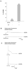
Elevation of oocyte Ca2+ does not mediate LH stimulation of meiotic resumption. A. EGTA does not inhibit the LH response in follicle-enclosed oocytes. Follicle-enclosed oocytes were injected with EGTA buffer (10 mM EGTA in the cytoplasm, free Ca2+ ~30 nM), and exposed to 1 μg/ml LH for 3–4 hours. The oocytes were then isolated, to determine %GVBD. The numbers in parentheses indicate the number of oocytes counted for each point, combining the results of 3 experiments. Column 2 is statistically different from column 1 (p < 0.0001), but is not different from column 3 of Fig. 2 (p = 0.6) (Fisher’s exact test). In a separate series of experiments, we found that injection of 10 mM of the EGTA buffer did not cause a signficant increase in meiotic resumption (20% GVBD, n = 15). B. EGTA is an effective inhibitor of Ca2+ elevation in isolated mouse oocytes. Isolated oocytes expressing the m1 acetylcholine receptor were injected with 20 μM calcium green dextran (upper trace), or 20 μM calcium green dextran + 10 mM of EGTA buffer (lower trace). Calcium green fluorescence was recorded before and after addition of 10 μM acetylcholine. C. EGTA is an effective inhibitor of Ca2+ elevation in antral follicle-enclosed mouse oocytes. Antral follicle-enclosed oocytes were injected with 1 μM caged IP3 and 20 μM calcium green 10 kDa-dextran, with or without 10 mM EGTA buffer. 3–4 hours later, IP3 was uncaged by exposing the follicle-enclosed oocytes to 330 ± 40 nm light (UV), for a period of 2 sec. In oocytes that had not been injected with EGTA, uncaging of IP3 resulted in an increase in calcium green fluorescence (n = 6 oocytes). In oocytes that had been injected with 10 mM EGTA buffer, 6/9 oocytes showed no detectable increase in calcium green fluorescence in response to uncaging IP3, and 3 others showed a small increase (~10%, or in one case ~20%, of that seen without EGTA). Note that the measurement systems used for B and C differed, so the Y axes cannot be directly compared.
Figure Four
Figure Four
Deleterious effect of BAPTA on oocyte structure. Mouse oocytes, which were held in meiotic arrest with 250 μM dbcAMP, were injected with BAPTA (upper panel) or EGTA (lower panel), at a cytoplasmic concentration of 10 mM (~30 nM free Ca2+). The oocytes were photographed 3–4 hours after injection.
Figure Five
Figure Five
Some of the possible pathways by which luteinizing hormone (LH) action on the somatic cells of the mammalian ovarian follicle could decrease cAMP in the oocyte, leading to the progression of meiosis. Of the pathways shown, experiments described in this paper argue against those marked with X’s; others remain to be tested. R = receptor; GPR3 = G protein receptor 3; AC3 = adenylate cyclase 3; GC = guanylate cyclase; Gi, Gz, Gs, Gt = G protein isoforms; PDE1, PDE3A, PDE6 = cyclic nucleotide phosphodiesterase isoforms. Green arrows indicate stimuli that increase the activity or amount of the indicated molecule; red lines indicate stimuli that decrease the activity or amount of the molecule. Pathways occuring in the somatic cells of the follicle, including signaling through the LH receptor, production of EGF receptor ligands, and the possible role of a steroid, are not shown (see Introduction and Discussion).

References

    1. Adler EM, Augustine GJ, Duffy SN, Charlton MP. Alien intracellular calcium chelators attenuate neurotransmitter release at the squid giant synapse. J Neurosci. 1991;11:1496–1507. - PMC - PubMed
    1. Akam EC, Challiss RAJ, Nahorski SR. Gq/11 and Gi/o activation profiles in CHO cells expressing human muscarinic acetylcholine receptors: dependence on agonist as well as receptor-subtype. British J Pharmacol. 2001;132:950–958. - PMC - PubMed
    1. Andersen CB, Roth RA, Conti M. Protein kinase B/Akt induces resumption of meiosis in Xenopus oocytes. J Biol Chem. 1998;273:18705–18708. - PubMed
    1. Andersen CB, Sakaue H, Nedachi T, Kovacina KS, Clayberger C, Conti M, Roth RA. Protein kinase B/Akt is essential for the insulin- but not progesterone-stimulated resumption of meiosis in Xenopus oocytes. Biochem J. 2003;369:227–238. - PMC - PubMed
    1. Anderson E, Albertini DF. Gap junctions between the oocyte and companion follicle cells in the mammalian ovary. J Cell Biol. 1976;71:680–686. - PMC - PubMed

Publication types

MeSH terms

LinkOut - more resources