. 2006 Dec 1;300(1):252-66.
doi: 10.1016/j.ydbio.2006.07.044. Epub 2006 Aug 7.

The echinoderm adhesome

Affiliations

The echinoderm adhesome

Charles A Whittaker et al. Dev Biol. .

Abstract

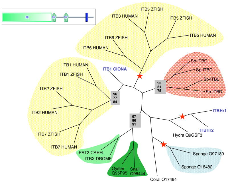
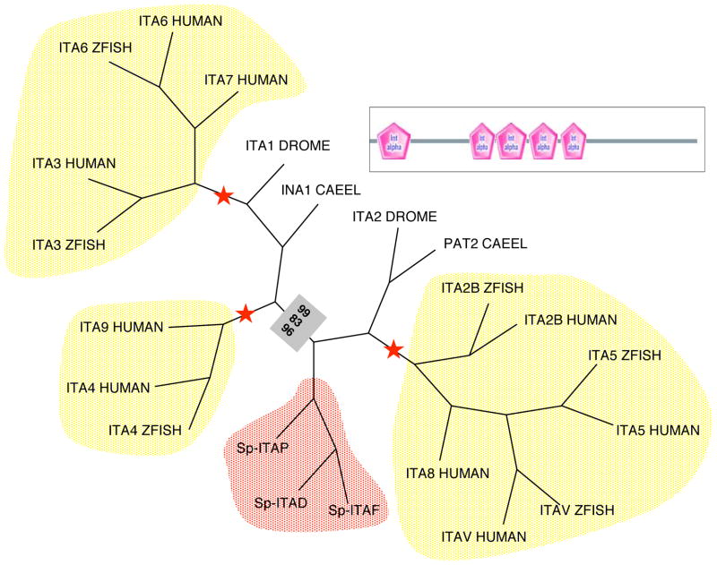
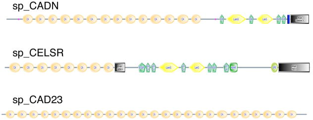
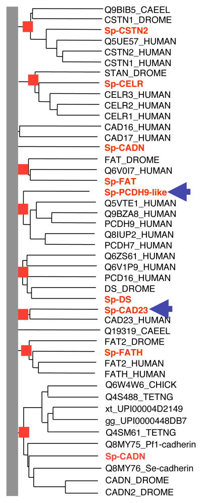
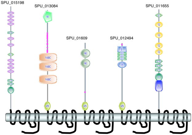
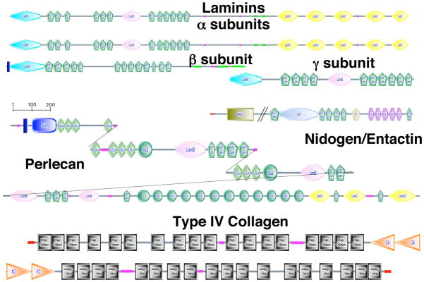
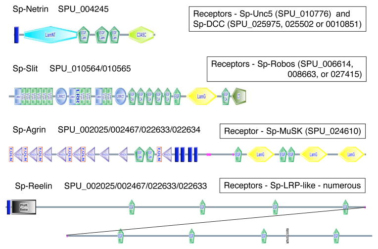
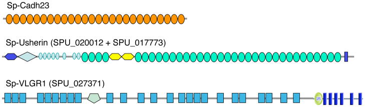
Although the development of sea urchin embryos has been studied extensively and clearly involves both cell adhesion and cell migration, rather little is known about the adhesion receptors and extracellular matrix molecules involved. The completion of the genome of Strongylocentrotus purpuratus allows a comprehensive survey of the complement of cell-cell and cell-matrix adhesion molecules in this organism. Furthermore, the phylogenetic position of echinoderms offers the opportunity to compare the complement of adhesion proteins between protostome and deuterostome invertebrates and between invertebrate and vertebrate deuterostomes. Many aspects of development and cell interactions differ among these different taxa and it is likely that analysis of the spectrum of adhesion receptors and extracellular matrix proteins can open up new insights into which molecules have evolved to suit particular developmental processes. In this paper, we report the results of an initial analysis along these lines. The echinoderm adhesome (complement of adhesion-related genes/proteins) is similar overall to that of other invertebrates although there are significant deuterostome-specific innovations and some interesting features previously thought to be chordate or vertebrate specific.


Figures

Figure 1
Figure 1. Phylogeny of Integrin β Subunits
Phylogenetic analysis of NPxY motif-containing β integrin subunits (i.e., excluding β4 and β8 from vertebrates) was performed using the PHYLIP programs fitch (distance-based), protpars (maximum parsimony) and proml (maximum likelihood). The aligned portion of the β subunits is indicated by the domain diagram. Red stars indicate 100% bootstrap support for the branch in each method. Bootstrap support out of 100 for stable branches is indicated within boxes: fitch results are on top, protpars in the middle and proml on the bottom. Clades of vertebrate betas are highlighted in yellow, echinoderm in red, porifera in blue and protostomes in shades of green (lophotrochozoans in dark green, ecdysozoans in light green). Urochordate beta integrins are in blue text; not shown; all members of the urochordate-specific clade of β subunits containing ITBHr1/2 (Ewan et al, 2005; our unpublished analyses). For information about the SMART domains presented in this figure, see Suppl. Figure 16.
Figure 2
Figure 2. Phylogeny of Integrin α Subunits
Phylogenetic analysis of selected sea urchin and other non-VWA domain-containing α integrin subunits was performed using the PHYLIP programs fitch (distance-based), protpars (maximum parsimony) and proml (maximum likelihood). The aligned portion of the α subunits is indicated by the domain diagram. Only three out of the eight sea urchin alpha gene predictions are included in these analyses because it was not possible to obtain sufficiently accurate gene models representing the other five. Red stars indicate 100% bootstrap support for the branch in each method. Bootstrap support out of 100 for stable branches is indicated within boxes: fitch results are on top, protpars in the middle and proml on the bottom. Note the echinoderm-specific clade (red), distinct from, but proximal with, the vertebrate RGD-specific clade (5/8/IIb). Other clades are the laminin-specific(3/6/7) and vertebrate-specific (4/9) subunits. For information about the SMART domains presented in this figure, see Suppl. Figure 16.
Figure 3
Figure 3. Representative Sea Urchin Cadherins
The figure shows domain structures for three of the sea urchin cadherins (see Suppl. Table 3 for the complete set). Sp-CELSR is an example of a structure shared with both major classes of bilateria (protostomes and deuterostomes). Sp-CADN is a cadherin with a structure common in protostomes, present in lower vertebrates (fish, amphibia and birds) but absent from mammals. Sp-CADH23 is an example of a deuterostome-specific cadherin with important functions in vertebrates (see text). For information about the SMART domains presented in this figure, see Suppl. Figure 16.
Figure 4
Figure 4. Phylogenetic Analysis of Sea Urchin Cadherins
Sea urchin cadherins were combined with insect, nematode and a subset of human cadherins. The excluded human cadherins were in clades that clearly lacked an echinoderm homolog; these included the desmosomal cadherins, vertebrate classical/Type-I cadherins, atypical/Type-II cadherins and the clustered protocadherins (Nollet et al., 2000). Information at the root of the tree was discarded for the purpose of presentation (grey bar). Clades with 100% bootstrap support are indicated by red boxes. The 8 named sea urchin cadherins are in red. Sp-CAD23 and Sp-PCDH9-like (indicated by blue arrows) are homologous to cadherins previously found only in chordates. The remaining sea urchin cadherins are homologous with protostome/deuterostome cadherins.
Figure 5
Figure 5. Representative Sea Urchin Adhesion GPCRs
The examples depict novel domain architectures not previously reported in this class of GPCRs, which is specific to deuterostomes. Examples with and without GPS cleavage domains are shown – all have 7tm-2 transmembrane segments similar to secretin-type receptors. The full set of sea urchin members of this family is listed in Supplementary Table 8. For information about the SMART domains presented in this figure, see Suppl. Figure 16.
Figure 6
Figure 6. Basement Membrane ECM Toolkit
To date, all metazoan genomes encode a basic set (“toolkit”) of extracellular matrix proteins that form the core of basement membranes. This set includes a pair of type IV collagen genes (collagen repeats are indicated by black boxes), typically arranged head-to-head and probably sharing a promoter, collagen XV/XVIII, a set of laminin genes (2α, 1β, 1γ), nidogen and perlecan. The sea urchin genome encodes a similar set as shown. Laminin and collagen genes are listed in Supplementary Tables 9 and 10. The nidogen gene comprises gene prediction, SPU_016055 plus a missing N-terminal NIDO domain, and the perlecan gene comprises gene predictions, SPU_000937; SPU_012324; SPU_026338; SPU_028620 (see Suppl. Table 2). In the present state of the genome assembly, it is not possible to derive complete gene predictions for these two genes but it seems clear that good orthologs do exist in the sea urchin genome. For information about the SMART domains presented in this figure, see Suppl. Figure 16.
Figure 7
Figure 7. Neural Adhesion Systems
The structures shown represent best estimates of the gene structures for four key ECM proteins involved in axonal guidance (netrin, slit), synapse formation (agrin) and neuroblast positioning (reelin). These predictions are based on the gene predictions listed, linkage in the current interim assembly and additional Genewise predictions on the scaffolds containing these predictions. Given the current state of the genome assembly and the presence of haplotype pairs, these predictions must be viewed as provisional and need confirmation by cDNA analyses. Nonetheless, it seems clear that good orthologs of each of these genes exist in the sea urchin genome. Also listed are the potential cellular receptors for each of these proteins. For information about the SMART domains presented in this figure, see Suppl. Figure 16.
Figure 8
Figure 8. Echinoderm Homologs of Human Deaf/Blindness Genes
The figure depicts the structures of three large membrane proteins encoded in the sea urchin genome, which are orthologs of human genes involved in organization of stereocilia in hair cells of the ear. As described in the text and listed in Table 2, the proteins encoded by these genes are part of an interacting network of membrane and cytoskeletal proteins that organize the stereocilia in the vertebrate ear. Their presence in sea urchins suggests that echinoderms may use a similar interacting network to organize cellular protrusions, perhaps involved in mechanosensation or other sensory functions. The number and position of the repeated FN3 domains in Sp-usherin and Calx_beta in Sp-VLGR are indicated by parentheses and subscripts. For information about the SMART domains presented in this figure, see Suppl. Figure 16.

References

    1. Adams JC, Lawler J. The thrombospondins. Int J Biochem Cell Biol. 2004;36:961–8. - PMC - PubMed
    1. Adato A, Lefevre G, Delprat B, Michel V, Michalski N, Chardenoux S, Weil D, El-Amraoui A, Petit C. Usherin, the defective protein in Usher syndrome type IIA, is likely to be a component of interstereocilia ankle links in the inner ear sensory cells. Hum Mol Genet. 2005a;14:3921–32. - PubMed
    1. Adato A, Michel V, Kikkawa Y, Reiners J, Alagramam KN, Weil D, Yonekawa H, Wolfrum U, El-Amraoui A, Petit C. Interactions in the network of Usher syndrome type 1 proteins. Hum Mol Genet. 2005b;14:347–56. - PubMed
    1. Behar O, Golden JA, Mashimo H, Schoen FJ, Fishman MC. Semaphorin III is needed for normal patterning and growth of nerves, bones and heart. Nature. 1996;383:525–8. - PubMed
    1. Birney E, Clamp M, Durbin R. GeneWise and Genomewise. Genome Res. 2004;14:988–95. - PMC - PubMed

Publication types

LinkOut - more resources