. 2006 Nov;142(3):1169-79.
doi: 10.1104/pp.106.082107. Epub 2006 Sep 1.

Characterization of a mitogen-activated protein kinase gene from cucumber required for trichoderma-conferred plant resistance

Affiliations

Characterization of a mitogen-activated protein kinase gene from cucumber required for trichoderma-conferred plant resistance

Michal Shoresh et al. Plant Physiol. 2006 Nov.

Abstract

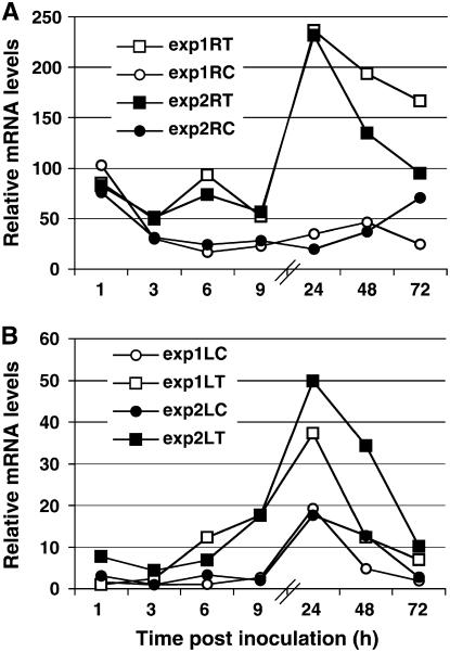
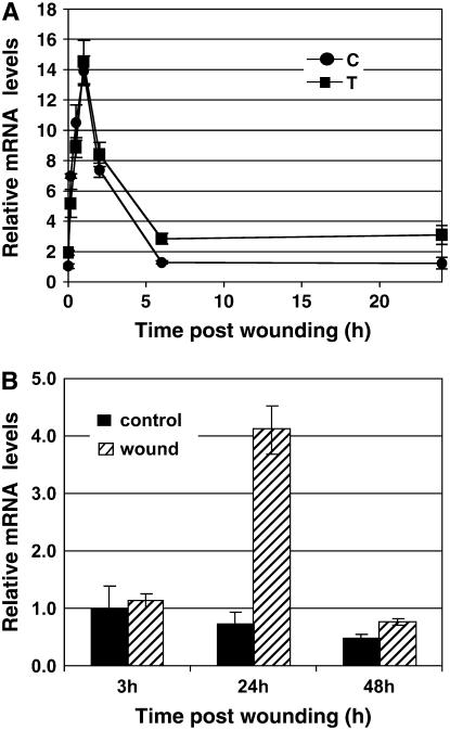
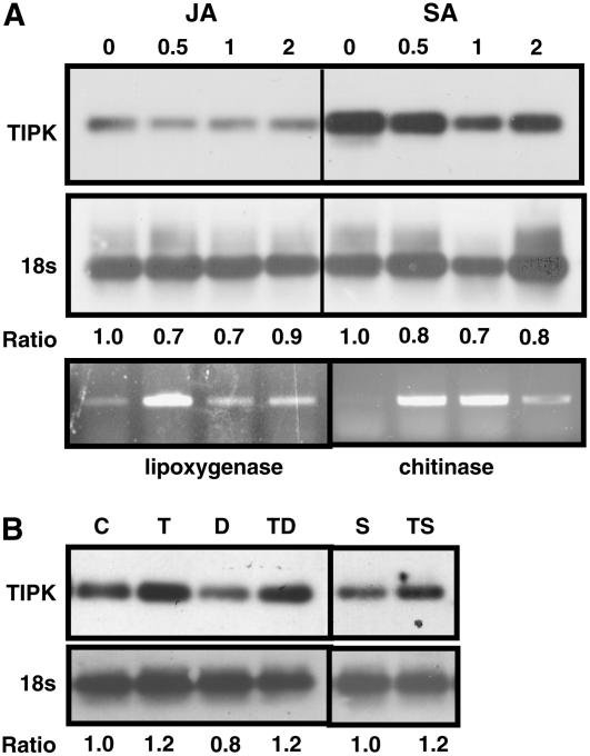
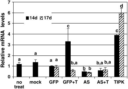
The fungal biocontrol agent Trichoderma asperellum has been recently shown to induce systemic resistance in plants through a mechanism that employs jasmonic acid and ethylene signal transduction pathways. Mitogen-activated protein kinase (MAPK) proteins have been implicated in the signal transduction of a wide variety of plant stress responses. Here we report the identification and characterization of a Trichoderma-induced MAPK (TIPK) gene function in cucumber (Cucumis sativus). Similar to its homologs, wound-induced protein kinase, MPK3, and MPK3a, TIPK is also induced by wounding. Normally, preinoculation of roots with Trichoderma activates plant defense mechanisms, which result in resistance to the leaf pathogen Pseudomonas syringae pv lachrymans. We used a unique attenuated virus vector, Zucchini yellow mosaic virus (ZYMV-AGII), to overexpress TIPK protein and antisense (AS) RNA. Plants overexpressing TIPK were more resistant to pathogenic bacterial attack than control plants, even in the absence of Trichoderma preinoculation. On the other hand, plants expressing TIPK-AS revealed increased sensitivity to pathogen attack. Moreover, Trichoderma preinoculation could not protect these AS plants against subsequent pathogen attack. We therefore demonstrate that Trichoderma exerts its protective effect on plants through activation of the TIPK gene, a MAPK that is involved in signal transduction pathways of defense responses.


Figures

Figure 1.
Figure 1.
Time course of TIPK gene expression. Expression was measured in roots (A) and leaves (B) of cucumber plants after Trichoderma inoculation of the root compartment (time zero) and normalized versus the control gene. Two experiments (white and black symbols) were conducted, each including approximately 20 plants per time point. Relative mRNA levels were determined by real-time PCR (see “Materials and Methods”). The internal sd values for each experiment were smaller than the size of the symbols. ▪ and □, Inoculated with Trichoderma; • and ○, control, mock-inoculated plants.
Figure 2.
Figure 2.
Genomic organization of TIPK gene. A, Schematic illustration of the TIPK gene as deduced from sequence and PCR analyses. B, Southern analysis of total cucumber DNA digested by the indicated restriction enzyme using TIPK cDNA as a probe. EcoRI digested DNA was made in separate membrane to get more clear data (faint bands correspond to residuals of partial digestion).
Figure 3.
Figure 3.
Protein-sequence alignment of the TIPK sequence (CsTIPK) with its homologs from Arabidopsis (AtMPK3; gi21431794), tobacco (NtWIPK; gi18143321), and parsley (PcMPK3a; gi2231034). Fully conserved residues are indicated by black boxes; gray and white colors represent similar and different amino acids, respectively.
Figure 4.
Figure 4.
Time course of TIPK gene expression. A, Expression was measured in wounded leaves of cucumber plants after wounding (time zero), 96 h post Trichoderma inoculation into the root compartment, and normalized versus the control gene. Relative levels of TIPK mRNA were determined by real-time PCR (see “Materials and Methods”). Symbol at each time point represents the average of 10 plants ± se. •, Non-Trichoderma inoculated and wounded; ▪, inoculated with Trichoderma and wounded. B, Expression was measured in the nonwounded upper leaves of wounded plants (hatched boxes) and control nonwounded plants (black boxes). The presented values are means (±se, n = 4).
Figure 5.
Figure 5.
RT-PCR analysis of the TIPK gene and the control gene 18S (A) after JA or SA root treatments at concentrations of 0, 0.5, 1, and 2 mm each, and after DIECA (inhibitor of jasmonate biosynthesis) or STS (inhibitor of ethylene action) root treatments at concentrations of 100 μm and 0.25 mm, respectively (B). PCR was conducted for 20 cycles for the TIPK gene, 25 cycles for lipoxygenase and chitinase genes, and 18 cycles for the 18S gene to keep the amplification within the linear region of the reaction. For each gene, we first normalized the intensity of the band versus its corresponding 18S band. We then calculated the ratio between each hormonal treatment to the control (no hormonal treatment). The ratio, therefore, indicates the expression of each gene as compared to control treatments. C, Control; T, Trichoderma inoculation; D, DIECA treatment; TD, Trichoderma inoculation and DIECA treatment; S, STS treatment; TS, Trichoderma inoculation and STS treatment.
Figure 6.
Figure 6.
Relative expression levels of TIPK postpathogen challenge. Expression was measured in roots (hatched boxes) and leaves (black boxes) of cucumber plants after the following treatments: −T−P, Control (mock inoculations); +T−P, Trichoderma inoculation in the root compartment, at time zero; −T+P, Psl infection of cotyledons at time 48 h; +T+P, Trichoderma inoculation at time zero and Psl infection at time 48 h. Roots and leaves for expression measurements were harvested 48 h after time of Psl infection. These experiments were repeated three times with approximately 15 plants/treatment. The presented values are means of all plants in each treatment (±se).
Figure 7.
Figure 7.
Expression of TIPK gene via ZYMV-AGII. A, Schematic presentation of the AGII genome. AGII noncoding (stippled) and coding (white boxes) regions, and the inserted foreign sequences (TIPK, AS, and GFP) are shown. Arrows indicate NIa protease, involved in proteolysis of the foreign gene products. NIa cleavage sites are indicated by /. Amino acid sequences corresponding to the NIa protease recognition motif are indicated in bold. B, Analysis by RT-PCR of AGII-GFP, AGII-AS, and AGII-TIPK viral RNA accumulation 14 and 17 dpi. Total RNA was extracted from infected and noninfected plants and subjected to RT-PCR with primers flanking the insertion point. The expected sizes of the PCR fragments are: AGII-GFP, 1,180 bp; AGII-AS, 680 bp; and AGII-TIPK, 1,480 bp.
Figure 8.
Figure 8.
Virus localization in leaves. Visualization of GFP fluorescence in leaves of AGII-GFP-infected plants and mock-infected plants (bottom) compared with the leaves (top), 17 d postinfection.
Figure 9.
Figure 9.
Relative expression levels of TIPK in AGII-construct-infected plants. Expression was measured in leaves of cucumber plants 14 d (black boxes) and 17 d (hatched boxes) post-AGII-construct infection. Whenever plants were also treated with Trichoderma, it was done 24 h prior to time point of 14 dpi. The number of plants in each treatment were as follows: no treatment, 14 dpi, six plants; mock inoculation, 14 dpi, 10 plants; AGII-GFP infected (GFP), 14 dpi, 12 plants, and 17 dpi, 20 plants; AGII-GFP infected with Trichoderma inoculation (GFP + T), 14 dpi, six plants and 17 dpi, six plants; AGII-AS infected (AS), 14 dpi, 16 plants and 17 dpi, 22 plants; AGII-AS infected with Trichoderma inoculation (AS + T), 14 dpi, six plants and 17 dpi, six plants; AGII-TIPK infected (TIPK), 14 dpi, six plants and 17 dpi, six plants. The presented values are means (±se). Different letters indicate statistically significant differences between treatments by one-way ANOVA data analysis and Tukey-Kramer for comparing means (α = 0.05).
Figure 10.
Figure 10.
Quantification of Psl multiplication in AGII-construct-treated plants. Cotyledons of 10-d-old cucumbers grown in the hydroponic system were mechanically inoculated with AGII-virus construct and after 13 d inoculated with Trichoderma. Challenge was performed 48 h post Trichoderma inoculation. Leaves were harvested 72 h postchallenge with Psl. The treatments and the number of repeats in each treatment were as follows: AGII-AS challenged with Psl, n = 12; AGII-AS preinoculated with Trichoderma and challenged with Psl, n = 20; AGII-GFP challenged with Psl, n = 23; AGII-GFP preinoculated with Trichoderma and challenged with Psl, n = 26; AGII-TIPK challenged with Psl, n = 18. The presented values are means (±se). Different letters indicate statistically significant differences between treatments by one-way ANOVA data analysis and Tukey-Kramer for comparing means (α = 0.05).

Similar articles

Cited by

References

    1. Abeles FB, Morgan PW, Saltveit ME Jr (1992) Ethylene in Plant Biology, Ed 2. Academic Press, San Diego
    1. Aly R, Mansour F, Abo Much F, Edelstein M, Gal-On A (2005) A novel approach to spider mite control based on expression of sarcotoxin IA peptide via ZYMV-AGII vector in squash plants. Phytoparasitica 33: 177–186
    1. Arazi T, Shiboleth YM, Gal-On A (2001) A nonviral peptide can replace the entire N terminus of zucchini yellow mosaic potyvirus coat protein and permits viral systemic infection. J Virol 75: 6329–6336 - PMC - PubMed
    1. Asai T, Tena G, Plotnikova J, Willmann MR, Chiu WL, Gomez-Gomez L, Boller T, Ausubel FM, Sheen J (2002) MAP kinase signalling cascade in Arabidopsis innate immunity. Nature 415: 977–983 - PubMed
    1. Bögre L, Ligterink W, Meskiene I, Barker PJ, Heberle-Bors E, Huskisson NS, Hirt H (1997) Wounding induces the rapid and transient activation of a specific MAP kinase pathway. Plant Cell 9: 75–83 - PMC - PubMed

Publication types

MeSH terms

Associated data