. 2006 Nov;188(22):7731-41.
doi: 10.1128/JB.00761-06. Epub 2006 Sep 1.

Interaction between coat morphogenetic proteins SafA and SpoVID

Affiliations

Interaction between coat morphogenetic proteins SafA and SpoVID

Teresa Costa et al. J Bacteriol. 2006 Nov.

Abstract

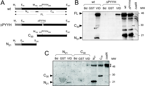
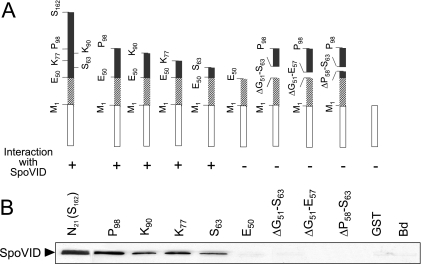
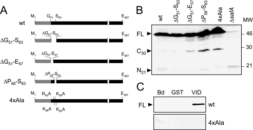
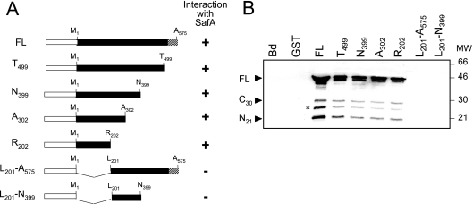
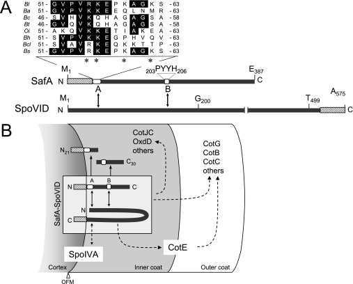
Morphogenetic proteins such as SpoVID and SafA govern assembly of the Bacillus subtilis endospore coat by guiding the various protein structural components to the surface of the developing spore. Previously, a screen for peptides able to interact with SpoVID led to the identification of a PYYH motif present in the C-terminal half of the SafA protein and to the subsequent demonstration that SpoVID and SafA directly interact. spoVID and safA spores show deficiencies in coat assembly and are lysozyme susceptible. Both proteins, orthologs of which are found in all Bacillus species, have LysM domains for peptidoglycan binding and localize to the cortex-coat interface. Here, we show that the interaction between SafA and SpoVID involves the PYYH motif (region B) but also a 13-amino-acid region (region A) just downstream of the N-terminal LysM domain of SafA. We show that deletion of region B does not block the interaction of SafA with SpoVID, nor does it bring about spore susceptibility to lysozyme. Nevertheless, it appears to reduce the interaction and affects the complex. In contrast, lesions in region A impaired the interaction of SafA with SpoVID in vitro and, while not affecting the accumulation of SafA in vivo, interfered with the localization of SafA around the developing spore, causing aberrant assembly of the coat and lysozyme sensitivity. A peptide corresponding to region A interacts with SpoVID, suggesting that residues within this region directly contact SpoVID. Since region A is highly conserved among SafA orthologs, this motif may be an important determinant of coat assembly in the group of Bacillus spore formers.


Figures

FIG. 1.
FIG. 1.
Interaction of the N terminus of SafA with GST-SpoVID. (A) The 387-residue-long wild-type (wt) SafA protein and SafA variants deleted for residues 203 to 206 (ΔPYYH), for the N-terminal first 163 residues (C164-387, corresponding to the SafAC30 form), or missing the last C-terminal 225 residues (N1-162, corresponding to the SafAN21 form). The striped pattern represents the LysM domain (first 50 residues) of SafA. Other relevant residues are also indicated. SafA accumulates in sporulating cells in three main forms: SafAFL (45 kDa), a C-terminal form (SafAC30, 30 kDa), and an N-terminal form (SafAN21, 21 kDa). (B and C) Interaction of immobilized GST-SpoVID with wild-type SafA and SafAΔPYYH (B) and with the SafAC30 and SafAN21 forms (C) present in extracts at hour 4 of sporulation. The extracts were incubated with GST-SpoVID (VID for simplicity), with GST bound to glutathione-Sepharose beads, or with the beads alone (Bd). Pulled-down proteins were resolved by SDS-PAGE and immunoblotted with an anti-SafA antibody. The positions of SafAFL, SafAC30, and SafAN21 (black arrowheads), of cross-reactive species (asterisks), and of molecular mass markers (in kilodaltons) are indicated. The last three lanes in panel C show the immunoblot analysis of the extracts from strains producing SafAN21 or SafAC30, used in the pull-down assays, and of a ΔsafA mutant, with the anti-SafA antibody.
FIG. 2.
FIG. 2.
Deletion mapping of region A in SafA. (A) The various GST-SafA fusions used, with the white box representing GST (not to scale) and the striped pattern (residues 1 to 50) the LysM motif. The plus or minus symbol below each diagram indicates an interacting or noninteracting fusion protein, respectively. (B) Results of pull-down assays using truncated forms of GST-SafA and SpoVID. A cell extract prepared from an E. coli strain overproducing native SpoVID was incubated with the various purified GST-SafA truncated forms containing the following residues, as indicated: 1 to 162 (S162), 1 to 98 (P98), 1 to 90 (K90), 1 to 77 (K77), 1 to 63 (S63), 1 to 50 (E50), 1 to 98 but excluding residues 51 to 63 (ΔG51-S63), 1 to 98 but excluding residues 51 to 57 (ΔG51-E57), and 1 to 98 but excluding residues 51 to 63 (ΔP58-S63). The same extracts were incubated with GST bound to glutathione-Sepharose beads or with beads alone (Bd), as controls. Pulled-down proteins were resolved by SDS-PAGE and immunoblotted with an anti-SpoVID antibody (see Materials and Methods). The position of SpoVID is indicated by the black arrowhead.
FIG. 3.
FIG. 3.
Accumulation of SafA variants in sporulating B. subtilis and analysis of their interaction with GST-SpoVID. (A) The 387-amino-acid wild-type (wt) SafA protein and SafA variants deleted for residues 51 to 63 (ΔG51-S63), 51 to 57 (ΔG51-E57), and 58 to 63 (ΔP58-S63) or with alanine substitutions of residues R55, K56, F62, and K59 (4×Ala). The LysM domain (residues 1 to 50) in SafA is indicated by the striped pattern. (B) Accumulation of SafA variants in cell extracts prepared from cultures of B. subtilis at hour 4 of sporulation. The proteins were resolved by SDS-PAGE and immunoblotted with an anti-SafA antibody. The positions of the full-length (FL), SafAC30, and SafAN21 forms of SafA (black arrowheads), of a cross-reactive species (asterisk), and of molecular mass markers (in kilodaltons) are indicated. (C) Interaction of SafA (wild type) or the SafA4×Ala variant present in extracts from sporulating B. subtilis with GST-SpoVID (VID). Cell extracts prepared from wild-type B. subtilis and from strains producing SafA+ or SafA4×Ala were incubated with GST-SpoVID or GST immobilized on glutathione-Sepharose beads or with the beads alone (Bd), and the pulled-down proteins were resolved by SDS-PAGE and immunoblotted with an anti-SafA antibody (see Materials and Methods). The position of SafAFL is indicated by the black arrowhead.
FIG. 4.
FIG. 4.
SafA interacts with a defined region of SpoVID. (A) Schematic representation of fusions of the 575-residue-long SpoVIDFL protein (FL), as well as forms of SpoVID containing residues 1 to 499 (T499), 1 to 399 (N399), 1 to 302 (A302), 1 to 202 (R201), 201 to 575 (L201-A575), and 201 to 399 (L201- N399), to GST. The white box represents GST (not to scale), and the LysM motif (residues 525 to 575) corresponds to the striped pattern. The plus or minus symbol on the right side of each diagram indicates a protein interacting or not interacting with SafA, respectively. (B) Results of pull-down assays performed with the various purified truncated forms of SpoVID fused to GST and extracts from wild-type B. subtilis AOB90 (Table 1) prepared at hour 4 of sporulation. The immunoblot was probed with an anti-SafA antibody. The same extracts were also incubated with immobilized GST or with glutathione beads alone (Bd). The black arrowheads indicate the positions of the full-length, SafAC30, and SafAN21 forms of SafA, and the asterisk indicates a degradation product of SafA. Molecular mass markers (in kilodaltons) are also indicated.
FIG. 5.
FIG. 5.
Spores of region A mutants have an altered coat and impaired germination. (A) Coomassie-stained gel of SDS-PAGE-resolved coat protein extracts prepared from purified spores of the wild type (wt) (lane 1) and the SafAΔG51-S63 (lane 2), SafAΔG51-E57 (lane 3), SafAΔP58-S63 (lane 4), SafA4×Ala (lane 5), and ΔsafA (lane 6) mutants. Open arrowheads point to bands (a to d) that were excised and subjected to mass spectrometry analysis (see Materials and Methods). The positions of bands corresponding to the 66-kDa form of CotB and to the 32- and 36-kDa forms of CotG are indicated by black arrowheads. (B) Germination responses to AGFK of purified spores of the wild type (closed diamonds) and the SafA4×Ala (closed squares), SafAΔG51-S63 (open squares), SafAΔG51-E57 (closed triangles), SafAΔP58-S63 (crosses), and ΔsafA mutants (open circles). Germination was monitored by following the decrease in absorbance at 580 nm over time (in minutes) and is shown as the percentage of the OD580 at the beginning of the assay.
FIG. 6.
FIG. 6.
Region A is important for the SpoVID-dependent localization of SafA. (A) Cells expressing a functional fusion of SafA to GFP (SafA-GFP) or SafAΔG51-S63-GFP in the wild type (wt) (panels a, c, e, and g) or in a spoVID background (panels b, d, f, and h) were observed by fluorescence microscopy at 2.5 h (panels a to d) and 4 h (panels e to h) after the onset of sporulation in DSM. Overlay images of GFP fluorescence and phase-contrast microscopy are shown in all panels. White arrowheads point to the region showing fluorescence in a selected cell, and the position of the prespore (as determined by DAPI chromosomal staining; not shown) is shown in the schematic representation of the same cell included in each panel. The scale bar represents 1 μm. (B) Quantification of GFP decoration patterns. A minimum of 75 cells were scored for fluorescence patterns designed by spot, septum, or cap at hours 2.5 and 4 in cultures of both the wild type and spoVID mutant expressing either SafA-GFP or SafAΔG51-S63-GFP: AH4102 (SafA-GFP; black bars), AH4103 (SafAΔG51-S63-GFP; striped bars), AH4107 (SafA-GFP spoVID; white bars), and AH4108 (SafAΔG51-S63-GFP spoVID; gray bars).
FIG. 7.
FIG. 7.
Interaction of SafA with SpoVID and role of the complex in spore coat assembly. (A) Structure of SafA and SpoVID, with the LysM motifs (striped pattern) shown in each protein, as well as regions A (residues 51 to 63) and B (PYYH motif) in SafA (white boxes). Also shown is an alignment of the sequence of region A with the following SafA orthologs: Bs, B. subtilis 168 (accession number NP_390662); Bl, B. licheniformis ATCC14580 (YP_080055); Bt, B. thuringiensis serovar konkukian strain 97-27 (YP-038478); Ba, B. anthracis “Ames ancestor” (YP_021306); Bc, B. cereus ATCC 10987 (NP_980805); Bcl, B. clausii KSM-K16 (YP_175046); Bh, B. halodurans C-125 (NP_242087); Oi, Oceanobacillus iheyensis HTE831 (NP_692961). Identical residues are highlighted, and asterisks indicate the alanine substitutions in SafA4×Ala. (B) Model for the localization of SafA and SpoVID. Both SafA and SpoVID localize at the cortex/coat interface, delimited by the outer forespore membrane (OFM). Targeting of SpoVID to this region requires SpoIVA and may involve a direct interaction between the two proteins. SafA is initially targeted to the cortex/coat interface independently of SpoVID, a step that may involve its LysM domain. In a second stage, SafA interacts with SpoVID via regions A and B to form the SpoVID-SafA complex (shown within a box), which allows SafA to encircle the spore. SafA or the SpoVID-SafA complex controls the assembly of a subset of inner and outer coat proteins (i.e., CotE controlled), and SpoVID is essential for maintaining the coat anchored to the cortex, in part because of its requirement for keeping CotE at the inner coat/outer coat interface. Solid arrows indicate a direct interaction, whereas broken arrows indicate direct or indirect interactions.

Similar articles

Cited by

References

    1. Bailey-Smith, K., S. J. Todd, T. W. Southworth, J. Proctor, and A. Moir. 2005. The ExsA protein of Bacillus cereus is required for assembly of coat and exosporium onto the spore surface. J. Bacteriol. 187:3800-3806. - PMC - PubMed
    1. Bateman, A., and M. Bycroft. 2000. The structure of a LysM domain from E. coli membrane-bound lytic murein transglycosylase D (MltD). J. Mol. Biol. 299:1113-1119. - PubMed
    1. Bauer, T., S. Little, A. G. Stöver, and A. Driks. 1999. Functional regions of the Bacillus subtilis spore coat morphogenetic protein CotE. J. Bacteriol. 181:7043-7051. - PMC - PubMed
    1. Beall, B., A. Driks, R. Losick, and C. P. Moran, Jr. 1993. Cloning and characterization of a gene required for assembly of the Bacillus subtilis spore coat. J. Bacteriol. 175:1705-1716. - PMC - PubMed
    1. Birkeland, N. K. 1994. Cloning, molecular characterization, and expression of the genes encoding the lytic functions of lactococcal bacteriophage phi LC3: a dual lysis system of modular design. Can. J. Microbiol. 40:658-665. - PubMed

Publication types

MeSH terms

LinkOut - more resources