. 2006 Nov;5(11):1857-65.
doi: 10.1128/EC.00244-06. Epub 2006 Sep 1.

Glutathione reductase and a mitochondrial thioredoxin play overlapping roles in maintaining iron-sulfur enzymes in fission yeast

Affiliations

Glutathione reductase and a mitochondrial thioredoxin play overlapping roles in maintaining iron-sulfur enzymes in fission yeast

Ji-Yoon Song et al. Eukaryot Cell. 2006 Nov.

Abstract

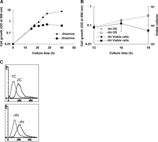
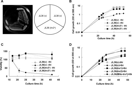
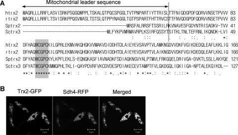
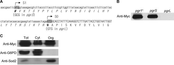
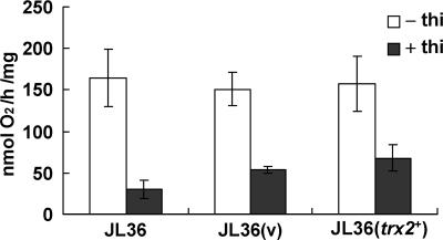
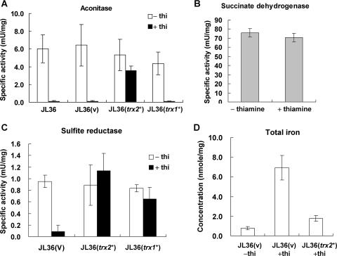
In the fission yeast Schizosaccharomyces pombe, the pgr1+ gene encoding glutathione (GSH) reductase (GR) is essentially required for cell survival. Depletion of GR caused proliferation arrest at the G1 phase of the cell cycle under aerobic conditions. Multicopy suppressors that restore growth were screened, and one effective suppressor was found to be the trx2+ gene, encoding a mitochondrial thioredoxin. This suggests that GR is critically required for some mitochondrial function(s). We found that GR resides in both cytosolic and organellar fractions of the cell. Depletion of GR lowered the respiration rate and the activity of oxidation-labile Fe-S enzymes such as mitochondrial aconitase and cytosolic sulfite reductase. Trx2 did not reverse the high ratio of oxidized glutathione to GSH or the low respiration rate observed in GR-depleted cells. However, it brought the activity of oxidation-labile Fe-S enzymes to a normal level, suggesting that the maintenance of Fe-S enzymes is a critical factor in the survival of S. pombe. The activity of succinate dehydrogenase, an oxidation-insensitive Fe-S enzyme, however, was not affected by GR depletion, suggesting that GR is not required for the biogenesis of the Fe-S cluster. The total iron content was greatly increased by GR depletion and was brought to a nearly normal level by Trx2. These results indicate that the essentiality of GR in the aerobic growth of S. pombe is derived from its role in maintaining oxidation-labile Fe-S enzymes and iron homeostasis.


Figures

FIG. 1.
FIG. 1.
Effect of GR deficiency on the growth, viability, and cell cycle arrest in S. pombe. (A) A seed culture of JL36 (nmt1-pgr1) cells grown overnight was inoculated to an OD595 of 0.01 in EMM in the presence (square) or absence (diamond) of 10 μM thiamine. The growth was monitored at 30°C by measuring the OD595. Growth analyses were carried out at least three times independently for each strain, and a representative growth curve for each condition is shown. (B) At around the time of growth arrest (13, 16, and 19 h postinoculation), cells were diluted and plated onto EMM plates in triplicate to monitor viable cell counts. (C) At the arrested time point (16 h postinoculation), cells were harvested and fixed with ethanol. After removing RNA, the remaining nucleic acid was stained with PI and subjected to fluorescence-activated cell sorter analysis. x and y axes represent fluorescence (DNA content) and cell number, respectively. The upper panel represents patterns for control cells of 1C or 2C DNA content. The lower panel represents results for JL36 cells grown in the absence (−) and presence (+) of thiamine (thi).
FIG. 2.
FIG. 2.
Suppression of GR deficiency by multicopy trx2+. (A) The trx2+ gene was subcloned into pREP1. JL38 cells harboring the control vector (pREP1) (v) and JL36 (nmt1-pgr1) cells harboring the control vector or pREP1-trx2+ were streaked onto an EMM plate containing 10 μM thiamine and incubated for 4 days at 30°C. (B) JL36 cells containing plasmid pREP1 (v) or pREP1-trx2+ (trx2+) were incubated in EMM in the presence (filled marks and solid lines) or absence (open marks and dotted lines) of 10 μM thiamine as described in the legend of Fig. 1. The growth was monitored by measuring the OD595. Growth analyses were carried out at least three times independently for each strain, and the growth curves from a single experiment are shown. (C) Cells were diluted and plated onto EMM plates in triplicate to monitor viable cell counts. Viability was presented as the percentage of viable cells compared to the total cell number calculated from the OD595. (D) To examine the effect of Trx1, JL36 cells containing plasmid pREP1 (v), pREP1-trx1+ (trx1), pREP1-trx2+ (trx2), or pREP1-Mito-trx1 (Mito-trx1) were incubated in EMM in the presence (filled marks and solid lines) or absence (open marks and dotted lines) of 10 μM thiamine as described in the legend of Fig. 1. The growth was monitored by measuring the OD595. Growth analyses were carried out at least three times, and representative growth curves from a single experiment are shown.
FIG. 3.
FIG. 3.
Subcellular localization of Trx2. (A) Amino acid sequences of mitochondrial thioredoxins from human, rat, S. pombe, and Saccharomyces cerevisiae were compared by CLUSTAL W. The positions of identical and similar amino acids are marked with asterisks and dots, respectively. The active site is shaded. The vertical line represents the plausible cleavage site for the mitochondrial leader peptide. (B) Cells containing an integrated copy of the RFP-tagged sdh4+ gene for mitochondrial succinate dehydrogenase were transformed with a pREP42EGFP-C-based recombinant plasmid containing the GFP-tagged trx2+ gene. Cells were grown in EMM to the stationary phase. Fluorescent images were visualized by using an LSM510 MLD confocal microscope (Carl-Zeiss).
FIG. 4.
FIG. 4.
Determination of the translational start site and subcellular localization of GR. (A) The nucleotide sequence around the previously reported translational start site of the pgr1+ gene (S0) is shown with its amino acid sequence. The deduced amino acid sequence translated from another in-frame ATG site (S1) is indicated with italics. The two potential translational start codons were mutated to create pgrS (S1 Met to Ala) and pgrL (S0 Met to Val) variants. (B) The nine-Myc-tagged wild-type pgr1+ ORF and its translation initiation mutants (pgrS and pgrL) were integrated into the chromosome in JL36 cells. The cells were incubated in EMM containing thiamine and harvested at an OD595 of 0.5. Total cell extracts were prepared, and Pgr1-Myc protein was detected by Western blotting. (C) Cell extracts were prepared from the total (Tot), cytosolic (Cyt), and organellar (Org) fractions of cells containing the integrated nine-Myc-tagged wild-type pgr1+ ORF. In each fraction, Pgr1-Myc, G6PD, and Sod2 (mitochondrial Mn-SOD) were detected by Western blotting.
FIG. 5.
FIG. 5.
Respiration rate of GR-deficient cells. JL36 cells with or without pREP1 (v) or pREP1-trx2+ (trx2+) were incubated in EMM in the presence (filled bars) or absence (open bars) of 10 μM thiamine as described in the legend of Fig. 1. Near the point of growth arrest (at an OD595 of about 0.5), cells were harvested, and the respiration rate was measured using an oxygen monitor. Average values with standard deviations from four independent experiments are presented.
FIG. 6.
FIG. 6.
Measurement of iron-sulfur enzymes and total iron. Cells were grown as described in the legend of Fig. 5. (A) Aconitase activity was measured for organellar cell extracts from JL36 cells with or without the parental pREP1 vector (v), the cloned trx2+ gene (trx2+), or the cloned trx1+ gene (trx1+) on pREP1. (B) Succinate dehydrogenase activity was measured for organellar extracts prepared from JL36 cells. (C) Sulfite reductase activity was measured using total cell extracts from JL36 cells containing the parental vector (v), the cloned trx2+ gene (trx2+), or the cloned trx1+ gene (trx1+). (D) Measurement of total iron concentration. Dried cells were prepared from JL36 cells containing either the parental vector (v) or the cloned trx2+ gene (trx2+). Total iron concentration was measured by ICP-AES. Average values with standard deviations from at least three independent experiments are presented.

References

    1. Alfa, C., P. Fantes, J. Hyams, M. Mcleod, and E. Warbrick. 1993. Experiments with fission yeast: a laboratory course manual. Cold Spring Harbor Laboratory Press, Cold Spring Harbor, N.Y.
    1. Beer, S. M., E. R. Taylor, S. E. Brown, C. C. Dahm, N. J. Costa, M. J. Runswick, and M. P. Murphy. 2004. Glutaredoxin 2 catalyzes the reversible oxidation and glutathionylation of mitochondrial membrane thiol proteins: implications for mitochondrial redox regulation and antioxidant defense. J. Biol. Chem. 279:47939-47951. - PubMed
    1. Beinert, H., R. H. Holm, and E. Münck. 1997. Iron-sulfur clusters: nature's modular, multipurpose structures. Science 277:653-659. - PubMed
    1. Beinert, H., and P. J. Kiley. 1999. Fe-S proteins in sensing and regulatory functions. Curr. Opin. Chem. Biol. 3:152-157. - PubMed
    1. Cadenas, E., and K. J. A. Davies. 2000. Mitochondrial free radical generation, oxidative stress, and aging. Free Radic. Biol. Med. 29:222-230. - PubMed

Publication types

MeSH terms

LinkOut - more resources