. 2006 Oct;5(10):1635-47.
doi: 10.1128/EC.00210-06. Epub 2006 Sep 1.

The SH3/PH domain protein AgBoi1/2 collaborates with the Rho-type GTPase AgRho3 to prevent nonpolar growth at hyphal tips of Ashbya gossypii

Affiliations

The SH3/PH domain protein AgBoi1/2 collaborates with the Rho-type GTPase AgRho3 to prevent nonpolar growth at hyphal tips of Ashbya gossypii

Philipp Knechtle et al. Eukaryot Cell. 2006 Oct.

Abstract

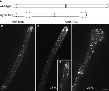
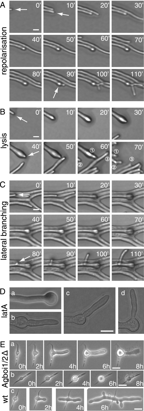
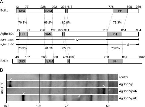
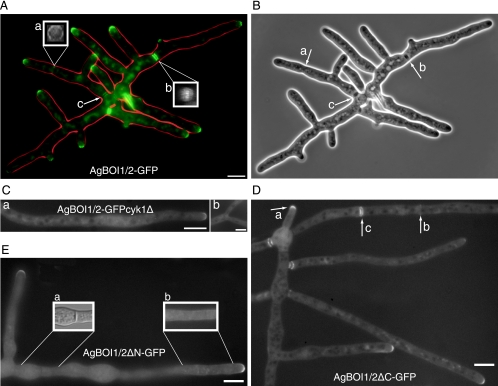
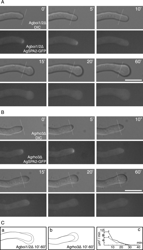
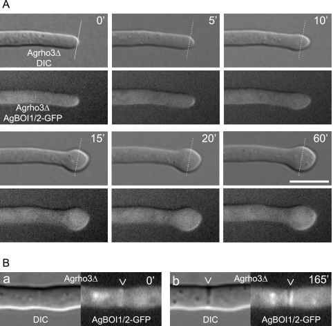
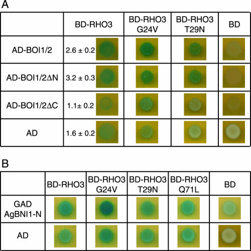
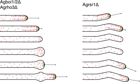
Unlike most other cells, hyphae of filamentous fungi permanently elongate and lack nonpolar growth phases. We identified AgBoi1/2p in the filamentous ascomycete Ashbya gossypii as a component required to prevent nonpolar growth at hyphal tips. Strains lacking AgBoi1/2p frequently show spherical enlargement at hyphal tips with concomitant depolarization of actin patches and loss of tip-located actin cables. These enlarged tips can repolarize and resume hyphal tip extension in the previous polarity axis. AgBoi1/2p permanently localizes to hyphal tips and transiently to sites of septation. Only the tip localization is important for sustained elongation of hyphae. In a yeast two-hybrid experiment, we identified the Rho-type GTPase AgRho3p as an interactor of AgBoi1/2p. AgRho3p is also required to prevent nonpolar growth at hyphal tips, and strains deleted for both AgBOI1/2 and AgRHO3 phenocopied the respective single-deletion strains, demonstrating that AgBoi1/2p and AgRho3p function in a common pathway. Monitoring the polarisome of growing hyphae using AgSpa2p fused to the green fluorescent protein as a marker, we found that polarisome disassembly precedes the onset of nonpolar growth in strains lacking AgBoi1/2p or AgRho3p. AgRho3p locked in its GTP-bound form interacts with the Rho-binding domain of the polarisome-associated formin AgBni1p, implying that AgRho3p has the capacity to directly activate formin-driven actin cable nucleation. We conclude that AgBoi1/2p and AgRho3p support polarisome-mediated actin cable formation at hyphal tips, thereby ensuring permanent polar tip growth.


Figures

FIG. 1.
FIG. 1.
Spherical enlargement of hyphal tips in Agboi1/2Δ. (A) Schematic representation of the tip polarization defect in Agboi1/2Δ. (B to E) Rhodamine-phalloidin stainings of wild-type and Agboi1/2Δ hyphae. Panels B, C, and E represent single hyphae from >20-h cultures, whereas panel D represents a hypha from a <16-h culture. More actin patches accumulate at hyphal tips in mature hyphae than in hyphae at younger stages. As the signal intensity of accumulated actin patches cross-fades the signal from the weaker actin cables, the actin cable network is better seen in younger hyphae. Refer also to Fig. 1A in reference . Bar, 10 μm.
FIG. 2.
FIG. 2.
Spherical enlargement and repolarization of Agboi1/2Δ hyphae (A to D) and polarity establishment in germinating Agboi1/2Δ spores (E). The three sequences of images in panels A, B, and C represent examples of fast-elongating hyphae at the edge of a colony grown on solid full medium for 3 days at 29°C. Images were acquired every 10 min, and the elapsed time is indicated for each frame. (A) Hypha elongating with 120 μm/h until the tip starts enlarging (second frame). After 80 min the hypha continues to grow in the original polarity axis (white arrow) followed by a tip branching within 10 to 15 min. This series of events was observed in 11 of 15 repolarizing hyphae; only the respective time intervals varied. (B) Enlargement of a hypha (second frame), a repolarization in the original polarity axis after 40 min, and tip lysis 20 to 30 min after repolarization. For better orientation, three hyphae of the frames at 60 and 70 min are numbered. Hypha “1” lysed between the two acquisitions and was pushed backwards; only the most apical part can be seen. Hypha “2” formed an apical branch, and hypha “3” was delocalized due to the abrupt lysis of hypha “1”. Two of the 15 repolarizing hyphae lysed and two continued to grow with 100 to 120 μm/h during the observation period. (C) Enlargement at the tip of a fast-growing hypha, which did not repolarize within 2 h but established a new lateral branch subapically (white arrow, frame 80 min). Four of five nonrepolarizing hyphae developed a lateral branch close to the swollen tip, and one lysed during swelling. (D) Treatment of wild-type hyphae with latrunculin A. Mycelia cultured from spores in liquid full medium for 18 h were treated with 20 μM latrunculin A for 20 min, resulting in more than 99% spherically enlarged tips (a). The drug was then washed out, and the mycelia were incubated on solid full medium. After 3 h, 77% of hyphae were repolarized. (b and c) Tips with a repolarization angle between 180° and 145°. (d) Repolarization angle between 145° and 90°. Bar, 10 μM. (E) The two sequences a and b represent examples of germinating Agboi1/2Δ spores at 37°C. (c) A wild-type spore at 37°C. Images were acquired every 2 h, and the elapsed time is indicated for each frame. Germination starts in the middle of the needle-shaped spores (time = 0 h). (a) The germ bubble establishes a polar growth site (2 h) and a germ tube, the first hypha, emerges (4 h). The germ tube enlarges at the tip (6 h) and subsequently lyses (8 h). (b) The germling does not establish a polar growth site, and lysis occurs at the stage of a germ bubble (8 h). The wild type (c) establishes two polar growth sites from the germ bubble in the first 4 h and initiates lateral branches. Bars, 10 μm. wt, wild type. latA, latrunculin A.
FIG. 3.
FIG. 3.
Alignment and expression of AgBoi1/2p. (A) Alignment of A. gossypii AgBoi1/2p with S. cerevisiae Boi1p and Boi2p. Individual domains are marked with boxes, and the identity between corresponding domains is indicated (generated with the application “needle” from “EMBOSS” [43]). The sequences of the partial proteins AgBoi1/2pΔN and AgBoi1/2pΔC are indicated below the AgBoi1/2p sequence, with the thin lines marking the respective deletions. (B) Western blot of wild-type AgBoi1/2p and truncated proteins. Proteins from 16-h cultures were extracted, separated on an SDS-polyacrylamide gel electrophoresis gel, transferred to a membrane, and hybridized with an anti-GFP antibody. Molecular masses from a standard protein marker are indicated in kilodaltons. The molecular mass of the GFP epitope tag with a linker is 28 kDa. A degradation product was detected for AgBoi1/2pΔC at ∼65 kDa.
FIG. 4.
FIG. 4.
Localization of AgBoi1/2p. (A) Localization of wild-type AgBoi1/2p. Localization of the GFP signal is represented in green, and the hyphal periphery is in red. AgBoi1/2p-GFP is detected as a crescent at hyphal tips and as two different structures at sites of septation, either as a single ring at presumptive sites of septation (a) or as a double disc in mature septa (b). The two inserts (a and b) show three-dimensional reconstructed views of the indicated septal localizations. For each insert, a series of images was acquired along the z axis, and the planes were 90° rotated along the vertical axis. This gives a view along the hyphal tube onto the septum. (c) In mature septa, AgBoi1/2-GFP localization diminishes. (B) Phase-contrast image of that shown in panel A. Presumptive sites of septation identified from AgBoi1/2p-GFP ring localization are not yet visible (a), and mature septa identified from AgBoi1/2p-GFP disc localization are visible as black structures spanning the hyphae (b and c). (C) Localization of AgBoi1/2p-GFP in an Agcyk1Δ background. Septa are absent in the Agcyk1Δ strain, and AgBoi1/2p-GFP signal was observed neither in subapical regions (a) nor at sites of apical branching (b) where they are found in wild type. Bar, 10 μm. (D) Localization of AgBoi1/2pΔC. AgBoi1/2pΔC-GFP localizes at hyphal tips (a) and at septa as single-ring (b) or double-disc (c) structures as seen for the wild type. (E) Localization of AgBoi1/2pΔN. AgBoi1/2pΔN-GFP localizes only to hyphal tips and not to septa. (a) Nomarski illumination of the hyphal segment below. In the middle a septum is clearly visible but a GFP signal is lacking at the corresponding site. The GFP signal also appears to localize weakly over the entire cortex, as seen on insert b, which represents a brighter scaling of a single plane from the hyphal segment below. Bar, 10 μm.
FIG. 5.
FIG. 5.
AgSpa2p-GFP localization during spherical enlargement in Agboi1/2Δ and Agrho3Δ. (A and B) The respective top images represents the Nomarski acquisitions, the bottom images the fluorescence acquisitions; the time is indicated in minutes. AgSpa2p-GFP localizes to hyphal tips during polarized growth phases, as indicated in the two first acquisitions (t = 0; t = 5). Lines in the respective frame indicate the tip extension during the first 5 min. AgSpa2p-GFP is lost while the tip is not yet enlarged (t = 10). Enlargement starts in the following frame (t = 15) and lasts for 45 min (t = 20 to 60). Due to different growth media, temperatures, and developmental stages, the extension rate is decreased compared to mature hyphae of the wild type on full medium at 30°C. The entire observation lasted for more than 120 min. Bar, 10 μm. (C) The hyphal cortex prior to enlargement at 10 min and fully enlarged at 60 min for Agboi1/2Δ (a) and Agrho3Δ (b), respectively. (c) The time-dependent decrease of surface expansion inAgboi1/2Δ and Agrho3Δ is indicated. The time corresponds to that for panels A and B; an asterisk indicates the last frame with polarized AgSpa2p.
FIG. 6.
FIG. 6.
AgBoi1/2p-GFP localization in Agrho3Δ. (A) The respective top images represent the Nomarski acquisitions, the bottom images the fluorescence acquisitions; the time is indicated in minutes. AgBoi1/2p-GFP localizes to hyphal tips during polarized growth phases as indicated in the two first acquisitions (t = 0; t = 5). When the hyphal tip starts to enlarge, AgBoi1/2p-GFP is distributed over the enlarging cortex at the hyphal tip. (B) Localization of AgBoi1/2p-GFP in Agrho3Δ at septal sites. AgBoi1/2p-GFP is established as a ring (a) and develops to a double-disc structure (b). The presumptive site of septation identified from AgBoi1/2p-GFP ring localization is not yet visible in Nomarski (a), whereas mature septa identified from AgBoi1/2p-GFP disc localization become visible as black structures spanning the hyphae (b).
FIG. 7.
FIG. 7.
AgBoi1/2p-AgRho3p-AgBni1p two-hybrid interactions. (A) AgBoi1/2p-AgRho3p interaction. In the left column, the (truncated) AgBoi1/2p fused to the activation domain and the activation domain alone are listed, and in the top row the AgRho3p fused to the binding domain and the binding domain alone are listed. (B) AgRho3p-AgBni1p interaction. In the left column the AgBni1 protein fused to the activation domain (47) and the activation domain alone are listed. In the top row, the AgRho3p fused to the binding domain and the binding domain alone are listed. In each image 5 μl of a yeast culture of strain PJ69-4a with an optical density at 600 nm of 0.1 and transformed with the corresponding plasmids encoding the fusions proteins was spotted, incubated overnight, overlaid with X-Gal, and incubated for 16 to 24 h. Blue color indicates interaction. The quantitative determination of the β-galactosidase activity is given in arbitrary units. Error is the standard error of the mean. AD, activation domain; BD, binding domain.
FIG. 8.
FIG. 8.
Schematic comparison of polarisome loss in Agrsr1Δ, Agboi1/2Δ, and Agrho3Δ strains. The contours of hyphae are depicted in black, the polarisome is depicted in green, actin cables are depicted as red lines, and actin patches are depicted as red dots. Arrows indicate the growth speed of hyphae. The developmental pattern is given in descending order. The initial pattern with the first three hyphae is comparable between Agrsr1Δ and Agboi1/2Δ/Agrho3Δ. In Agboi1/2Δ/Agrho3Δ the hyphal tip enlarges, which is not observed for Agrsr1Δ. Repolarization in Agboi1/2Δ and Agrho3Δ maintains the axis of growth, whereas in Agrsr1Δ resumption of growth occurred in a frequently uncoordinated manner to the previous axis of polarity.

Similar articles

Cited by

References

    1. Adamo, J. E., G. Rossi, and P. Brennwald. 1999. The Rho GTPase Rho3 has a direct role in exocytosis that is distinct from its role in actin polarity. Mol. Biol. Cell 10:4121-4133. - PMC - PubMed
    1. Adams, A. 1998. Methods in yeast genetics: a Cold Spring Harbor Laboratory course manual. Cold Spring Harbor Laboratory Press, Cold Spring Harbor, N.Y.
    1. Akada, R., J. Yamamoto, and I. Yamashita. 1997. Screening and identification of yeast sequences that cause growth inhibition when overexpressed. Mol. Gen. Genet. 254:267-274. - PubMed
    1. Altmann-Johl, R., and P. Philippsen. 1996. AgTHR4, a new selection marker for transformation of the filamentous fungus Ashbya gossypii, maps in a four-gene cluster that is conserved between A. gossypii and Saccharomyces cerevisiae. Mol. Gen. Genet. 250:69-80. - PubMed
    1. Ashby, S. F., and W. Nowell. 1926. The fungi of stigmatomycosis. Ann. Botany 40:69-84.

Publication types

LinkOut - more resources