. 2006 Sep 1;20(17):2450-64.
doi: 10.1101/gad.382806.

A novel genetic hierarchy functions during hypaxial myogenesis: Pax3 directly activates Myf5 in muscle progenitor cells in the limb

Affiliations

A novel genetic hierarchy functions during hypaxial myogenesis: Pax3 directly activates Myf5 in muscle progenitor cells in the limb

Lola Bajard et al. Genes Dev. .

Abstract

We address the molecular control of myogenesis in progenitor cells derived from the hypaxial somite. Null mutations in Pax3, a key regulator of skeletal muscle formation, lead to cell death in this domain. We have developed a novel allele of Pax3 encoding a Pax3-engrailed fusion protein that acts as a transcriptional repressor. Heterozygote mouse embryos have an attenuated mutant phenotype, with partial conservation of the hypaxial somite and its myogenic derivatives, including some hindlimb muscles. At these sites, expression of Myf5 is compromised, showing that Pax3 acts genetically upstream of this myogenic determination gene. We have characterized a 145-base-pair (bp) regulatory element, at -57.5 kb from Myf5, that directs transgene expression to the mature somite, notably to myogenic cells of the hypaxial domain that form ventral trunk and limb muscles. A Pax3 consensus site in this sequence binds Pax3 in vitro and in vivo. Multimers of the 145-bp sequence direct transgene expression to sites of Pax3 function, and an assay of its activity in the chick embryo shows Pax3 dependence. Mutation of the Pax3 site abolishes all expression controlled by the 145-bp sequence in transgenic mouse embryos. We conclude that Pax3 directly regulates Myf5 in the hypaxial somite and its derivatives.


Figures

Figure 1.
Figure 1.
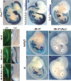
The Pax3Pax3–En-IRESnlacZ allele: strategy and phenotypes. (A) General strategy of Pax3–En-IRESnlacZ targeting into the Pax3 locus. (Pax3–En) cDNA sequence encoding the Pax3–Engrailed fusion protein; (IRES) internal ribosome entry site; (nlacZ) reporter encoding β-galactosidase (β-gal) with a nuclear localization sequence (n); (GFP) green fluorescent protein; (Puro pA) puromycin selection cassette; (loxP) target sites for Cre recombinase; (FRT) target for Flip recombinase. (BG′) X-gal staining of Pax3nlacZ /+ control embryos (B,E,E′), Pax3 loss-of-function mutants, Pax3nlacZ/Sp embryos, where Sp stands for the natural Splotch mutation in Pax3 (C,F,F′), Pax3Pax3–En-IRESnlacZ /+ (Pax3–En/+) embryos where one allele encodes the dominant-negative factor Pax3–En (D,G,G′) at E11.5 (B–D) and forelimbs (outlined) (E–G) and hindlimbs (E′–G′) at E12.5. (B–D) Black arrowheads indicate the hypoglossal cord; white arrows show the normal localization of hindlimb muscle masses. (H–J) View of the thoracic somites, stained by X-gal, of Pax3nlacZ /+ (H), Pax3nlacZ/nlacZ (I), and Pax3Pax3–En-IRESnlacZ /+ (J) embryos at E10.5. (K–M) Coimmunohistochemistry on transverse sections (level of section boxed in HJ) of thoracic somites showing the hypaxial region of the dermomyotome Pax3nlacZ /+ (K), Pax3nlacZ/nlacZ (L), and Pax3Pax3–En-IRESnlacZ /+ (M) embryos at E10.5, using antibodies against β-gal (Pax3, red) and activated caspase 3, which marks dying cells (green). Nuclei are shown by DAPI staining (blue).
Figure 2.
Figure 2.
Impaired hypaxial Myf5 expression in the presence of Pax3–En. (A,B) X-gal staining of Pax3nlacZ /+ (A) and Pax3Pax3–En-IRESnlacZ /+ (B) embryos at E9.5. (C,D) Whole-mount in situ hybridization using a Myf5 antisense probe on Pax3nlacZ /+(C) and Pax3Pax3–En-IRESnlacZ /+ (D) embryos at E9.5. The black arrows point to the hypaxial somite. (E,F) X-gal staining of thoracic somites at E10.5 of Pax3nlacZ /+ (E) and Pax3Pax3–En-IRESnlacZ /+ (F) embryos. (G–N) Whole-mount in situ hybridization of thoracic somites of Pax3nlacZ /+ (G–M) or Pax3Pax3–En-IRESnlacZ /+ (H–N) embryos at E10.5 using riboprobes recognizing Myf5 (G,H), Mrf4 (I,J), MyoD (K,L), or Sim1 (M,N) transcripts. (O,P) Whole-mount in situ hybridization at forelimb level of Pax3 nlacZ/+ (O) or Pax3Pax3–En-IRESnlacZ /+(P) embryos at E11.5, hybridized with a riboprobe for c-met transcripts. (Q–S) Coimmunohistochemistry on transverse sections of thoracic somites from Pax3nlacZ/+ (Q), Pax3nlacZ/nlacZ (R), or Pax3Pax3–En-IRESnlacZ/+ (S) embryos at E10.5, using antibodies recognizing Myf5 (green) and β-gal (red).
Figure 3.
Figure 3.
Expression of Myf5 in the limb depends on Pax3. (A–J) Coimmunohistochemistry on transverse sections of hind-limbs from wild-type (WT) (A–G) or Pax3Pax3–En-IRESnlacZ/+ (H–J) embryos at E11.5, using antibodies recognizing Pax3 (red) (B,D,F,G,I,J) or Myf5 (green) (C,D,E,G,H,J). DAPI stainings are shown in A, E, and H. EJ are close-ups of the dorsal muscle masses. (K) Quantification of the percentage of Pax3-positive (Pax3+) cells coexpressing Myf5 in hindlimbs from wild-type (WT) or Pax3–En-expressing embryos at E11.5. (L,M) Dorsal views of X-gal-stained hindlimbs of Pax3nlacZ/+ (L) and Pax3Pax3–En-IRESnlacZ/+ (M) embryos at E11.5. (N–Q) Dorsal views of hindlimbs from Pax3nlacZ/+ (L,N,P) or Pax3Pax3–En-IRESnlacZ/+ (M,O,Q) embryos at E11.5, stained by whole-mount in situ hybridization using riboprobes recognizing Myf5 (N,O) or MyoD (P,Q) transcripts.
Figure 4.
Figure 4.
A 145-bp element, located at −57.5 kb upstream of Myf5, directs expression in somites, trunk, and limb muscles. (A) Schematic representation of the 145 baMyf5nlacZ transgene. A 145-bp element located at –57.5 kb from the Myf5 gene was cloned upstream of a 3-kb fragment that includes the Myf5 promoter and a branchial arch (ba) element as an internal control (not drawn to scale). (BE,G,H) Whole-mount X-gal staining of 145 baMyf5nlacZ transgenic embryos at E10.5 (B), E11.5 (C,G), E12.5 (D), E13.5 (H), and E15.5 (E). (I) Whole-mount X-gal staining of a control embryo at E13.5 expressing a y240Myf5nlacZ transgene that recapitulates the complete Myf5 expression pattern. (F) Section of a thoracic somite from a transgenic embryo at E11.5, showing labeled myogenic progenitor cells in the epithelial structure of the hypaxial dermomyotome (also inset), as well as scattered β-gal+ cells, in the myotome (bracket). White arrowheads indicate the hypaxial domain of the somite (C,G) or one of its derivatives: the rectus abdominis muscle (D,E,H,I). Black arrowheads point to distal limb muscles, present in the control embryo (I), but lacking in D, E, and H. The gray arrowheads in B, C, and G point to the hypoglossal cord. Labeling in the neural tube (B) as well as the branchial arches is due to the Myf5 promoter region (Summerbell et al. 2000), present in the transgenes. In the transgenic line, ectopic expression in dorsal root ganglia is also seen. (ba) Branchial arches; (fl) forelimb; (hl) hindlimb. B–E are from a transgenic mouse line; G and H are transitory Fo transgenic embryos (see Table 1 for numbers); I is from the y240Myf5nlacZ mouse line (Hadchouel et al. 2000).
Figure 5.
Figure 5.
The 145-bp sequence contains a putative Pax3-binding site to which Pax3 binds in vitro and in vivo. (A) The 145-bp sequence in the mouse genome and comparison with a homologous region of the human and chick genomes, with nonconserved bases in gray. There is a high degree of conservation with both human (95.2%) and chick (83.5%) sequences. A putative Pax3-binding site is framed. (B) Comparison of the putative Pax3-binding site with the consensus described by Epstein et al. (1996). Bases mutated in ΔPax3 are indicated in bold type. (C) Autoradiography of an EMSA experiment showing the binding of a His-tagged Pax3 protein to a radio-labeled DNA probe of 27 bp including the Pax3 site from the 145-bp sequence (1), with increasing doses of unlabeled probe (24), or a probe with the Pax3 site mutated (ΔPax3) (57) or with an antibody to Pax3 (8) or to Pax7 (9). The bands shown in brackets probably represent interactions between the probe and incomplete Pax3 translation products that would contain the 5′ sequence with the DNA-binding domain but not the 3′ sequence that is recognized by the antibody. (D) ChIP analysis of the binding of Pax3 to the 145-bp sequence in vivo was performed with chromatin prepared from either wild-type or P34 transgenic embryos (without head and internal organs) at E11.5. Histograms indicate the fold enrichment with the 145-bp sequence (145), compared with an unrelated control sequence, X3 (Navarro et al. 2005), after ChIP with a Pax3 antibody. P34 embryos, which express a transgene regulated by Pax3-binding sites (Relaix et al. 2004), provide a positive control (P34). Results are the average of three PCR assays per sample, in two independent experiments. (D′) Example of a ChIP experiment showing the binding of Pax3 (black) or of a control NG2 antibody (gray) expressed as the percent of DNA immunoprecipitated, using primers for the 145-bp sequence (145), for control sequences at 200 kb 5′ (5′) or at 2.3 kb 3′ (3′) from it, and for the X3 sequence.
Figure 6.
Figure 6.
The 145-bp element responds to Pax3 through a site that is essential for activity in vivo. (A–C) X-gal staining at E11.5 of a Pax3nlacZ/+ embryo (A), or transgenic embryos with either (145)6 tknlacZ (B) or (145)2 tknlacZ transgenes (C). (D–I) Electroporation experiments in the chick neural tube. D, F, and H are in ovo views of GFP expression driven by pCIG control (D) or pCIG-Pax3 (F,H) plasmids that encode this reporter, 24 h after electroporation. E, G, and I show X-gal staining of the embryos shown in D, F, and H, which have been coelectroporated with the −58/−57 baMyf5nlacZ transgene (−58/−57) (E,G) or a −58/−57ΔPax3 baMyf5nlacZ transgene (−58/ −57 ΔPax3), in which the Pax3 site in the 145-bp sequence has been mutated (I). (J–M) X-gal staining of transgenic mouse embryos at E11.5 expressing −58/−57 baMyf5nlacZ (J), −58/−57ΔPax3 baMyf5nlacZ (K), −58/−57 tknlacZ (L), or −58/−57ΔPax3 tknlacZ (M) transgenes. Embryos shown in B, C, JM are transitory transgenics. (ba) Branchial arch derivatives; (DRG) dorsal root ganglia; (fl) forelimb; (hl) hindlimb; (hc) hypoglossal cord; (NT) neural tube; (TG) trigeminal ganglia. (See Tables 1 and 2 and Material and Methods for numbers of transgenic and electroporated embryos, respectively.)

References

    1. Ben-Yair R., Kalcheim C. Lineage analysis of the avian dermomyotome sheet reveals the existence of single cells with both dermal and muscle progenitor fates. Development. 2005;132:689–701. - PubMed
    1. Bladt F., Riethmacher D., Isenmann S., Aguzzi A., Birchmeier C. Essential role for the c-met receptor in the migration of myogenic precursor cells into the limb bud. Nature. 1995;376:768–771. - PubMed
    1. Borycki A.G., Li J., Jin F., Emerson C.P., Epstein J.A. Pax3 functions in cell survival and in pax7 regulation. Development. 1999;126:1665–1674. - PubMed
    1. Braun T., Rudnicki M.A., Arnold H.H., Jaenisch R. Targeted inactivation of the muscle regulatory gene Myf-5 results in abnormal rib development and perinatal death. Cell. 1992;71:369–382. - PubMed
    1. Brohmann H., Jagla K., Birchmeier C. The role of Lbx1 in migration of muscle precursor cells. Development. 2000;127:437–445. - PubMed

Publication types

MeSH terms