. 2006 Sep 1;20(17):2465-78.
doi: 10.1101/gad.381806.

The zinc-finger factor Insm1 (IA-1) is essential for the development of pancreatic beta cells and intestinal endocrine cells

Affiliations

The zinc-finger factor Insm1 (IA-1) is essential for the development of pancreatic beta cells and intestinal endocrine cells

Mathias S Gierl et al. Genes Dev. .

Abstract

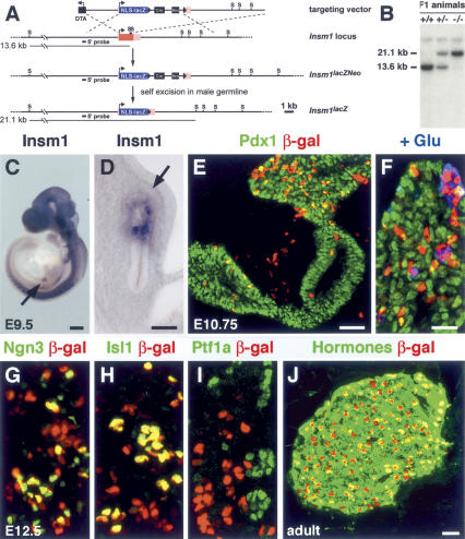
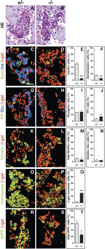
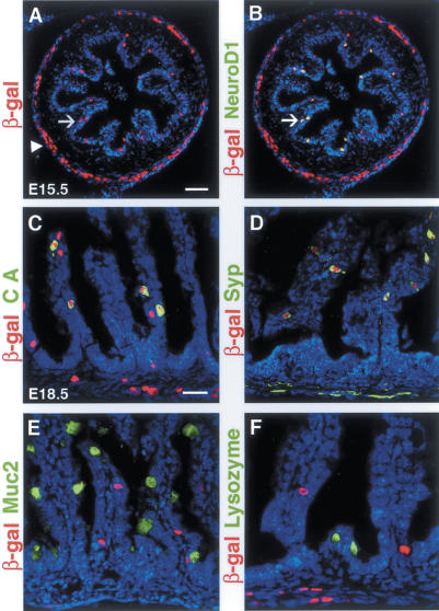
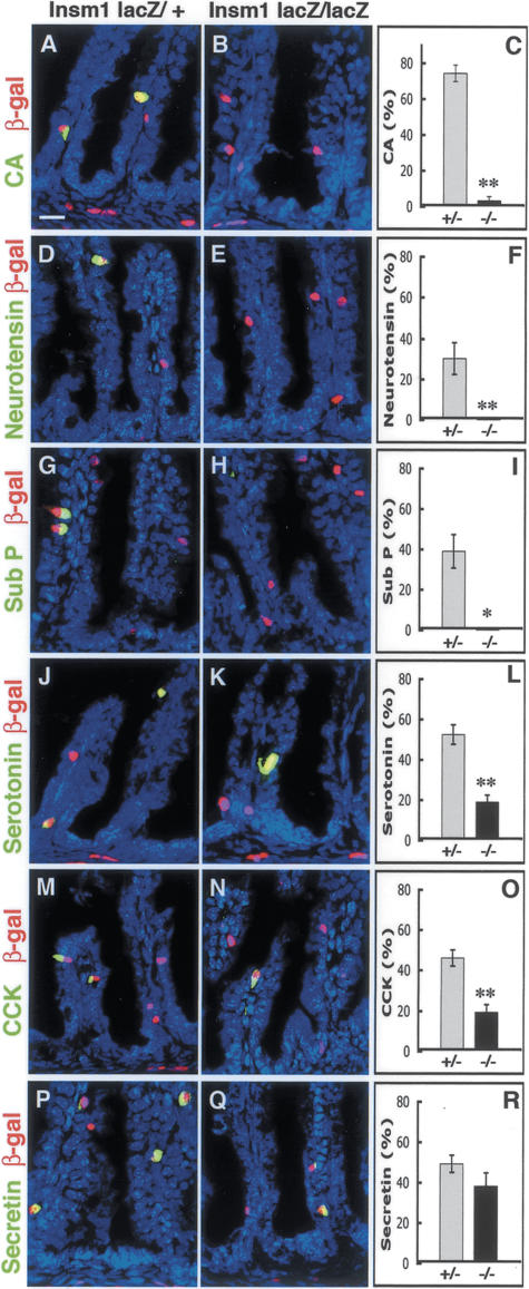
The pancreatic and intestinal primordia contain epithelial progenitor cells that generate many cell types. During development, specific programs of gene expression restrict the developmental potential of such progenitors and promote their differentiation. The Insm1 (insulinoma-associated 1, IA-1) gene encodes a Zinc-finger factor that was discovered in an insulinoma cDNA library. We show that pancreatic and intestinal endocrine cells express Insm1 and require Insm1 for their development. In the pancreas of Insm1 mutant mice, endocrine precursors are formed, but only few insulin-positive beta cells are generated. Instead, endocrine precursor cells accumulate that express none of the pancreatic hormones. A similar change is observed in the development of intestine, where endocrine precursor cells are formed but do not differentiate correctly. A hallmark of endocrine cell differentiation is the accumulation of proteins that participate in secretion and vesicle transport, and we find many of the corresponding genes to be down-regulated in Insm1 mutant mice. Insm1 thus controls a gene expression program that comprises hormones and proteins of the secretory machinery. Our genetic analysis has revealed a key role of Insm1 in differentiation of pancreatic and intestinal endocrine cells.


Figures

Figure 1.
Figure 1.
Generation of the Insm1lacZ mouse strain and Insm1 expression in the pancreas. (A) Schematic representation of the targeting vector, the wild-type Insm1 locus, and the mutated Insm1 allele, before and after removal of the self-excision neomycin (cre, neo) cassette. Coding (red) and noncoding (pink) Insm1 sequences, NLS-lacZ (blue), DTA, the self-excision neomycin cassette, loxP (arrowhead), and StuI (S) restriction sites are depicted. Black lines indicate the predicted fragment sizes obtained after StuI digestion of genomic DNA. A black bar shows the 5′ sequence used as a probe for Southern analyses shown in B. (B) Southern blot analyses of StuI-digested genomic DNA from wild-type, Insm1lacZ/+, and Insm1lacZ/Insm1lacZ mice. (C,D) In situ hybridization of E9.5 mouse embryos using an Insm1-specific probe; Insm1 is expressed in the dorsal pancreatic bud (arrows) and the developing central and peripheral nervous system. (E,F) Immunohistological analysis of the developing pancreas of Insm1lacZ/+ embryos at E10.75 using antibodies against β-galactosidase (red) and Pdx1 (green) (E), and β-galactosidase (red), Pdx1 (green), and glucagon (blue) (F). (G–I) Immunohisto-logical analysis of the dorsal pancreas of Insm1lacZ/+ embryos at E12.5 using antibodies against β-galactosidase (red), and Ngn3 (green) (G), Isl1 (green) (H), and Ptf1a (green) (I). Coexpression of β-galactosidase with proteins that mark endocrine cells (Isl1, Ngn3, glucagon) is observed; in contrast, Ptf1a is present in exocrine cells and is not coexpressed with β-galactosidase. (J) Immunohistological analysis of the pancreas of adult Insm1lacZ/+ mice using antibodies against β-galactosidase (red) and a mixture of antibodies directed against insulin, glucagon, PP, somatostatin, and ghrelin (green). Bars: C, 400 μm; D, 50 μm; E,F,J, 20 μm.
Figure 2.
Figure 2.
Impaired differentiation of α and β cells in the pancreas of Insm1 mutant mice. Immunohistological analysis of the developing dorsal pancreas of Insm1lacZ/+ and Insm1lacZ/Insm1lacZ mice at E12.5 (A–C,G–I) and E15.5 (D–F,J–L); the genotypes are indicated by +/− and −/−, respectively. The antibodies used were directed against β-galactosidase (red) and insulin (green) (A,B,D,E), and β-galactosidase (red) and glucagon (green) (G,H,J,K). (C,I) Proportions of β-galactosidase+ cells that express insulin (C) and glucagon (I) in the dorsal pancreas in Insm1lacZ/+ (gray column) and in Insm1lacZ/Insm1lacZ (black column) mice at E12.5. (F,L) Proportions of β-galactosidase+ cells that express insulin (F) or glucagon (L) in the dorsal (D) and ventral (V) pancreas of Insm1lacZ/+ and Insm1lacZ/ Insm1lacZ mice at E15.5. Asterisks indicate p-values of <0.01. Bar, 20 μm.
Figure 3.
Figure 3.
Changed expression of transcription factors in the pancreas of Insm1 mutant mice. Immunohistological analysis of transcription factors in the developing dorsal pancreas of Insm1lacZ/+ and Insm1lacZ/Insm1lacZ mice at E15.5. The following antibodies were used: (A–F) Pdx1 (green), β-galactosidase (red), and insulin (blue). (G,H) MafA (green) and β-galactosidase (red). (I–N) MafB (green), β-galactosidase (red), and glucagon (blue). (O,P) Ptf1a (green) and β-galactosidase (red). Bar, 20 μm.
Figure 4.
Figure 4.
Islet histology and hormone expression in the pancreas of Insm1 mutant mice. Histological and immunohisto-logical analyses of the developing pancreas of Insm1lacZ/+ and Insm1lacZ/Insm1lacZ mice at E18.5; the genotypes are indicated by +/− and −/−, respectively. (A,B) Hematoxylin/eosin (HE) staining of semi-thin sections of the pancreas. Islets are present in Insm1lacZ/Insm1lacZ mice but display a changed nuclear density. Immunohistological analysis of the pancreas using antibodies directed against β-galactosidase (shown in red). In addition, antibodies directed against insulin (blue) and somatostatin (green) (C,D), glucagon (blue) and PP (green) (G,H), and ghrelin (green) (K,L) were used. (O,P) Immunohistology using rabbit anti-insulin, rabbit anti-glucagon, rabbit anti-somatostatin, rabbit anti-PP, and rabbit anti-ghrelin (green), and goat anti-β-galactosidase (red) antibodies simultaneously. Note that many β-galactosidase+ cells exist in the pancreas of Insm1lacZ/ Insm1lacZ embryos that do not express any of these five hormones. (R,S) Immunohistological analysis of the pancreas using antibodies against β-galactosidase (shown in red) and IAPP (green). The proportions of the β-galactosidase+ cells that express insulin (E), somatostatin (F), glucagon (I), PP (J), ghrelin (M), ghrelin but not glucagon (N), any of the five hormones (Q), or IAPP (T) are displayed. Single and double asterisks indicate p-values of <0.01 and <0.001, respectively. Bars, 20 μm.
Figure 5.
Figure 5.
Insm1 expression in the developing intestine. Immunohistological analysis of the developing intestine of Insm1lacZ/+ mice at E15.5 (A,B) and E18.5 (C–F). (A) β-Galactosidase (red) was observed in the intestinal epithelium (arrow) and the enteric nervous system (arrowhead) of Insm1lacZ/+ mice. (B) β-Galactosidase (red) is coexpressed with NeuroD1 (green) in entero-endocrine cells located in the intestinal epithelium (arrows), but not in the enteric nervous system (arrow-head); coexpressing cells appear yellow, an overlap of the red and green fluorescence. In the intestinal epithelium, all cells that contained chromogranin A (CA, green) (C) or synaptophysin (Syp, green) (D) coexpressed β-galactosidase. In contrast, Mucin2 (Muc2, green) (E) or lysozyme (green) (F) proteins were not observed in the β-galactosidase+ (red) cells. Sections were counterstained with TOTO-3 (blue). Bars: A, 50 μm; C, 20 μm.
Figure 6.
Figure 6.
Insm1 is essential for the differentiation of enteroendocrine cells. Immunohistological analysis of the intestine of Insm1lacZ/+ and Insm1lacZ/Insm1lacZ mice at E18.5, using antibodies directed against chromogranin A (A,B), neurotensin (D,E), substance P (G,H), serotonin (J,K), CCK (M,N), and secretin (P,Q) (all shown in green); in addition, antibodies against β-galactosidase (shown in red) were used. The proportion of β-galactosidase+ cells that express chromogranin A (C), neurotensin (F), substance P (I), serotonin (L), CCK (O), and secretin (R) are displayed. Sections were counterstained with TOTO-3 (blue). Single and double asterisks indicate p-values of <0.005 and <0.0001, respectively. Bar, 20 μm.

References

    1. Ahlgren U., Jonsson J., Jonsson L., Simu K., Edlund H. β-Cell-specific inactivation of the mouse Ipf1/Pdx1 gene results in loss of the β-cell phenotype and maturity onset diabetes. Genes & Dev. 1998;12:1763–1768. - PMC - PubMed
    1. Apelqvist A., Li H., Sommer L., Beatus P., Anderson D.J., Honjo T., Hrabe de Angelis M., Lendahl U., Edlund H. Notch signalling controls pancreatic cell differentiation. Nature. 1999;400:877–881. - PubMed
    1. Artner I., Le Lay J., Hang Y., Elghazi L., Schisler J.C., Henderson E., Sosa-Pineda B., Stein R. MafB: An activator of the glucagon gene expressed in developing islet α-and β-cells. Diabetes. 2006;55:297–304. - PubMed
    1. Bell G.I., Polonsky K.S. Diabetes mellitus and genetically programmed defects in β-cell function. Nature. 2001;414:788–791. - PubMed
    1. Breslin M.B., Zhu M., Notkins A.L., Lan M.S. Neuroendocrine differentiation factor, IA-1, is a transcriptional repressor and contains a specific DNA-binding domain: Identification of consensus IA-1 binding sequence. Nucleic Acids Res. 2002;30:1038–1045. - PMC - PubMed

MeSH terms