. 2006 Sep;188(18):6580-91.
doi: 10.1128/JB.00771-06.

Identification of an essential gene of Listeria monocytogenes involved in teichoic acid biogenesis

Affiliations

Identification of an essential gene of Listeria monocytogenes involved in teichoic acid biogenesis

Iharilalao Dubail et al. J Bacteriol. 2006 Sep.

Abstract

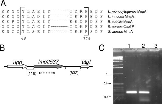
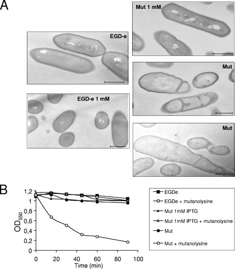
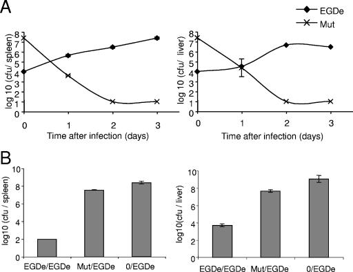
Listeria monocytogenes is a facultative intracellular gram-positive bacterium responsible for severe opportunistic infections in humans and animals. We had previously identified a gene encoding a putative UDP-N-acetylglucosamine 2-epimerase, a precursor of the teichoic acid linkage unit, in the genome of L monocytogenes strain EGD-e. This gene, now designated lmo2537, encodes a protein that shares 62% identity with the cognate epimerase MnaA of Bacillus subtilis and 55% identity with Cap5P of Staphylococcus aureus. Here, we addressed the role of lmo2537 in L. monocytogenes pathogenesis by constructing a conditional knockout mutant. The data presented here demonstrate that lmo2537 is an essential gene of L. monocytogenes that is involved in teichoic acid biogenesis. In vivo, the conditional mutant is very rapidly eliminated from the target organs of infected mice and thus is totally avirulent.


Figures

FIG. 1.
FIG. 1.
Schematic organization of the lmo2537 locus. (A) Alignment of the B. subtilis MnaA domains comprising T69 and P374 with their counterparts in S. aureus, L. monocytogenes, and L. innocua. (B) lmo2537 gene diagram. Arrows indicate the orientation and approximate sizes of the open reading frames. Parenthetic numbers give the sizes (in base pairs) of the intergenic regions flanking lmo2537. upp, uracil phosphoribosyltransferase; lmo2537, UDP-GlcNAc 2-epimerase; atpI, ATP synthase subunit I. The dotted line flanked by black triangles below lmo2537 indicates the positions of the primers used in the RT-PCR analysis. (C) Tris-acetate-EDTA-agarose gel electrophoresis of transcripts amplified by RT-PCR. Numbers on the left correspond to the sizes (in kilobases) on the DNA ladder. Lanes: 1, PCR on control DNA; 2, RT-PCR plus RNA; 3, RT-PCR without RT.
FIG. 2.
FIG. 2.
Growth properties of the conditional mutant. (A) Growth kinetics in broth at 30°C (in BHI for EGD-e and in BHI-Cm supplemented with various concentrations of IPTG for EGDpLIV and EGDΔlmo2537/pLivlmo2537). (B) Growth at 30°C on BHI plates, supplemented with IPTG 1 mM (upper panel) or without IPTG (lower panel). In both cases, the bacteria were initially grown to stationary phase in liquid (in BHI for EGD-e [WT] and in BHI-Cm containing IPTG 1 mM for EGDΔlmo2537/pLivlmo2537 [Mut]). Mut, EGDΔlmo2537/pLivlmo2537.
FIG. 3.
FIG. 3.
(A) Transmission electron microscopy. The left panels show EGD-e grown in BHI with or without 1 mM IPTG; the right panels show the conditional mutant (Mut) grown in BHI with either 1 mM or no IPTG. (B) Sensitivity to mutanolysin. Bacteria were incubated with mutanolysin (50 U/ml [final concentration]) at 30°C for 90 min. At selected intervals, the OD550 of the suspensions was determined.
FIG. 4.
FIG. 4.
Intracellular behavior of EGDΔlmo2537/pLivlmo2537. (A) The intracellular growth of the conditional mutant was compared to that of EGD-e in J774 macrophages. Invasion and intracellular multiplication was monitored in J774 macrophages at a bacterium/macrophage ratio of 10:1 over a 6-h period in the presence of gentamicin at 50 μg/ml (final concentration) as described previously (27). At selected intervals, infected J774 cells were lysed, and the titer of viable bacteria was determined by spreading cells onto BHI plates containing (in all cases) 1 mM IPTG. Mut+IPTG:cells+IPTG, EGDΔlmo2537/pLivlmo2537 was grown in BHI containing 1 mM, and infection was performed in the presence of 1 mM IPTG. Mut+IPTG:cells−IPTG, EGDΔlmo2537/pLivlmo2537 was grown in BHI containing 1 mM, and infection was performed in the absence of IPTG. Mut-IPTG:cells−IPTG, EGDΔlmo2537/pLivlmo2537 was grown in BHI containing 10 μM IPTG, and infection was performed in the absence of IPTG. (B) Fluorescence analyses. Infections of J774 macrophage-like cells were performed at a bacterium/macrophage ratio of 10:1. At 30 min (a) or 4 h (b) postinfection, J774 macrophages infected by EGD-e expressing GFP under plmo2537 promoter control were collected and processed for fluorescence analysis. (C) Real-time PCR. The induction ratio of lmo2537 transcription in EGD-e was quantified under two growth conditions: (i) in infected J774 macrophage-like cells after 30 min and 4 h of infection or (ii) in DMEM or BHI broth.
FIG. 5.
FIG. 5.
Kinetics of survival in infected organs and immunization assay. (A) The kinetics of bacterial growth were monitored in mice infected with either EGD-e (⧫) or the conditional mutant (×). Mice were inoculated intravenously with 0.5 ml of bacterial suspension (containing 104 bacteria for EGD-e or 2 × 10⁷ bacteria for the conditional mutant). Bacterial survival in the spleen (left) and liver (right) was monitored over a 3-day period. Values and error bars represent, respectively, the means of the number (in log10) of bacteria per organ (five mice per point at day 1, 2, and 3) and the standard deviations for each point. (B) Protection. After immunization and challenge, the bacterial counts were determined in the spleen (left) and the liver (right) 3 days after the challenge. EGDe/EGDe, mice immunized with 104 EGD-e bacteria and challenged with 105 EGD-e; Mut/EGDe, mice immunized with 107 EGDΔlmo2537/pLivlmo2537 and challenged with 105 EGD-e; and 0/EGDe, nonimmunized mice control group challenged with 105 EGD-e.

Similar articles

Cited by

References

    1. Abachin, E., C. Poyart, E. Pellegrini, E. Milohanic, F. Fiedler, P. Berche, and P. Trieu-Cuot. 2002. Formation of d-alanyl-lipoteichoic acid is required for adhesion and virulence of Listeria monocytogenes. Mol. Microbiol. 43:1-14. - PubMed
    1. Ames, B. N. 1966. Assay of inorganic phosphate, total phosphate, and phosphatases. Methods Enzymol. 8:115-118.
    1. Appelberg, R. 2006. Macrophage nutriprive antimicrobial mechanisms. J. Leukoc. Biol. - PubMed
    1. Autret, N., and A. Charbit. 2005. Lessons from signature-tagged mutagenesis on the infectious mechanisms of pathogenic bacteria. FEMS Microbiol. Rev. 29:703-717. - PubMed
    1. Autret, N., I. Dubail, P. Trieu-Cuot, P. Berche, and A. Charbit. 2001. Identification of new genes involved in the virulence of Listeria monocytogenes by signature-tagged transposon mutagenesis. Infect. Immun. 69:2054-2065. - PMC - PubMed

Publication types

MeSH terms

LinkOut - more resources