. 2006 Sep;188(18):6669-79.
doi: 10.1128/JB.00631-06.

Inactivation of Rv2525c, a substrate of the twin arginine translocation (Tat) system of Mycobacterium tuberculosis, increases beta-lactam susceptibility and virulence

Affiliations

Inactivation of Rv2525c, a substrate of the twin arginine translocation (Tat) system of Mycobacterium tuberculosis, increases beta-lactam susceptibility and virulence

Brigitte Saint-Joanis et al. J Bacteriol. 2006 Sep.

Abstract

The twin arginine translocation (Tat) system is used by many bacteria to export fully folded proteins containing cofactors. Here, we show genetically that this system is essential for Mycobacterium tuberculosis, as the tatAC operon and tatB genes could be inactivated only in partially diploid strains. Using comparative genomics, the rv2525c gene of M. tuberculosis was identified as encoding a histidine-rich protein, with a twin arginine signal peptide, and orthologous genes were shown to be present in several but not all actinobacterial species. Conservation of this gene by Mycobacterium leprae, which has undergone reductive evolution, suggested an important role for rv2525c. An rv2525c knockout mutant was constructed, and biochemical analysis indicated that the mature Rv2525c protein is secreted. Upon exposure to antituberculous drugs, rv2525c expression is significantly up-regulated together with those of other genes involved in cell wall biogenesis. Phenotypic comparison of the mutant with the parental strain revealed an increase in susceptibility to some beta-lactam antibiotics and, despite slower growth in vitro, enhanced virulence in both cellular and murine models of tuberculosis. The Tat system thus contributes in multiple ways to survival of the tubercle bacillus.


Figures

FIG. 1.
FIG. 1.
Organization of Mycobacterium tuberculosis tat genes. The genetic organization of M. tuberculosis tat genes is compared with that of E. coli. The percentage of identity between the corresponding homologous Tat proteins is indicated. In M. tuberculosis, tatA and tatC are separated by an intergenic region of 16 bp.
FIG. 2.
FIG. 2.
Allelic replacement at the tatAC and tatB loci. (A) PCR analysis using genomic DNAs from the strain H37Rv (lanes Rv), from the diploid strain H37Rv/pYUB412::IE214 (lane Di), and from the mutant Hygr Kanr Sucr strain (lane 3) as targets and primers 17 and 18 (Table 1) in all reactions. Lane (−) corresponds to the negative control without target DNA. (B) Southern blot analysis of genomic DNAs from H37Rv (lane Rv), from the diploid strain H37Rv/pYUB412::IE214 (lane Di), and from the mutant Hygr Kanr Sucr strain (lane 3) digested by SmaI and probed with the 4-kb BamHI fragment extracted from pPR27-tatA,C carrying the disrupted tatA gene. (C) PCR analysis using genomic DNAs from the diploid strain H37Rv/pYUB412::IE254 (lane Di) and from the mutant Hygr Kanr Sucr strains (lanes 1 to 3) as targets and primers 23 and 24 (Table 2) in all reactions. Lane (−) corresponds to the negative control without target DNA. (D) Southern blot analysis of genomic DNAs from the diploid strain H37Rv/pYUB412::IE254 (lane Di) and from the mutant Hygr Kanr Sucr strains (lanes 2 and 3) digested by SmaI and probed with the 4-kb BamHI fragment extracted from pPR27-tatB carrying the disrupted tatB gene. Mw, molecular weight.
FIG. 3.
FIG. 3.
CLUSTAL alignment of rv2525c orthologs. Conserved residues discussed in the text and the likely signal peptidase cleavage site are indicated by arrowheads. Abbreviations: Mm3975c, Mycobacterium marinum; MU4189c, Mycobacterium ulcerans; Rv2525c; M. tuberculosis; ML1190, M. leprae; MAP2334c, Mycobacterium avium subsp. paratuberculosis; nfa47650, Nocardia farcinica. Stars denote identical residues; periods and colons indicate functional similarity.
FIG. 4.
FIG. 4.
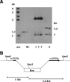
Analysis of the rv2525c knockout mutant. (A) Southern blot analysis of genomic DNA from pPR27-rv2525c transformants of M. tuberculosis Kanr Sucr strains (lanes 1 to 4) and from H37Rv (lane Rv). DNA was digested by SmaI and hybridized with the radiolabeled plasmid pPR27-rv2525c. Lane 4 characterizes the knockout mutant. (B) SmaI restriction profile of the disrupted rv2525c locus.
FIG. 5.
FIG. 5.
Evidence for loss of Rv2525c from mutant. (A) SELDI analysis of proteins from bacterial supernatants onto CM10 ProteinChip array. Concentrated supernatants (10 μg) from late-log-phase cultures of M. tuberculosis, H37Rv (top panel) and the H37Rv rv2525c knockout mutant (bottom panel), were analyzed on CM10 arrays at pH 4. (B) Immunoblotting, using an anti-Rv2525c polyclonal antibody, of 10 μg of supernatants from late-log-phase cultures of M. tuberculosis H37Rv (H37Rv), the H37Rv rv2525c knockout mutant (KO), and the complemented strain (KO+) and of 1 μg of purified recombinant Rv2525c protein (Rv2525).
FIG. 6.
FIG. 6.
Growth of strains in bone marrow macrophages. (A) Growth of M. tuberculosis H37Rv (•), the H37Rv rv2525c knockout mutant KO (▪), and the complemented rv2525c knockout mutant strain KO+ (♦) in C57BL/6 murine bone marrow-derived resting macrophages. (B) Growth of M. tuberculosis H37Rv (•), the H37Rv rv2525c knockout mutant KO (▪), and the complemented rv2525c knockout mutant strain KO+ (♦) in C57BL/6 murine bone marrow-derived activated macrophages. Data are the mean and standard deviation of triplicate measurements and are representative of two independent experiments. Differences in CFU means with H37Rv- and KO-infected cells were analyzed by ANOVA (NS, not significant; **, P > 0.01).
FIG. 7.
FIG. 7.
Results of virulence studies of mice. (A) Growth of M. tuberculosis H37Rv (black bars), the H37Rv rv2525c knockout mutant (gray bars), and the complemented rv2525c knockout mutant strain (white bars) in BALB/c mouse organs following infection via the aerosol route. (B) Growth of M. tuberculosis H37Rv (black bars), the H37Rv rv2525c knockout mutant (gray bars), and the complemented rv2525c knockout mutant strain (white bars) in SCID mouse organs following intravenous infection with 106 CFU. For each time point of experiments A and B, data are the mean ± standard deviation from three to four mice per group. (C) Percent survival of cohorts of 10 SCID mice infected with 106 CFU of either M. tuberculosis H37Rv (solid line) or the H37Rv rv2525c knockout mutant (broken line). Data are the mean and standard deviation of CFU measured from four animals per group. Differences in CFU means with the H37Rv- and KO-infected groups were analyzed by ANOVA (*, P > 0.05; **, P > 0.01).

Similar articles

Cited by

References

    1. Andersen, P. 1991. Proteins released from Mycobacterium tuberculosis during growth. Infect. Immun. 59:1905-1910. - PMC - PubMed
    1. Bange, W. R., F. M. Collins, and W. R. Jacobs, Jr. 1999. Survival of mice infected with Mycobacterium smegmatis containing large DNA fragments from Mycobacterium tuberculosis. Tuber. Lung Dis. 79:171-180. - PubMed
    1. Berks, B. C., F. Sargent, and T. Palmer. 2000. The Tat protein export pathway. Mol. Microbiol. 35:260-274. - PubMed
    1. Bogsch, E. G., F. Sargent, N. R. Stanley, B. C. Berks, C. Robinson, and T. Palmer. 1998. An essential component of a novel bacterial protein export system with homologues in plastids and mitochondria. J. Biol. Chem. 273:18003-18006. - PubMed
    1. Boshoff, H. I., T. G. Myers, B. R. Copp, M. R. McNeil, M. A. Wilson, and C. E. Barry III. 2004. The transcriptional responses of Mycobacterium tuberculosis to inhibitors of metabolism: novel insights into drug mechanisms of action. J. Biol. Chem. 279:40174-40184. - PubMed

Publication types

MeSH terms

LinkOut - more resources