. 2006 Sep 12;114(11):1159-68.
doi: 10.1161/CIRCULATIONAHA.106.637124. Epub 2006 Sep 4.

Foxo transcription factors blunt cardiac hypertrophy by inhibiting calcineurin signaling

Affiliations

Foxo transcription factors blunt cardiac hypertrophy by inhibiting calcineurin signaling

Yan G Ni et al. Circulation. .

Abstract

Background: Cellular hypertrophy requires coordinated regulation of progrowth and antigrowth mechanisms. In cultured neonatal cardiomyocytes, Foxo transcription factors trigger an atrophy-related gene program that counters hypertrophic growth. However, downstream molecular events are not yet well defined.

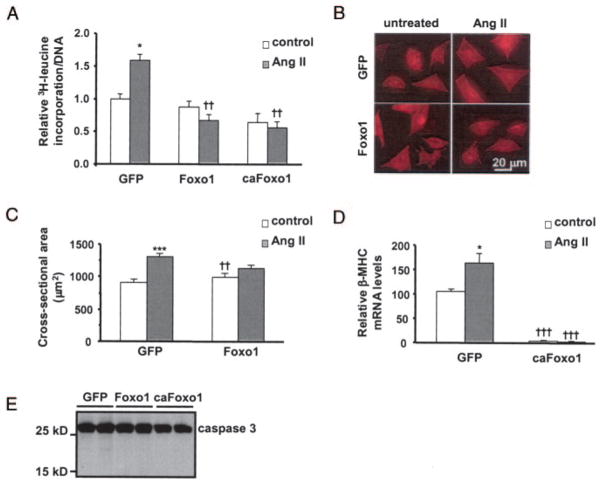
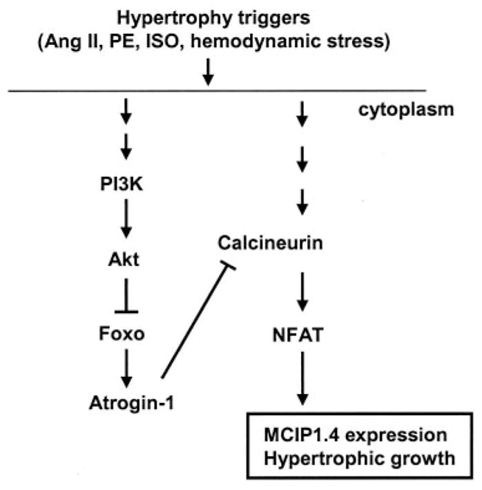
Methods and results: Here, we report that expression of either Foxo1 or Foxo3 in cardiomyocytes attenuates calcineurin phosphatase activity and inhibits agonist-induced hypertrophic growth. Consistent with these results, Foxo proteins decrease calcineurin phosphatase activity and repress both basal and hypertrophic agonist-induced expression of MCIP1.4, a direct downstream target of the calcineurin/NFAT pathway. Furthermore, hearts from Foxo3-null mice exhibit increased MCIP1.4 abundance and a hypertrophic phenotype with normal systolic function at baseline. Together, these results suggest that Foxo proteins repress cardiac growth at least in part through inhibition of the calcineurin/NFAT pathway. Given that hypertrophic growth of the heart occurs in multiple contexts, our findings also suggest that certain hypertrophic signals are capable of overriding the antigrowth program induced by Foxo. Consistent with this, multiple hypertrophic agonists triggered inactivation of Foxo proteins in cardiomyocytes through a mechanism requiring the PI3K/Akt pathway. In addition, both Foxo1 and Foxo3 are phosphorylated and consequently inactivated in hearts undergoing hypertrophic growth induced by hemodynamic stress.

Conclusions: This study suggests that inhibition of the calcineurin/NFAT signaling cascade by Foxo and release of this repressive action by the PI3K/Akt pathway are important mechanisms whereby Foxo factors govern cell growth in the heart.


Figures

Figure 1
Figure 1
Activation of Foxo1 inhibits endogenous calcineurin activity. Myocytes were infected with adenovirus as indicated. A, At 48 hours after infection, cells were harvested and calcineurin phosphatase activity was measured. B and C, Western blots and densitometric analysis of calcineurin, MCIP1.1, and MCIP1.4 in whole-cell lysates harvested 48 hours after infection. D, mRNA levels of atrogin-1 and GAPDH were detected by RT-PCR 24 hours after infection. E, Schematic of the MCIP1.4-luciferase construct. F, Myocytes were coinfected with adenovirus encoding MCIP1.4-luc plus GFP or caFoxo1-GFP and processed for luciferase activity. G, Cells were infected with adenovirus (48 hours), and MCIP1.4 and GAPDH mRNA levels were determined by real-time PCR. Data are mean±SEM (n=6 to 8). *P≤0.05; **P≤0.01; ***P≤0.001.
Figure 2
Figure 2
Foxo1 activity inhibits Ang-II–induced hypertrophic growth in cardiomyocytes. A, Myocytes were cultured and infected with adenovirus and treated with Ang II (48 hours). Cells were then assayed for [3H]leucine incorporation normalized to cellular DNA content. B, Representative fields of cardiomyocytes stained with an α-actinin antibody. C, Quantification of cell cross-sectional area from experiments shown in B. Sixty-five to 75 randomly selected cells from each group were measured. D, β-MHC mRNA levels were measured by real-time PCR. E, Western blots of caspase 3 in whole-cell lysates harvested from myocytes 48 hours after adenovirus infection (A, D, n=8; C, n=65 to 75). *P≤0.05, ***P≤0.001 vs untreated control; ††P≤0.01, †††P≤0.001 vs respective GFP control group.
Figure 3
Figure 3
Activation of Foxo1 blunts agonist-induced MCIP1.4 expression. Serum-starved myocytes were infected and treated with IGF-1 (A, B) or Ang II (C, D), and the protein levels of calcineurin and MCIP1 were analyzed (n=6). *P≤0.05 vs untreated control; ††P≤0.01 vs respective GFP control group.
Figure 4
Figure 4
Foxo3-null mice are hypertrophic. A and B, Western blot analysis of MCIP1.4 protein levels in extracts from sham-operated or TAB LVs from Foxo3–knockout (ko) or wild-type (wt) mice (n=6 to 8). **P≤0.01 vs sham-operated; †P≤0.05 vs wt sham group. C, Heart weight/body weight (HW/BW; left) and heart weight/tibia length (HW/T; right) ratios of control, sham-operated, or TAB mice 3 weeks after surgery. Male Foxo3-null mice and wild-type littermates were studied at 12 weeks.
Figure 5
Figure 5
Foxo1 and Foxo3 are inactivated in load-induced hypertrophy. A, Protein extracts from sham-operated or TAB LVs 3 weeks after surgery were separated by SDS-PAGE and probed with antibodies against phosphorylated or total Foxo1 and Foxo3. B, Mean values of phosphorylated Foxo/total Foxo ratios from sham- or TAB-treated groups were normalized to the mean value of the phosphorylated Foxo/total Foxo from the sham group (n=4 to 5). C, Time course of Foxo phosphorylation in hypertrophic hearts. LV protein extracts from sham-operated (S) or TAB (T) mice were probed with antibodies against phosphorylated or total Foxo protein. *P≤0.05, **P≤0.01 vs sham group.
Figure 6
Figure 6
Multiple hypertrophic ligands inactivate Foxo in cardiomyocytes. A–D, Western blots and densitometric analysis of levels of phosphorylated Foxo1 and phosphorylated Foxo3. Serum-starved cardiomyocytes were treated with Ang II, PE (10 μmol/L), or ISO (10 nmol/L) for indicated durations. E, Ang II induces Foxo1-GFP nuclear export. Myocytes were infected, serum starved overnight, and then treated with vehicle or Ang II for 30 minutes. Hoechst 33334 was used for nuclear staining. F, Hypertrophic ligands induce endogenous Foxo3 translocation. Serum-starved myocytes were treated with Ang II or PE. Nuclear and cytoplasmic extracts were analyzed for total Foxo3, GAPDH, or lamin A/C. G, Ang II decreases atrogin-1 expression. Serum-starved myocytes were treated with Ang II and assayed for atrogin-1 and GAPDH mRNA levels by RT-PCR (n=6). Con indicates vehicle-treated control; N, nuclear fraction; and C, cytoplasmic fraction. *P≤0.05 vs untreated control group.
Figure 7
Figure 7
Ang II induces Foxo phosphorylation through the PI3K/Akt pathway. A–D, Western blots and densitometric analysis of levels of phosphorylated Foxo1 and phosphorylated Foxo3. Serum-starved cardiomyocytes were treated with Ang II, PE (10 μmol/L), or ISO (10 nmol/L) for indicated durations. E–G, LY294002 blocks Ang II–induced increases in Foxo phosphorylation (E, F) and Foxo1-GFP nuclear export (G). Serum-starved neonatal rat ventricular myocytes were preincubated with LY294002 (1 hour) before treatment with Ang II (30 minutes). H, I, Ang II–induced phosphorylation of both Foxo and Akt was blocked by LY294002 but not by inhibitors of other kinases. Western blots (H) and densitometric analysis (I) of levels of phosphorylated Foxo1 and phosphorylated Foxo3 (n=4). *P≤0.05, ***P≤0.001 vs vehicle-treated or Ang II–alone controls.
Figure 8
Figure 8
Role of Foxo proteins in cardiac hypertrophy. Neuro-hormonal activation or hemodynamic stress induces Foxo phosphorylation and consequent Foxo inhibition through the PI3K/Akt pathway. Activation of Foxo inhibits hypertrophic growth, at least in part, by blunting calcineurin-mediated pro-growth pathways.

Similar articles

Cited by

References

    1. Accili D, Arden KC. FoxOs at the crossroads of cellular metabolism, differentiation, and transformation. Cell. 2004;117:421–426. - PubMed
    1. Tran H, Brunet A, Griffith EC, Greenberg ME. The many forks in FOXO’s road. Sci STKE. 2003:RE5. - PubMed
    1. Stitt TN, Drujan D, Clarke BA, Panaro F, Timofeyva Y, Kline WO, Gonzalez M, Yancopoulos GD, Glass DJ. The IGF-1/PI3K/Akt pathway prevents expression of muscle atrophy-induced ubiquitin ligases by inhibiting FOXO transcription factors. Mol Cell. 2004;14:395–403. - PubMed
    1. Sandri M, Sandri C, Gilbert A, Skurk C, Calabria E, Picard A, Walsh K, Schiaffino S, Lecker SH, Goldberg AL. Foxo transcription factors induce the atrophy-related ubiquitin ligase atrogin-1 and cause skeletal muscle atrophy. Cell. 2004;117:399–412. - PMC - PubMed
    1. Frey N, Katus HA, Olson EN, Hill JA. Hypertrophy of the heart: a new therapeutic target? Circulation. 2004;109:1580–1589. - PubMed

Publication types

MeSH terms

Substances