Comparative Study
. 2006 Oct 9;95(7):896-905.
doi: 10.1038/sj.bjc.6603341. Epub 2006 Sep 5.

Efficient loading of dendritic cells following cryo and radiofrequency ablation in combination with immune modulation induces anti-tumour immunity

Affiliations
Comparative Study

Efficient loading of dendritic cells following cryo and radiofrequency ablation in combination with immune modulation induces anti-tumour immunity

M H M G M den Brok et al. Br J Cancer. .

Abstract

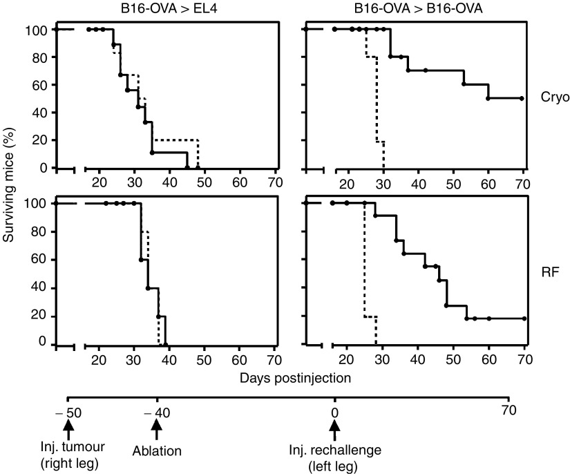
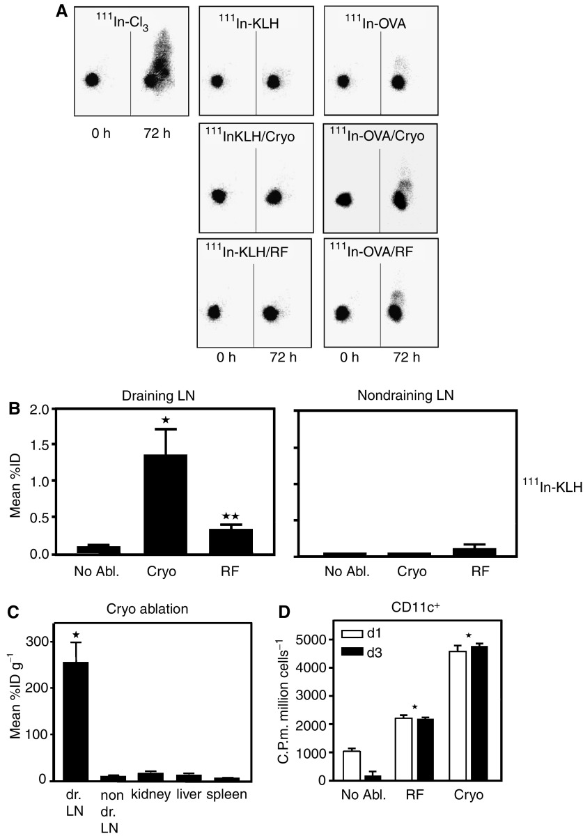
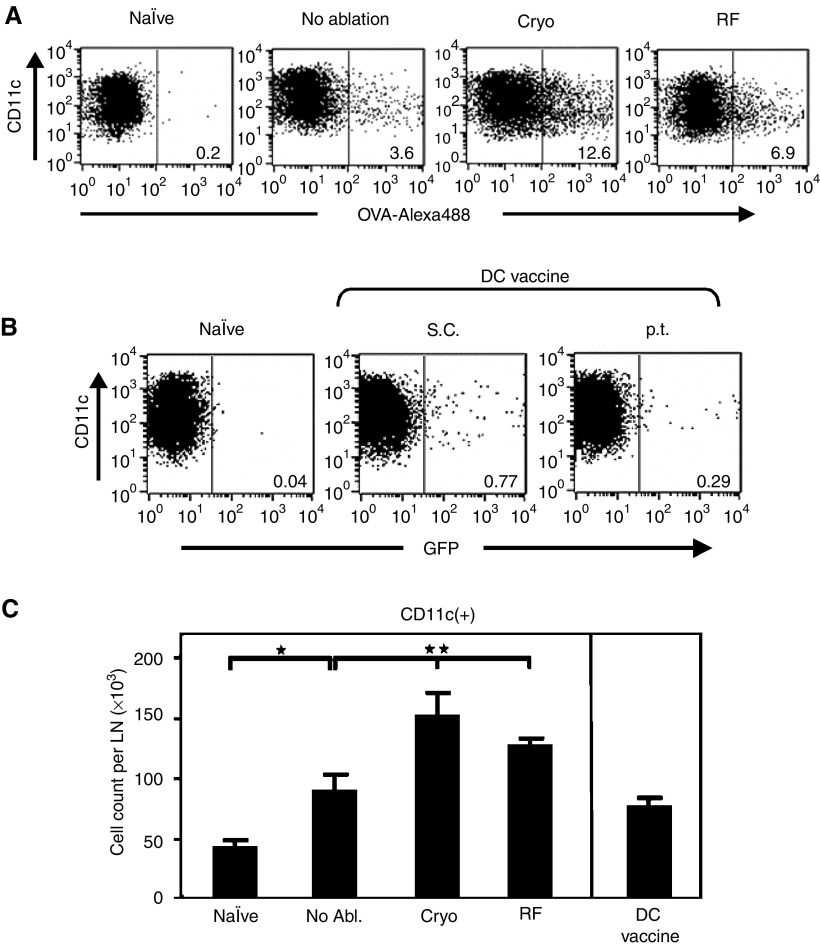
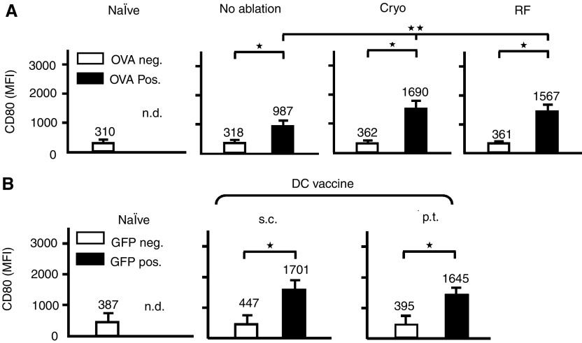
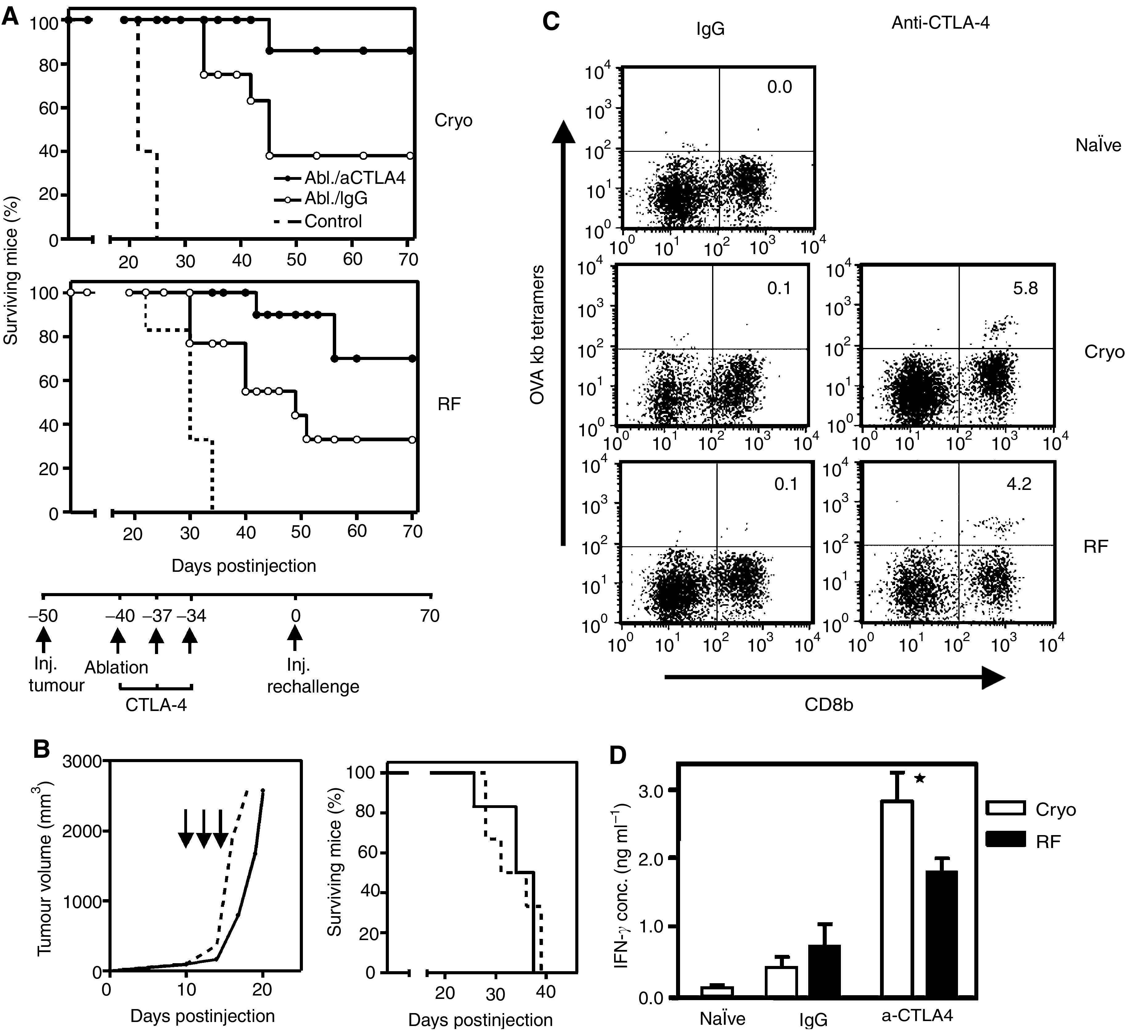
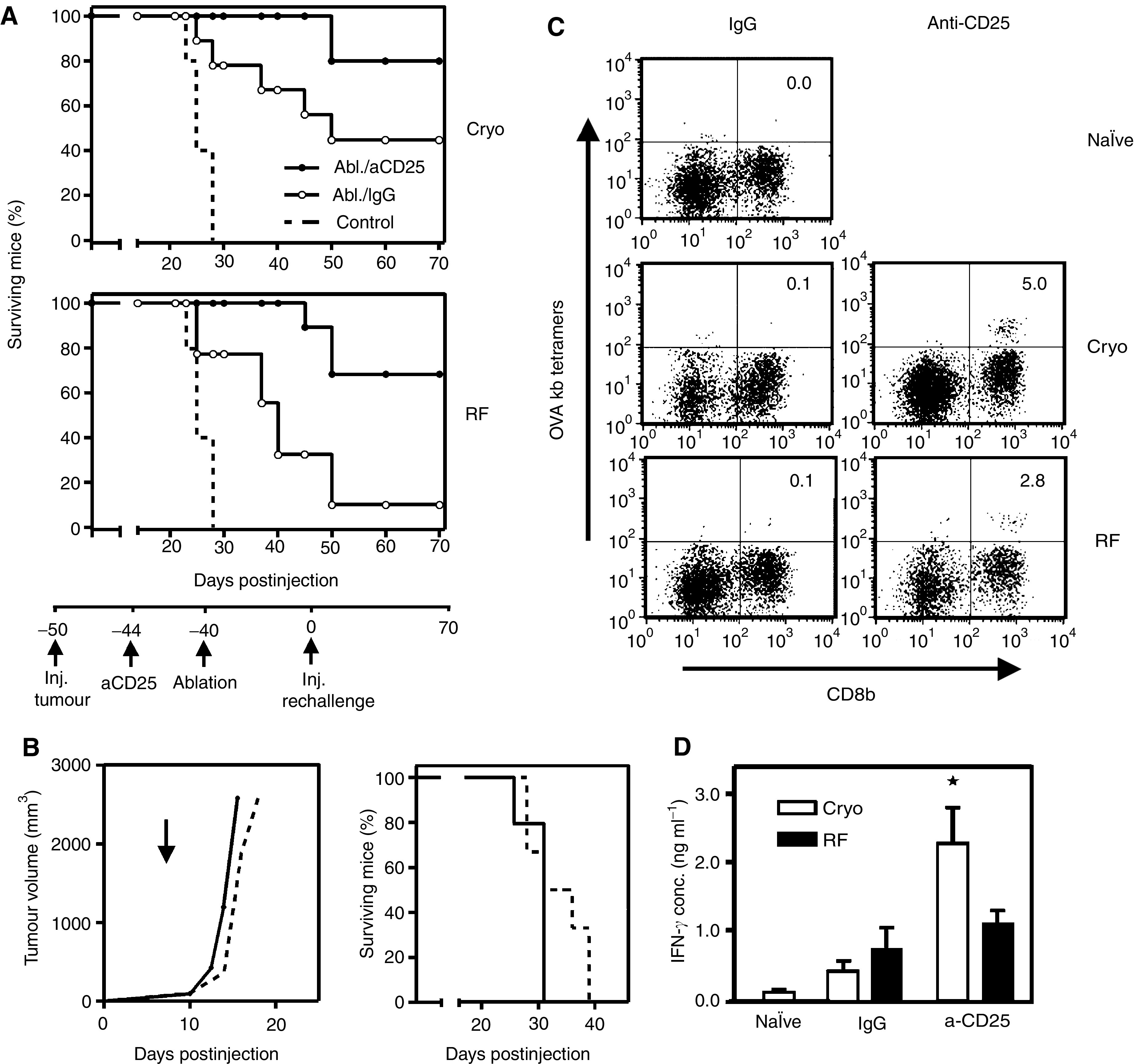
Dendritic cells (DC) are professional antigen-presenting cells that play a pivotal role in the induction of immunity. Ex vivo-generated, tumour antigen-loaded mature DC are currently exploited as cancer vaccines in clinical studies. However, antigen loading and maturation of DC directly in vivo would greatly facilitate the application of DC-based vaccines. We formerly showed in murine models that radiofrequency-mediated tumour destruction can provide an antigen source for the in vivo induction of anti-tumour immunity, and we explored the role of DC herein. In this paper we evaluate radiofrequency and cryo ablation for their ability to provide an antigen source for DC and compare this with an ex vivo-loaded DC vaccine. The data obtained with model antigens demonstrate that upon tumour destruction by radiofrequency ablation, up to 7% of the total draining lymph node (LN) DC contained antigen, whereas only few DC from the conventional vaccine reached the LN. Interestingly, following cryo ablation the amount of antigen-loaded DC is almost doubled. Analysis of surface markers revealed that both destruction methods were able to induce DC maturation. Finally, we show that in situ tumour ablation can be efficiently combined with immune modulation by anti-CTLA-4 antibodies or regulatory T-cell depletion. These combination treatments protected mice from the outgrowth of tumour challenges, and led to in vivo enhancement of tumour-specific T-cell numbers, which produced more IFN-gamma upon activation. Therefore, in situ tumour destruction in combination with immune modulation creates a unique, 'in situ DC-vaccine' that is readily applicable in the clinic without prior knowledge of tumour antigens.


Figures

Figure 1
Figure 1
Anti-tumour immunity following radiofrequency or cryo ablation. Mice with established B16-OVA melanomas (5–7 mm) were ablated by cryo (cryo, upper panels) or radiofrequency ablation (RF, lower panels). Forty days later, a re-challenge with 15 × 103 EL4 (left panels) or 15 × 103 B16-OVA cells (right panels) was given s.c. in the contra-lateral leg. Figures depict survival curves demonstrating specificity of the response and growth reduction/protection after ablation. As a control, tumour growth was monitored by injection of the same tumour dose into naïve mice (dotted lines). T=0 corresponds to the time of injection of the tumour re-challenge. P<0.005 for both B16 lines vs control. One out of four representative experiments is shown (n=5–11).
Figure 2
Figure 2
Preferential uptake of tumour-derived antigens by LN DCs. (A) To study the fate of tumour antigens after ablation, 20 μCi of 111In-Cl3 (left panel), 111In-KLH (middle panels) or 111In-OVA (right panels) was injected into established B16-OVA tumours (5–7 mm). Tumours were left untreated or ablated by cryo ablation (cryo) or radiofrequency ablation (RF) directly after these injections. γ-Camera imaging was performed at the indicated time points. For 111In-Cl3-injected mice the contours, tumour and liver are visible. One representative mouse out of three is shown. (B and C) Biodistribution of 111In-KLH was determined in dissected LNs and organs of mice injected i.t. 1 day before. Tumours were either left untreated or ablated directly after KLH injection. Radioactivity values from LN's of four mice per group are presented as mean percentages of injected dose with s.d., whereas the values for the organs are also corrected for weight. Mice in panel C received cryo ablation, but comparable results were obtained with RF ablation. (D) Lymph node suspensions from non-ablated and ablated mice (five mice pooled per group) that received i.t. 111In-KLH were subjected to magnetic bead sorting of CD11c(+) cells. After sorting at days 1 and 3 after ablation, the cell-associated radioactivity was measured in the CD11c(+) and CD11c(−) (not shown) cell fraction. Values are presented as counts per minute, corrected for 1 × 10⁶ cells and natural decay, with s.d. from triplicates. *=P<0.005 compared to no ablation. In all figures one out of three representative experiments is shown.
Figure 3
Figure 3
Increased numbers of antigen-positive DC in draining LN following ablation. (A) Fluorescence-activated cell sorting analysis of CD11c(+) DC isolated from pooled LN suspensions of naïve, tumour-bearing or tumour-ablated mice (n=6 per group). Mice received 20 μg ovalbumin conjugated to Alexa-488 (OVA-Alexa488) i.t. just before the time point of ablation. Two days after the indicated treatments, CD11c(+) DC were isolated from draining LNs, stained for CD11c (clone HL3), gated and plotted for OVA-Alexa488 content. Values shown are percentages of OVA-Alexa488(+) cells within the CD11c(+) fraction. (B) Bone marrow dendritic cells were cultured from GFP-transgenic mice, loaded with peptides and matured ex vivo with LPS. Dendritic cells (1 × 10⁶ ) were injected p.t. in tumour-bearing mice or s.c. into naïve mice. Two days later CD11c(+) DC were isolated from pooled LN suspensions (n=6 per group), stained, gated and plotted for GFP. (C) Absolute CD11c(+) cell count per LN. Data are obtained from experiments described in A and B and presented as means with s.d. from three independent experiments. As controls for all experiments, naïve mice were used that did not receive any injection (naïve). *=P<0.005 vs naïve, **=P<0.01 both vs no ablation.
Figure 4
Figure 4
Ablation induces maturation of antigen-positive DC. OVA-Alexa488-positive or -negative CD11c(+) cells (A), or GFP-positive or negative CD11c(+) cells (B), obtained in the experiments shown in Figure 3, were analysed separately for expression of the maturation marker CD80. Values indicated are mean MFI's with s.d. from three independent experiments. *=P<0.005, **=P<0.05 both vs no ablation.
Figure 5
Figure 5
Anti-CTLA-4 improves therapeutic outcome of ablation. 0, 3 and 6 days after ablation of B16-OVA tumours, mice were injected i.p. with 200 μg anti-CTLA-4 antibodies or control IgG. (A) Forty days following ablation of tumour-bearing mice receiving antibodies, a tumour re-challenge was performed as described before. Figures depict survival curves demonstrating growth reduction/protection after ablation plus CTLA-4 treatment. As a control, tumour growth was monitored by injection of the same tumour dose into naïve mice (dotted lines). T=0 corresponds to the time of injection of the tumour re-challenge. P<0.05 for CTLA-4 vs IgG in both cryo and RF figures. One out of three representative experiments is shown (n=5–11). (B) Control experiment showing that CTLA-4 treatment by itself is insufficient to eradicate the primary tumour or re-challenges. Mice with established B16OVA tumours (5–7 mm) were injected with 200 μg anti-CTLA-4 antibodies at days 10, 13 or 16 after tumour inoculation (solid line, arrows) or PBS (dotted line). Next, tumour growth was monitored in time (left panel). CTLA-4 treatment or PBS given 40 days before a B16OVA challenge (15 × 103 cells) did not affect survival of the mice (right panel). (C) At day 10 after ablation, a mix of LN and spleen cells was obtained from mice treated as indicated. T cells were harvested from spleen and LN and restimulated with irradiated, IFN-γ-treated B16-OVA cells and IL-2 for 10 days, followed by staining with OVA tetramers (Kb) and anti-CD8b. Depicted numbers represent the percentages of tetramer-positive cells within the CD8b+ population. (D) T cells from the same bulk cultures were used for restimulation with B16OVA cells. Supernatant from these cultures was harvested 24 h later and analysed for IFN-γ content by standard ELISA methods. Shown are means with s.d. from triplicates, *=P<0.05 vs IgG. Experiments shown in figures (BD) were repeated twice with comparable results.
Figure 6
Figure 6
Depletion of regulatory T cells improves therapeutic outcome of ablation. Four days before ablation of B16-OVA tumours, mice were injected i.p. with 200 μg anti-CD25 antibodies or control IgG. (A) Forty days following ablation of tumour-bearing mice receiving antibodies, a tumour re-challenge was performed as described before. Figures depict survival curves demonstrating growth reduction/protection after ablation plus Treg depletion. As a control, tumour growth was monitored by injection of the same tumour dose into naïve mice (dotted lines). T=0 corresponds to the time of injection of the tumour re-challenge. P<0.05 for aCD25 vs IgG in both cryo and RF figures. One out of three representative experiments is shown (n=5–9). (B) Control experiment showing that Treg depletion by itself is insufficient to eradicate the primary tumour or re-challenges. Mice with established B16OVA tumours (5–7 mm) were injected with 200 μg anti-CD25 antibodies (solid line, arrow) or PBS (dotted line). Next, tumour growth was monitored in time (left panel). Treg depletion or PBS given 40 days before a B16OVA challenge (15 × 103 cells) did not affect survival of the mice (right panel). (C) At day 10 after ablation, a mix of LN and spleen cells was obtained from mice treated as indicated. T cells were harvested from spleen and LN and restimulated with irradiated, IFN-γ-treated B16-OVA cells and IL-2 for 10 days, followed by staining with OVA tetramers (Kb) and anti-CD8b. Depicted numbers represent the percentages of tetramer-positive cells within the CD8b+ population. (D) T cells from the same bulk cultures were used for restimulation with B16OVA cells. Supernatant from these cultures was harvested 24 h later and analysed for IFN-γ content by standard ELISA methods. Shown are means with s.d. from triplicates, *=P<0.05 vs IgG. Experiments shown in figures (BD), were repeated twice with comparable results.

References

    1. Adams S, O'Neill DW, Bhardwaj N (2005) Recent advances in dendritic cell biology. J Clin Immunol 25: 87–98 - PubMed
    1. Banchereau J, Briere F, Caux C, Davoust J, Lebecque S, Liu YJ, Pulendran B, Palucka K (2000) Immunobiology of dendritic cells. Annu Rev Immunol 18: 767–811 - PubMed
    1. Banchereau J, Palucka AK (2005) Dendritic cells as therapeutic vaccines against cancer. Nat Rev Immunol 5: 296–306 - PubMed
    1. Chen L, Ashe S, Brady WA, Hellstrom I, Hellstrom KE, Ledbetter JA, McGowan P, Linsley PS (1992) Costimulation of antitumor immunity by the B7 counterreceptor for the T lymphocyte molecules CD28 and CTLA-4. Cell 71: 1093–1102 - PubMed
    1. Coulie PG, van der Bruggen P (2003) T-cell responses of vaccinated cancer patients. Curr Opin Immunol 15: 131–137 - PubMed

Publication types