. 2006 Sep 12;45(36):10815-27.
doi: 10.1021/bi060782u.

Structure of the DPS-like protein from Sulfolobus solfataricus reveals a bacterioferritin-like dimetal binding site within a DPS-like dodecameric assembly

Affiliations

Structure of the DPS-like protein from Sulfolobus solfataricus reveals a bacterioferritin-like dimetal binding site within a DPS-like dodecameric assembly

George H Gauss et al. Biochemistry. .

Abstract

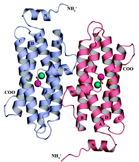
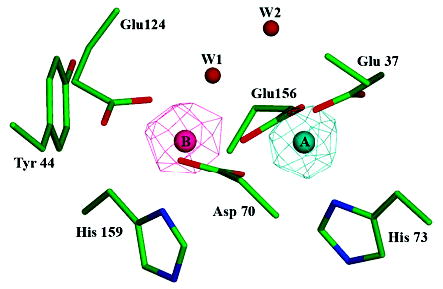
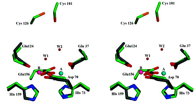
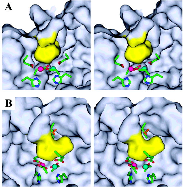
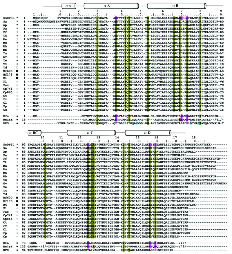
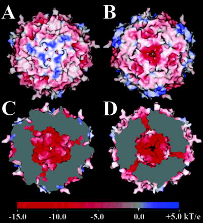
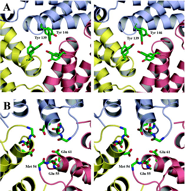
The superfamily of ferritin-like proteins has recently expanded to include a phylogenetically distinct class of proteins termed DPS-like (DPSL) proteins. Despite their distinct genetic signatures, members of this subclass share considerable similarity to previously recognized DPS proteins. Like DPS, these proteins are expressed in response to oxidative stress, form dodecameric cage-like particles, preferentially utilize H(2)O(2) in the controlled oxidation of Fe(2+), and possess a short N-terminal extension implicated in stabilizing cellular DNA. Given these extensive similarities, the functional properties responsible for the preservation of the DPSL signature in the genomes of diverse prokaryotes have been unclear. Here, we describe the crystal structure of a DPSL protein from the thermoacidophilic archaeon Sulfolobus solfataricus. Although the overall fold of the polypeptide chain and the oligomeric state of this protein are indistinguishable from those of authentic DPS proteins, several important differences are observed. First, rather than a ferroxidase site at the subunit interface, as is observed in all other DPS proteins, the ferroxidase site in SsDPSL is buried within the four-helix bundle, similar to bacterioferritin. Second, the structure reveals a channel leading from the exterior surface of SsDPSL to the bacterioferritin-like dimetal binding site, possibly allowing divalent cations and/or H(2)O(2) to access the active site. Third, a pair of cysteine residues unique to DPSL proteins is found adjacent to the dimetal binding site juxtaposed between the exterior surface of the protein and the active site channel. The cysteine residues in this thioferritin motif may play a redox active role, possibly serving to recycle iron at the ferroxidase center.


Figures

Figure 1
Figure 1
DPS-like protein from Sulfolobus solfataricus. The ribbon diagram depicts 2 of the 12 subunits in the SsDPSL dodecamer viewed from the exterior surface of the dodecamer. The overall fold of the SsDPSL subunit is most similar to that of authentic DPS molecules. It is composed of a 4-helix bundle (helices A, B, C, and D), common to members of the ferritin superfamily, decorated by two additional helices. The BC helix is found in all DPS structures to date, whereas the N-terminal helix (helix N) is observed only in the L. lactis DPS structure (). Each chain contains a ferroxidase site buried within the core of the four-helix bundle. In contrast, metal binding sites in authentic DPS dodecamers are found at the 2-fold interface. The ferroxidase site contains a mixture of metals at both the A- and B-sites (cyan and pink, respectively). The side chains for Cys101 and Cys126, which are adjacent to the ferroxidase site, are also shown in green (carbon) and orange (sulfur).
Figure 2
Figure 2
Bacterioferritin-like dimetal binding site. Residues Glu37, Asp70, His73, Glu124, Glu156, and His159 of SsDPSL form a dimetal binding site within the core of the four-helix bundle. Side chain atoms are colored in green (carbon), blue (nitrogen), and red (oxygen). Anomalous difference electron density maps (mesh) indicate asymmetric binding of the iron and zinc atoms, with Zn2+ binding preferentially to the A site (cyan) and Fe3+ to the B site (pink). The iron edge anomalous difference map is contoured at 8 σ (pink mesh), whereas the zinc edge anomalous difference map is contoured at 15 σ (cyan mesh). When contoured at lower levels, however, it is clear that each site contains a mixed population of both iron and zinc. Water W1 shows strong coordination to the B site metals but only weak coordination, at best, to the A site.
Figure 3
Figure 3
Superposition of the SsDPSL and E. coli bacterioferritin ferrioxidase sites. Residues composing the dimetal binding site of SsDPSL are superimposed on those of E. coli bacterioferritin (1BCF) (). Atom types are colored as in Figure 2; however, the carbon atoms of the bacterioferritin side chains are in gray. The cysteine pair of SsDPSL is also depicted and can be seen above the dimetal binding site. For clarity, only residues in SsDPSL are labeled.
Figure 4
Figure 4
Active site channel. (A) The ferroxidase center lies adjacent to a boot shaped, solvent filled channel that opens to the outside surface of the dodecamer. The van der Waals surface of the active site cavity (yellow) is shown as viewed from inside the monomer from a position behind the heel of the boot shaped cavity. The line of sight is approximately down the longitudinal axis of the four-helix bundle, looking toward the N-terminal end. In this orientation, the exterior of the dodecamer is up, and the internal cavity of the dodecamer is down. The conserved cysteine pair is hidden from view behind the upper surface of the active site channel (the tongue of the boot). The dimetal binding site lies beneath the channel (the heel of the boot). Metal sites A and B are depicted in cyan and pink, respectively. Active site residues are colored by atom: carbon, green; nitrogen, blue; oxygen, red and sulfur, orange. (B) The relative proximity of the cysteine pair (boot laces) to the dimetal binding site is more apparent in this view, in which the ferroxidase center has been rotated 60° about the vertical axis.
Figure 5
Figure 5
SsDPSL sequence alignments. Position specific iterated (PSI) BLAST (, ) was used to identify 26 SsDPSL homologues. The alignments reveal strong sequence conservation within the ferroxidase center and for the cysteine pair (Cys101 and Cys126). The Figure also presents structure based alignments between SsDPSL and structures representative of bacterioferritin (Bfn: E. coli, 1BCF (33)), DPS (DPS: E. coli, 1DPS (7)), and manganese catalase (MnCat: L. plantarum, 1JKU (35)) (bottom). Secondary structural elements for SsDPSL are indicated above the sequence. Conserved residues involved in metal coordination are found at structurally equivalent positions in the SsDPSL protein, E. coli bacterioferritin, and L. plantarum manganese catalase (pink) but not in E. coli DPS (1DPS). Conversely, the E. coli DPS metal binding site residues (blue) are not conserved in SsDPSL. Residues conserved among the DPSL homologues are highlighted in olive; this includes residues involved in metal coordination and the cysteine pair. The canonical dimetal carboxylate motif within helix B is D/ExxH, with two residues separating the acidic residue and the histidine. Two SsDPSL homologues do not satisfy this spacing; both Ap and Pa have 3 residues separating the acidic residue and the histidine at the first metal site. This and the presence of adjacent acidic residues result in local misalignment by ClustalW within helix B. However, the individual sequence alignments from PSI-BLAST do correctly align the D/ExxH motif in the B helix. Members of Pfam CD1052.2 used to seed the search are denoted as follows: SsDPSL, Sulfolobus solfataricus DPS-like protein; Pf, Pyrococcus furiosus DPS-like protein; Tm, Thermotoga maritima hypothetical protein TM0560; Mb, Methanosarcina barkeri ferritin-like protein; Ap, Aeropyrum pernix hypothetical protein APE1457; Pa, Pyrobaculum aerophilum hypothetical protein PAE2701; Tt, Thermoanaerobacter tengcongensis hypothetical protein TTE2230; Ct, Chlorobium tepidum hypothetical protein CT1328; Bt, Bacteroides thetaiotaomicron hypothetical protein BT3823. The 18 additional homologues identified by PSI-BLAST are from Sa, Sulfolobus acidocaldarius; Tk, Thermococcus kodakarensis; Ma, Methanosarcina acetivorans; Mm, Methanococcus maripaludis S2; Mh, Methanospirillum hungatei; Gv, Gloeobacter violaceus; Te, Thermoanaerobacter ethanolicus; Td, Thiomicrospira denitrificans; Bf600, Bacteroides fragilis gi:52217600; Bf175, Bacteroides fragilis gi:60494175; Pl, Pelodictyon luteolum; Pas, Prosthecochloris aestuarii; Cp741, Chlorobium phaeobacteroides gi:67939741; Cp865, Chlorobium phaeobacteroides gi:67934865; Pv, Prosthecochloris vibrioformis; Cl, Chlorobium limicola; Pp, Pelodictyon phaeoclathratiforme; and Cc, Chlorobium chlorochromatii. Symbols indicate taxa: (*) crenarchaeotes; (†) euryarchaeotes; (‡) thermotogales; (⋄) cyanobacteria; ((♦) eubacteria; (□) e-proteobacteria; (▪) CFB group bacteria; (○) green sulfur bacteria; (•) bacteria.
Figure 6
Figure 6
Surface electrostatic potential of the SsDPSL dodecamer. (A) Exterior surface surrounding the N-terminal 3-fold interface. The positive surface potential, indicated in blue, is imparted by basic residues present in the N-terminal helix and the N-terminus. Three small pores opening from the N-terminal channel are immediately adjacent to the center of symmetry. (B) Exterior surface potential surrounding the C-terminal 3-fold pore. The surrounding surface is acidic, with a strong negative potential (red). Relative to panel A, the dodecamer has been rotated 180° about the vertical axis. (C) Interior surface at the N-terminal 3-fold interface. Relative to panel A, the dodecamer has been rotated 180° about the vertical axis, and the clipping plane has been positioned to cut away a portion of the particle, revealing the negatively charged (red) internal surface surrounding the N-terminal pore (center). An oblique view of the surfaces lining the three symmetry related N-terminal pores is also apparent, seen as channels of negative potential (red) connecting the interior and exterior surfaces. Unlike the C-terminal channel (below), the N-terminal channels do not lie completely within the clipping plane; hence, a contiguous surface from the interior to the exterior of the dodecamer is not apparent. (D) Interior surface surrounding the C-terminal pore. Relative to panel B, the dodecamer has been rotated 180° about the vertical axis, and the clipping plane has been positioned to cut away a portion of the particle, revealing the negatively charged (red) internal surface surrounding the acidic C-terminal pore (center). An oblique view of the surfaces lining the three symmetry related C-terminal pores is also seen, with red channels of negative potential connecting the interior and exterior surfaces. Electrostatic potentials were generated with SPOCK (), using a probe radius of 1.4 Å, a temperature of 353 K, an ionic strength of 0.15 M, and protein and solvent dielectric constants of 4 and 80, respectively.
Figure 7
Figure 7
N- and C-terminal pores. Subunits related by the 3-fold symmetry of the particle are colored in yellow, pink, and blue. (A) N-terminal pore. Tyr139 forms a partial cap over the exterior end of the N-terminal channel, whereas the side chains of Tyr146 (behind Tyr139) help to form the walls of the channel. (B) C-terminal pore. Three successive layers of negative charge line the C-terminal pore. The first layer, on the exterior of the particle, is composed of three Glu55 side chains. The second layer comprises the carbonyl groups of Glu55 and Met54. The side chains of Met54 have been omitted for clarity. The third layer is contributed by the side chains of Glu61 on the interior of the dodecamer. In both views, the line of sight is along the 3-fold axis, looking from the exterior toward the interior. Atom colors are carbon, green; nitrogen, blue; and oxygen, red.

References

    1. Andrews SC, Robinson AK, Rodriguez-Quinones F. Bacterial iron homeostasis. FEMS Microbiol Rev. 2003;27:215–237. - PubMed
    1. Baaghil S, Lewin A, Moore GR, Le Brun NE. Core formation in Escherichia coli bacterioferritin requires a functional ferroxidase center. Biochemistry. 2003;42:14047–14056. - PubMed
    1. Ceci P, Cellai S, Falvo E, Rivetti C, Rossi GL, Chiancone E. DNA condensation and self-aggregation of Escherichia coli Dps are coupled phenomena related to the properties of the N-terminus. Nucleic Acids Res. 2004;32:5935–5944. - PMC - PubMed
    1. Zhao GH, Ceci P, Ilari A, Giangiacomo L, Laue TM, Chiancone E, Chasteen ND. Iron and hydrogen peroxide detoxification properties of DNA-binding protein from starved cells: A ferritin-like DNA-binding protein of Escherichia coli. J Biol Chem. 2002;277:27689–27696. - PubMed
    1. Ilari A, Latella MC, Ceci P, Ribacchi F, Su M, Giangiacomo L, Stefanini S, Chasteen ND, Chiancone E. The unusual intersubunit ferroxidase center of Listeria innocua Dps is required for hydrogen peroxide detoxification but not for iron uptake. A study with site-specific mutants. Biochemistry. 2005;44:5579–5587. - PubMed

Publication types

MeSH terms

Associated data