Comparative Study
. 2006 Sep 12;45(36):10998-1004.
doi: 10.1021/bi061098i.

Induced chirality of the light-harvesting carotenoid salinixanthin and its interaction with the retinal of xanthorhodopsin

Affiliations
Comparative Study

Induced chirality of the light-harvesting carotenoid salinixanthin and its interaction with the retinal of xanthorhodopsin

Sergei P Balashov et al. Biochemistry. .

Abstract

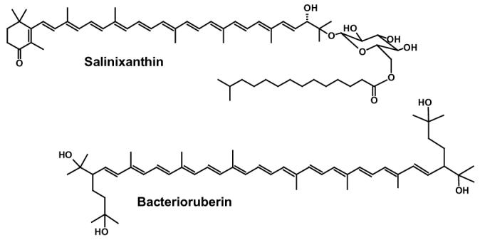
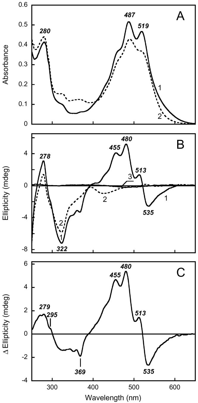
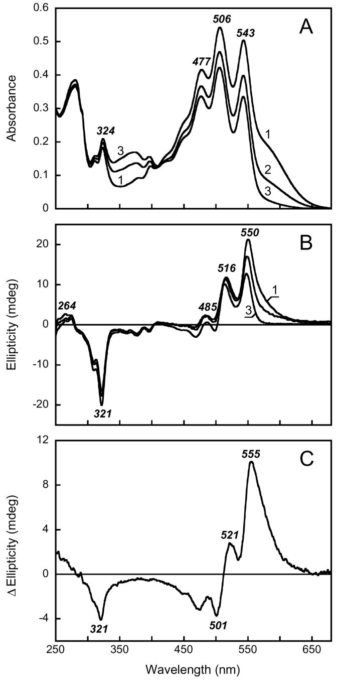
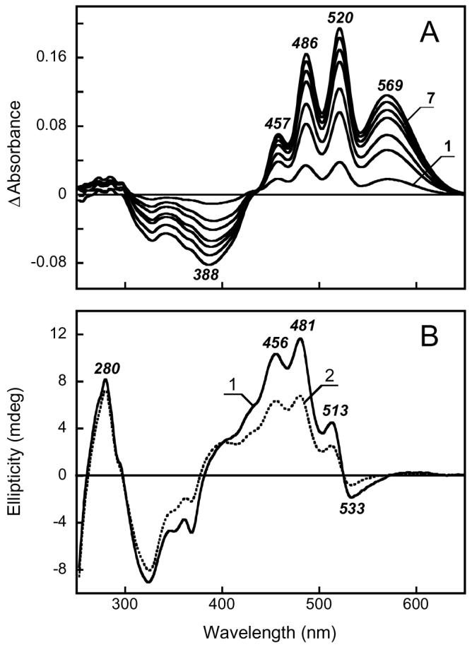
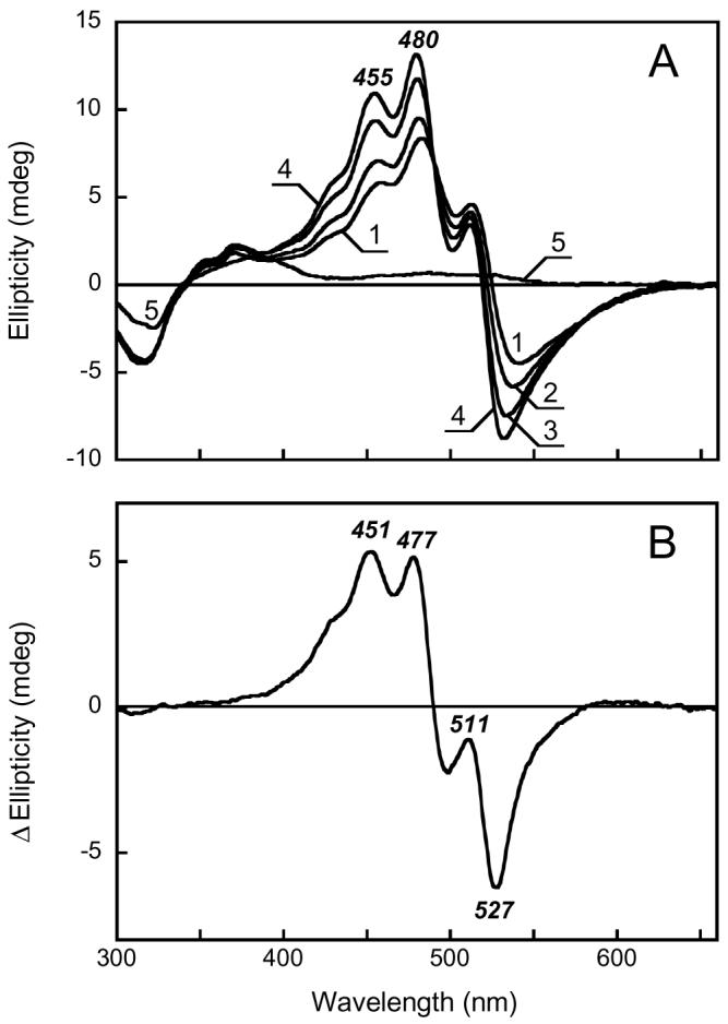
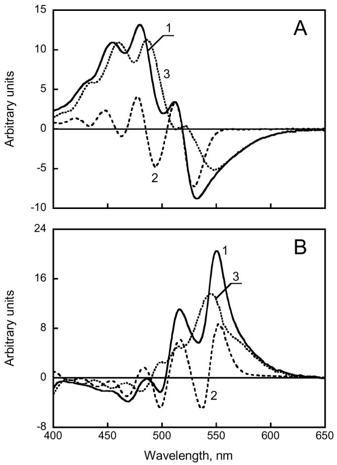
In xanthorhodopsin, a retinal protein-carotenoid complex of Salinibacter ruber, the carotenoid salinixanthin functions as a light-harvesting antenna in supplying additional excitation energy for retinal isomerization and proton transport. Another retinal protein, archaerhodopsin, has been shown to contain a carotenoid, bacterioruberin, but without an antenna function. We report here that the binding site confers a chiral geometry on salinixanthin in xanthorhodopsin and confirm that the same is true for bacterioruberin in archaerhodopsin. Cell membranes containing these rhodopsins exhibit CD spectra with sharp positive bands in the visible region where the carotenoids absorb, and in the case of xanthorhodopsin a negative band at 536 nm, as well as bands in the UV region. The carotenoid in ethanol has very weak optical activity in the visible region of the spectrum. Denaturation of the opsin upon deprotonation of the Schiff base at pH 12.5 eliminates the induced CD bands in both proteins. In one of these proteins, but not in the other, the carotenoid binding site depends entirely on the retinal. Hydrolysis of the retinal Schiff base of xanthorhodopsin with hydroxylamine eliminates the induced CD bands of salinixanthin. In contrast, hydrolysis of the Schiff base in archaerhodopsin does not abolish the CD bands of bacterioruberin. Thus, consistent with its antenna function, the carotenoid binding site interacts closely with the retinal only in xanthorhodopsin, and this interaction is the major source of the CD bands. In this protein, protonation of the counterion with a decrease in pH from 8 to 5 causes significant changes in the CD spectrum. The observed spectral features suggest that binding of salinixanthin in xanthorhodopsin involves the cyclohexenone ring of the carotenoid and its conformational heterogeneity is restricted.


Figures

Figure 1
Figure 1
Chemical structures of: salinixanthin (3) and bacterioruberin (28).
Figure 2
Figure 2
Absorption (A) and CD (B) spectra of xanthorhodopsin. Spectrum 1, suspension of Salinibacter ruber cell membranes containing xanthorhodopsin in 100 mM NaCl, pH 8.5; spectrum 2, after illumination at >550 nm for 3 hours in the presence of 200 mM hydroxylamine and 10 mg/ml of bovine serum albumin. Spectrum 3, salinixanthin in ethanol. C. Difference CD spectrum between unbleached and bleached cell membranes containing xanthorhodopsin: spectrum 1 minus spectrum 2 in Figure 1 B.
Figure 3
Figure 3
Absorption (A) and CD (B) spectra of archaerhodopsin. Spectra 1 through 3, initial spectrum of Halorubrum sp. membranes at pH 7.5 and after illumination for 2 and 9 hrs at >550 nm in the presence of 200 mM hydroxylamine and 10 mg/ml of bovine serum albumin, respectively. After 9 hrs of illumination (spectrum 3) the absorption band of the retinal chromophore decreased to less than 10% of initial value, as indicated by lowered absorption at 600 nm and laser flash induced absorption changes (not shown). C. Difference CD spectrum: spectrum 1 minus spectrum 3 in Fig. 2B.
Figure 4
Figure 4
A. Absorption changes observed upon reconstitution with all-trans retinal the Salinibacter ruber membranes bleached with hydroxylamine. Curve 1 through 7, spectra taken 2, 6, 10, 20, 30, 50, 90 min after addition of retinal. B. CD spectra of reconstituted membranes: 1, 100% reconstitution; 2, 50% reconstitution.
Figure 5
Figure 5
pH dependence of the CD spectrum of xanthorhodopsin. A. Purified xanthorhodopsin solubilized in 0.15% dodecylmaltoside at pH 4.5, 5.9, 6.7, 8.5, and 12.8, in spectra 1 through 5, respectively. B. Difference spectrum “pH 8.5 minus pH 4.5” (spectrum 4 minus spectrum 1 in Fig. 5A).
Figure 6
Figure 6
Decomposition of the CD spectra of xanthorhodopsin (A) and archaerhodopsin (B) to conservative and non-conservative components. The former was approximated by the first derivative of the absorption spectrum. A. Spectrum 1, CD spectrum of xanthorhodopsin solubilized in 0.15% dodecylmaltoside at pH 8.5; spectrum 2, first derivative of the absorption spectrum dA/dλ, multiplied by λ22dA/dλ = − dA/dν) and scaled to fit the amplitude of main peaks in the CD spectrum; spectrum 3, the difference “spectrum 1 minus spectrum 2”. B. Spectrum 1, CD spectrum of cell membranes containing archaerhodopsin; spectrum 2, first derivative multiplied by −λ2; spectrum 3, “spectrum 1 minus spectrum 2”.

Similar articles

Cited by

References

    1. Antón J, Rosselló-Mora R, Rodríguez-Valera F, Amann R. Extremely halophilic Bacteria in crystallizer ponds from solar salterns. Appl. Environ. Microbiol. 2000;66:3052–3057. - PMC - PubMed
    1. Antón J, Oren A, Benlloch S, Rodríguez-Valera F, Amann R, Rosselló-Mora R. Salinibacter ruber gen. nov., sp. nov., a novel, extremely halophilic member of the Bacteria from saltern crystallizer ponds. Int. J. Syst. Evol. Micrbiol. 2002;52:485–491. - PubMed
    1. Lutnaes BF, Oren A, Liaaen-Jensen S. New C-40-carotenoid acyl glycoside as principal carotenoid in Salinibacter ruber, an extremely halophilic eubacterium. J. Nat. Prod. 2002;65:1340–1343. - PubMed
    1. Balashov SP, Imasheva ES, Boichenko VA, Antón J, Wang JM, Lanyi JK. Xanthorhodopsin: A proton pump with a light-harvesting carotenoid antenna. Science. 2005;309:2061–2064. - PMC - PubMed
    1. Oesterhelt D, Stoeckenius W. Functions of a new photoreceptor membrane. Proc. Natl. Acad. Sci. U.S.A. 1973;70:2853–2857. - PMC - PubMed

Publication types