. 2006 Oct 27;281(43):32366-74.
doi: 10.1074/jbc.M605275200. Epub 2006 Sep 5.

The Actin cross-linking domain of the Vibrio cholerae RTX toxin directly catalyzes the covalent cross-linking of actin

Affiliations

The Actin cross-linking domain of the Vibrio cholerae RTX toxin directly catalyzes the covalent cross-linking of actin

Christina L Cordero et al. J Biol Chem. .

Abstract

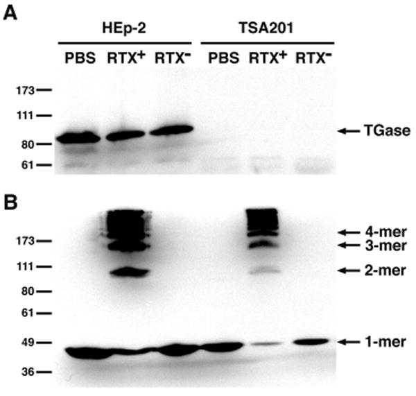
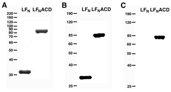
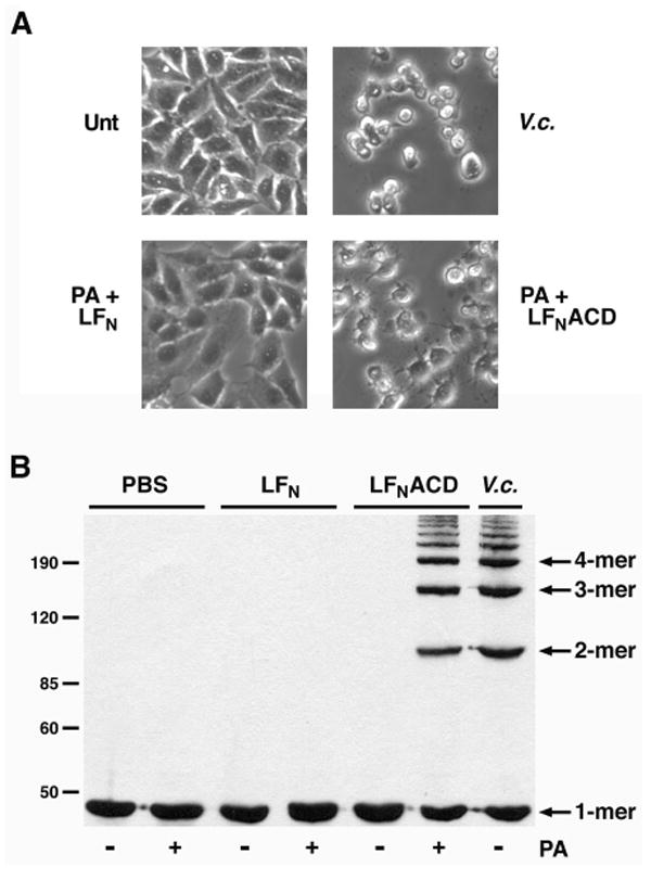
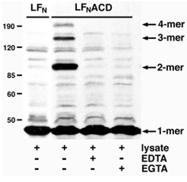
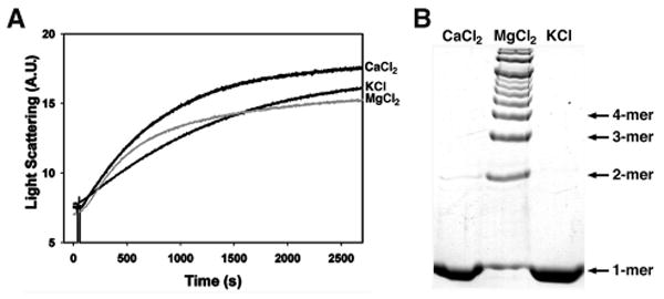
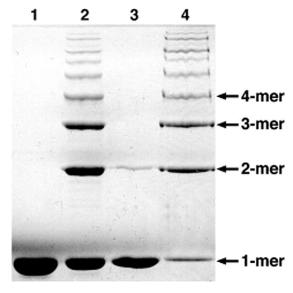
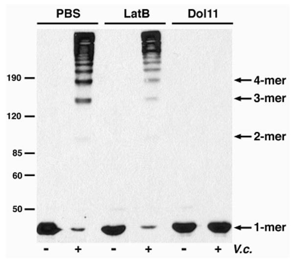
Vibrio cholerae is a Gram-negative bacterial pathogen that exports enterotoxins to alter host cells and to elicit diarrheal disease. Among the secreted toxins is the multifunctional RTX toxin, which causes cell rounding and actin depolymerization by covalently cross-linking actin monomers into dimers, trimers, and higher multimers. The region of the toxin responsible for cross-linking activity is the actin cross-linking domain (ACD). In this study, we further investigated the role of the ACD in the actin cross-linking reaction. We show that the RTX toxin cross-links actin independently of tissue transglutaminase, thus eliminating an indirect model of ACD activity. We demonstrate that a fusion protein of the ACD and the N-terminal portion of lethal factor from Bacillus anthracis (LF(N)ACD) has cross-linking activity in vivo and in crude cell extracts. Furthermore, we determined that LF(N)ACD directly catalyzes the formation of covalent linkages between actin molecules in vitro and that Mg(2+) and ATP are essential cofactors for the cross-linking reaction. In addition, G-actin is proposed as a cytoskeletal substrate of the RTX toxin in vivo. Future studies of the in vitro cross-linking reaction will facilitate characterization of the enzymatic properties of the ACD and contribute to our knowledge of the novel mechanism of covalent actin cross-linking.


Figures

FIGURE 1
FIGURE 1. The RTX toxin cross-links actin through a tissue TGase-independent mechanism
HEp-2 and TSA201 cells were incubated with either PBS-washed bacteria from an RTX-producing strain (RTX+), a strain harboring a deletion in the rtxA gene (RTX-), or PBS for 70 min. The cell pellets were boiled in SDS-PAGE sample buffer and lysates were electrophoresed on 8% SDS-polyacrylamide gels. Tissue TGase expression and actin cross-linking were detected by immunoblotting. The numbers at the left indicate the location of the Invitrogen Benchmark Pre-stained protein standards.
FIGURE 2
FIGURE 2. Characterization of LFNACD
LFN (32 kD) and LFNACD (81 kD) were purified by affinity chromatography on a nickel-chelating column and desalted by gel filtration. Purified proteins were loaded onto 10% SDS-polyacrylamide gels and either A, stained with Coomassie Blue, B, immunoblotted with a mouse monoclonal anti-His antibody, or C, immunoblotted with a rabbit polyclonal anti-ACD antibody. The numbers at the left correspond to the location of either the Invitrogen A, Benchmark Unstained or B, C, Benchmark Pre-stained protein standards.
FIGURE 3
FIGURE 3. LFNACD has actin cross-linking activity in vivo
HEp-2 cells were treated with PBS (Unt), exposed to PBS-washed V. cholerae strain KFV119 (V.c.), or intoxicated with 31.7 nM purified PA and either 13.6 nM LFN or LFNACD. A, Cell rounding was observed after 90 min, and phase contrast images were acquired at 200X magnification. B, Treated cells were harvested, lysates were separated on 8% SDS-polyacrylamide gels, and the formation of cross-linked actin species was monitored by immunoblotting. The protein standards are labeled on the left side.
FIGURE 4
FIGURE 4. Divalent cations are important for actin cross-linking in crude cell extracts
HEp-2 cells were sonicated in PBS and cleared by centrifugation at 1000 × g. 120 μg cell extract was incubated with 5 μg of purified LFN or LFNACD for 90 min. EDTA and EGTA were added to a final concentration of 5.0 mM where indicated. Actin cross-linking activity was monitored by immunoblotting. The location of the protein standards are indicated at the left.
FIGURE 5
FIGURE 5. LFNACD directly cross-links actin in the presence of Mg2+
A, 10 μM purified rabbit skeletal actin and 0.018 μM LFN or LFNACD were incubated for 20 min in Buffer B. The reactions were supplemented with 2.0 mM CaCl2, MgCl2, MnCl2, or ZnCl2 as indicated. B, 0.018 μM LFNACD, 10μM actin, and 2.0 mM MgCl2 were co-incubated and the reactions were terminated at designated time points. For both A and B, samples were boiled in SDS-PAGE sample buffer for 5 min, then loaded onto 8% SDS-polyacrylamide gels. Covalent actin cross-linking was detected by Coomassie Blue staining.
FIGURE 6
FIGURE 6. Mg2+ is essential for the ACD-catalyzed actin cross-linking reaction
10 μM G-actin in the A, absence or B, presence of 0.018 μM purified LFNACD was incubated with 2.0 mM CaCl2, 2.0 mM MgCl2, or 50mM KCl. A, Actin polymerization was observed by light scattering. B, Actin cross-linking was monitored in samples boiled in SDS-PAGE sample buffer and electrophoresed on 8% SDS-polyacrylamide gels. Actin species were visualized by staining with Coomassie Blue R-250.
FIGURE 7
FIGURE 7. The actin cross-linking reaction requires ATP as a cofactor
Purified LFNACD was incubated with (1) 30 μM Mg-ADP-actin or (2) 30 μM Mg-ATP-actin at a molar ratio of 1:500 in 2mM MgCl2, and actin cross-linking was assayed after 60 min. 10 μM actin, further purified by gel filtration, was added to LFNACD and Mg2+, supplemented with either (3) 0.5 mM AMP-PNP or (4) 0.5 mMATP, and the reaction was terminated after 12 min. All samples were analyzed by SDS-PAGE, and cross-linked actin species were detected by Coomassie Brilliant Blue R-250.
FIGURE 8
FIGURE 8. Dol11, not LatB, inhibits actin cross-linking in vivo
HEp-2 cells were pre-treated with either 5.0 μM LatB or 5.0 μM Dol11 for 90 min. Cells were incubated with V. cholerae strain KFV119 (V.c.) for an additional 90 min, harvested, and boiled in SDS-PAGE sample buffer. The lysates were separated on 8% SDS-polyacrylamide gels and covalent actin cross-linking was detected by immunoblot with an anti-actin antibody. Arrows at the left correspond to the protein standards.

References

    1. Kaper JB, Morris JGJ, Levine MM. Clin Microbiol Rev. 1995;8:48–86. - PMC - PubMed
    1. Lin W, Fullner KJ, Clayton R, Sexton JA, Rogers MB, Calia KE, Calderwood SB, Fraser C, Mekalanos JJ. Proc Natl Acad Sci USA. 1999;96:1071–1076. - PMC - PubMed
    1. Chow KH, Ng TK, Yuen KY, Yam WC. J Clin Microbiol. 2001;39:2594–2597. - PMC - PubMed
    1. Dalsgaard A, Serichantalergs O, Forslund A, Lin W, Mekalanos J, Mintz E, Shimada T, Wells JG. J Clin Microbiol. 2001;39:4086–4092. - PMC - PubMed
    1. Boardman BK, Satchell KJ. J Bacteriol. 2004;186:8137–8143. - PMC - PubMed

Publication types

MeSH terms

LinkOut - more resources