

Serodiagnosis using recombinant nipah virus nucleocapsid protein expressed in Escherichia coli
- PMID: 16954238
- PMCID: PMC1594737
- DOI: 10.1128/JCM.00693-06
Serodiagnosis using recombinant nipah virus nucleocapsid protein expressed in Escherichia coli
Abstract
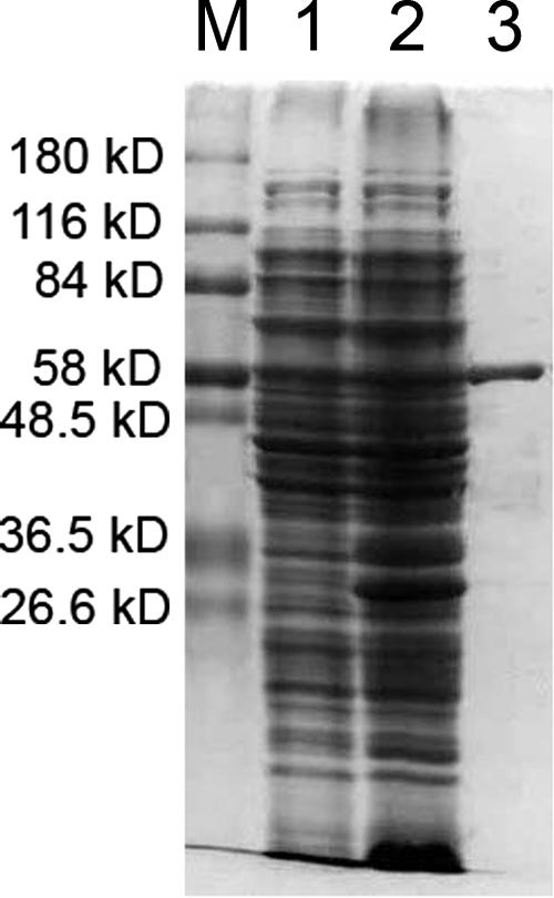
Nipah virus nucleocapsid (NiV-N) protein was expressed in Escherichia coli and purified by histidine tag-based affinity chromatography. An indirect immunoglobulin G (IgG) enzyme-linked immunosorbent assay (ELISA) for human and swine sera and an IgM capture ELISA for human sera were established using the recombinant NiV-N protein as an antigen. One hundred thirty-three suspected patient sera and 16 swine sera were used to evaluate the newly established ELISA systems in comparison with the CDC inactivated-virus-based ELISA systems. For the human sera, the NiV-N protein-based indirect IgG ELISA had a sensitivity of 98.6% and a specificity of 98.4%, and the NiV-N protein-based IgM capture ELISA had a sensitivity of 91.7% and a specificity of 91.8%, with reference to the CDC ELISA systems. The NiV-N-based IgM ELISA was found to be more sensitive than the inactivated-virus-based ELISA in that it captured eight additional cases. For the swine sera, the two test systems were in 100% concordance. Our data indicate that the Nipah virus nucleocapsid protein is a highly immunogenic protein in human and swine infections and a good target for serodiagnosis. Our NiV-N protein-based ELISA systems are useful, safe, and affordable tools for diagnosis of Nipah virus infection and are especially fit to be used in large-scale epidemiological investigations and to be applied in developing countries.
Figures
References
-
- Anonymous. 2004. Nipah virus outbreak(s) in Bangladesh, January-April 2004. Wkly. Epidemiol. Rec. 79:161-172. - PubMed
-
- Bradford, M. M. 1976. A rapid and sensitive method for the quantitation of microgram quantities of protein utilizing the principle of protein-dye binding. Anal. Biochem. 72:248-254. - PubMed
-
- Chua, K. B., W. J. Bellini, P. A. Rota, B. H. Harcourt, A. Tamin, S. K. Lam, T. G. Ksiazek, P. E. Rollin, S. R. Zaki, W. Shieh, C. S. Goldsmith, D. J. Gubler, J. T. Roehrig, B. Eaton, A. R. Gould, J. Olson, H. Field, P. Daniels, A. E. Ling, C. J. Peters, L. J. Anderson, and B. W. Mahy. 2000. Nipah virus: a recently emergent deadly paramyxovirus. Science 288:1432-1435. - PubMed
-
- Chua, K. B., C. L. Koh, P. S. Hooi, K. F. Wee, J. H. Khong, B. H. Chua, Y. P. Chan, M. E. Lim, and S. K. Lam. 2002. Isolation of Nipah virus from Malaysian Island flying-foxes. Microbes Infect. 4:145-151. - PubMed
Publication types
MeSH terms
Substances
LinkOut - more resources
Full Text Sources
