. 2006 Nov;26(22):8639-54.
doi: 10.1128/MCB.00816-06. Epub 2006 Sep 5.

Identification of yin-yang regulators and a phosphorylation consensus for male germ cell-associated kinase (MAK)-related kinase

Affiliations

Identification of yin-yang regulators and a phosphorylation consensus for male germ cell-associated kinase (MAK)-related kinase

Zheng Fu et al. Mol Cell Biol. 2006 Nov.

Abstract

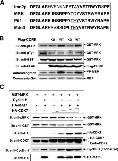
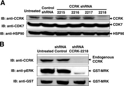
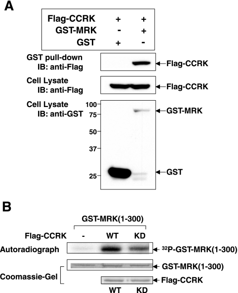
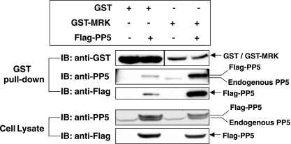
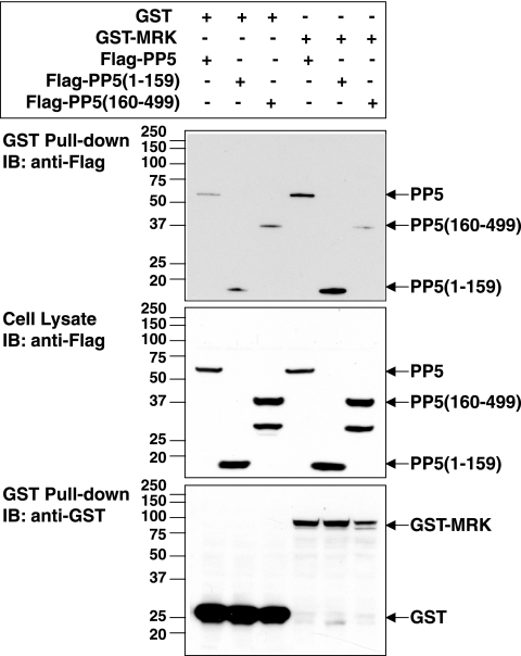
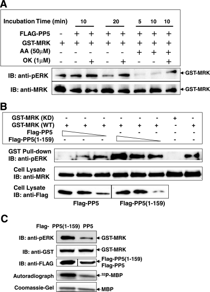
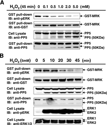
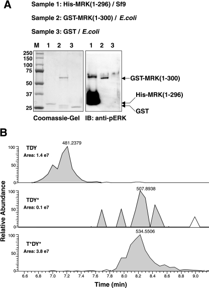
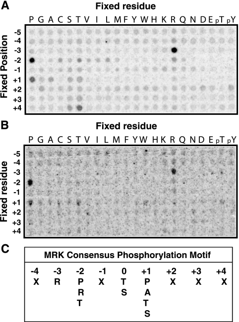
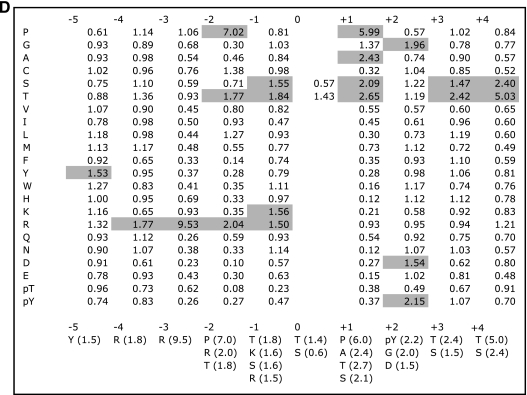
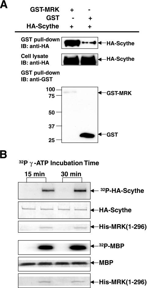
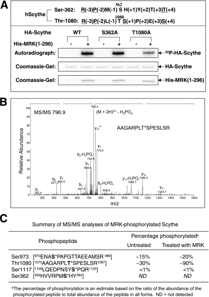
MAK (male germ cell-associated protein kinase) and MRK/ICK (MAK-related kinase/intestinal cell kinase) are human homologs of Ime2p in Saccharomyces cerevisiae and of Mde3 and Pit1 in Schizosaccharomyces pombe and are similar to human cyclin-dependent kinase 2 (CDK2) and extracellular signal-regulated kinase 2 (ERK2). MAK and MRK require dual phosphorylation in a TDY motif catalyzed by an unidentified human threonine kinase and tyrosine autophosphorylation. Herein, we establish that human CDK-related kinase CCRK (cell cycle-related kinase) is an activating T157 kinase for MRK, whereas active CDK7/cyclin H/MAT1 complexes phosphorylate CDK2 but not MRK. Protein phosphatase 5 (PP5) interacts with MRK in a complex and dephosphorylates MRK at T157 in vitro and in situ. Thus, CCRK and PP5 are yin-yang regulators of T157 phosphorylation. To determine a substrate consensus, we screened a combinatorial peptide library with active MRK. MRK preferentially phosphorylates R-P-X-S/T-P sites, with the preference for arginine at position -3 (P-3) being more stringent than for prolines at P-2 and P+1. Using the consensus, we identified a putative phosphorylation site (RPLT(1080)S) for MRK in human Scythe, an antiapoptotic protein that interacts with MRK. MRK phosphorylates Scythe at T1080 in vitro as determined by site-directed mutagenesis and mass spectrometry, supporting the consensus and suggesting Scythe as a physiological substrate for MRK.


Figures

FIG. 1.
FIG. 1.
CCRK, but not the Cdk7 complex, increases MRK phosphorylation in the TDY motif in cells. (A) Alignment of the T-loops of MRK, MAK, and their putative homologs in yeasts, Ime2p, Pit1, and Mde3. Identical amino acids are shown in bold type, and the TDY motif is underlined. (B and C) GST-MRK was coexpressed with either WT and KD Flag-CCRK (B) or recombinant Cdk7/cyclin H/MAT1 complex (C) in HEK293T cells. GST fusion proteins were pulled down by glutathione-Sepharose beads and analyzed for MRK phosphorylation in the TDY motif by Western blotting (immunoblotting [IB]) against the anti-phospho-ERK antibody (anti-pERK). The amounts of recombinant proteins on beads and in cell lysate were indicated by Western blotting. After coexpression with either WT or KD Flag-CCRK, GST-MRK bead samples were assayed for kinase activity in vitro using myelin basic protein as the substrate (B). Endo+Exo, endogenous and exogenous.
FIG. 2.
FIG. 2.
Downregulation of endogenous CCRK expression impairs phosphorylation of GST-MRK in the TDY motif. (A) HEK293T cells were infected with either four different lentiviral shRNAs targeting the CCRK gene or a nontarget control lentiviral shRNA. Two days after infection, cells were harvested and lysed. Expression of endogenous CCRK in whole-cell lysate was analyzed by Western blotting (immunoblotting [IB]) against the anti-CCRK antibody. Expression of CDK7 and Hsp90 was analyzed as controls. (B) HEK293T cells that were either uninfected or infected with nontarget control lentiviral shRNA or CCRK-specific lentiviral shRNA-2218 were transfected with GST-MRK. Two days after transfection, cells were harvested and lysed. Expression of CCRK and GST-MRK in whole-cell lysate was analyzed by Western blotting against the anti-CCRK and anti-GST antibodies, respectively. Phosphorylation of GST-MRK in the TDY motif was assessed by Western blotting against the anti-phospho-ERK antibody (anti-pERK).
FIG. 3.
FIG. 3.
Recombinant CCRK interacts with recombinant MRK in cells and phosphorylates recombinant MRK in vitro. (A) GST-MRK or GST was coexpressed with Flag-CCRK in HEK293T cells. GST fusion proteins were pulled down by glutathione-Sepharose beads and analyzed for association with Flag-CCRK by Western blotting (immunoblotting [IB]) against the anti-Flag antibody. The amounts of GST, GST-MRK, and Flag-CCRK in cell lysate were indicated by Western blotting against the anti-GST and anti-Flag antibodies. The positions of molecular mass markers (in kilodaltons) are shown to the left of the blot. (B) GST-MRK(1-300) purified from E. coli was assayed for in vitro phosphorylation by WT or KD Flag-CCRK purified from HEK293T cells. MRK phosphorylation (autoradiograph) and the amounts of substrates and kinases (Coomassie blue-stained gels) are shown.
FIG. 4.
FIG. 4.
Recombinant MRK interacts with both recombinant and endogenous PP5 in cells. GST-MRK or GST was expressed alone or coexpressed with Flag-PP5 in HEK293T cells. GST fusion proteins were purified on glutathione-Sepharose beads and analyzed for association with recombinant and/or endogenous PP5 by Western blotting (immunoblotting [IB]) against the anti-Flag and anti-PP5 antibodies, respectively. The amounts of GST fusion proteins on beads and the amounts of Flag-PP5 and endogenous PP5 in cell lysate were indicated by Western blotting.
FIG. 5.
FIG. 5.
The TPR domain of PP5 is required for the interaction between PP5 and MRK. GST-MRK or GST was coexpressed with Flag-tagged full-length PP5 or PP5(1-159), consisting of only the N-terminal TPR domain, or PP5(160-499), lacking the TPR domain, in HEK293T cells. GST fusion proteins were purified on glutathione-Sepharose beads and analyzed for association with Flag-PP5 as described in the legend to Fig. 4. Note that the anti-Flag antibody also recognized a 30-kDa band in addition to the 37-kDa band of Flag-PP5(160-499) (middle gel), which is probably a product of proteolysis or truncation in translation. IB, immunoblotting.
FIG. 6.
FIG. 6.
PP5 dephosphorylates MRK in the TDY motif in vitro and in vivo. (A) GST-MRK purified on glutathione-Sepharose beads from HEK293T cells was incubated with purified Flag-PP5 protein in the absence (−) or presence (+) of 50 μM arachidonic acid (AA) in vitro. For a control, 1 μM okadaic acid (OK) was added to inhibit PP5 phosphatase activity. After the reaction, the GST-MRK bead samples were analyzed for MRK phosphorylation in the TDY motif by Western blotting (immunoblotting [IB]) against the anti-phospho-ERK antibody (anti-pERK). The amount of GST-MRK on beads was indicated by Western blotting against the anti-MRK antibody. (B) GST-MRK was coexpressed with either the full-length Flag-PP5 or the catalytic inactive form Flag-PP5(1-159) in HEK293T cells. GST fusion proteins were purified on glutathione-Sepharose beads and analyzed for MRK phosphorylation in the TDY motif as described above for panel A. The amounts of GST-MRK, Flag-PP5, and Flag-PP5(1-159) in cell lysate were indicated by Western blotting against the anti-MRK and anti-Flag antibodies. KD GST-MRK was used as a negative control for TDY motif phosphorylation. (C) GST-MRK was coexpressed with either the full-length Flag-PP5 or the catalytic inactive form Flag-PP5(1-159) in HEK293T cells. GST fusion proteins were analyzed for MRK phosphorylation in the TDY motif as described above for panel B and for MRK kinase activity in vitro using MBP as the substrate.
FIG. 7.
FIG. 7.
Activation of endogenous PP5 by H2O2-induced oxidative stress causes dephosphorylation in the TDY motif of MRK. HEK293T cells transfected with GST-MRK were treated either with an increasing concentration of H2O2 (0 to 5 mM) for 45 min (A) or with 5 mM H2O2 for an increasing length of time (0 to 45 min) (B) before harvesting. GST fusion proteins were pulled down by glutathione-Sepharose beads and analyzed for MRK phosphorylation in the TDY motif by Western blotting (immunoblotting [IB]) against the anti-phospho-ERK antibody (pERK). The amounts of GST-MRK on beads were indicated by Western blotting against the anti-GST antibody. The amounts of endogenous full-length PP5 (55 kDa) and the C-terminally truncated form of PP5 (50 kDa) in cell lysate and on beads were indicated by the anti-PP5 antibody. For comparison, the TEY motif phosphorylation of ERK1 and ERK2 under oxidative stress was also analyzed by the anti-phospho-ERK antibody (B).
FIG. 8.
FIG. 8.
His-MRK(1-296) purified from Sf9 cells is highly active and is predominantly doubly phosphorylated in the TDY motif. His-MRK(1-296) was expressed in Sf9 cells and affinity purified. The purity was indicated on a Coomassie blue-stained SDS gel (A), and the state of the TDY motif phosphorylation was analyzed by Western blotting (immunoblotting [IB]) against the anti-phospho-ERK antibody (pERK) (A) and by mass spectrometry (B). The positions of molecular mass markers (M) are indicated (in kilodaltons) to the left of the gel. (B) Comparison of selected ion chromatograms from MS targeted analysis of tryptic peptides containing the TDY motif is shown. The peak area of each form of the TDY motif phosphorylation was calculated to indicate relative abundance. The control is GST-MRK(1-300) purified from E. coli that is weakly reactive to the anti-phospho-ERK antibody (A) and not detectably doubly phosphorylated in the TDY motif by mass spectrometry (17).
FIG. 9.
FIG. 9.
Determination of MRK phosphorylation specificity. Using a combinatorial peptide library method (25), we analyzed MRK phosphorylation specificity using purified His-MRK(1-296) protein from Sf9 cells (A) and purified GST-MRK(1-300) protein from E. coli (B). Note that the apparent preferences for peptides with fixed Ser and Thr residues may reflect phosphorylation at the fixed residue rather than at the central position and should be interpreted with caution. (C) The substrate phosphorylation consensus for MRK is R-P-X-S/T-P, with the preference for arginine at P−3 being more stringent than for prolines at P−2 and P+1. (D) Sequence preferences for phosphorylation by MRK. The amount of radioactivity incorporated into each peptide mixture was measured by a phosphorimager using ImageQuant software (Molecular Dynamics). Data are normalized so that the average value for the 20 natural amino acids within a given position is 1; thus, values greater than 1 indicate positive selection. Selectivity values are therefore given relative to the other residues at the same position. Note that cysteine selections should be treated more qualitatively because oxidation is likely to make quantitation of cysteine unreliable. Also note that the apparent preferences for peptides with fixed Ser and Thr residues may reflect phosphorylation at the fixed residue rather than at the central position and should be interpreted with caution. Values for preferred residues are shaded.
FIG. 9.
FIG. 9.
Determination of MRK phosphorylation specificity. Using a combinatorial peptide library method (25), we analyzed MRK phosphorylation specificity using purified His-MRK(1-296) protein from Sf9 cells (A) and purified GST-MRK(1-300) protein from E. coli (B). Note that the apparent preferences for peptides with fixed Ser and Thr residues may reflect phosphorylation at the fixed residue rather than at the central position and should be interpreted with caution. (C) The substrate phosphorylation consensus for MRK is R-P-X-S/T-P, with the preference for arginine at P−3 being more stringent than for prolines at P−2 and P+1. (D) Sequence preferences for phosphorylation by MRK. The amount of radioactivity incorporated into each peptide mixture was measured by a phosphorimager using ImageQuant software (Molecular Dynamics). Data are normalized so that the average value for the 20 natural amino acids within a given position is 1; thus, values greater than 1 indicate positive selection. Selectivity values are therefore given relative to the other residues at the same position. Note that cysteine selections should be treated more qualitatively because oxidation is likely to make quantitation of cysteine unreliable. Also note that the apparent preferences for peptides with fixed Ser and Thr residues may reflect phosphorylation at the fixed residue rather than at the central position and should be interpreted with caution. Values for preferred residues are shaded.
FIG. 10.
FIG. 10.
Recombinant MRK interacts with recombinant Scythe in cells and phosphorylates recombinant Scythe in vitro. (A) GST-MRK or GST was coexpressed with Flag-Scythe in HEK293T cells. GST fusion proteins were pulled down by glutathione-Sepharose beads and analyzed for association with Flag-Scythe by Western blotting (immunoblotting [IB]) against the anti-Flag antibody. The amounts of GST, GST-MRK and Flag-Scythe in cell lysate were indicated by Western blotting. The positions of molecular mass markers (in kilodaltons) are indicated to the left of the blot. (B) Flag-Scythe immunoprecipitated from HEK293T cells was assayed for phosphorylation by MRK in vitro using purified His-MRK(1-296) (top panel). MBP served as a positive-control substrate for MRK kinase activity (bottom panel).
FIG. 11.
FIG. 11.
Thr-1080 is an in vivo phosphorylation site located within the BAG domain of Scythe that can be phosphorylated by MRK in vitro. (A) In vitro kinase assay of two point mutations (S362A and T1080A) on Scythe by MRK. hScythe, human Scythe. (B) Tandem mass spectrometry (MS/MS) of the phosphorylated form of the RPLT1080SP tryptic peptide that is derived from in-gel digestion of affinity-purified Flag-Scythe (see Materials and Methods). (C) Summary of mass spectrometry analyses of the relative phosphorylation level of Scythe on several sites (shown in bold type with an asterisk) with and without MRK treatment in vitro. The in vitro kinase assay was done as described above for panel A, except that only nonradiolabeled ATP was used.

Similar articles

Cited by

References

    1. Abe, H., and C. Shimoda. 2000. Autoregulated expression of Schizosaccharomyces pombe meiosis-specific transcription factor Mei4 and a genome-wide search for its target genes. Genetics 154:1497-1508. - PMC - PubMed
    1. Abe, S., T. Yagi, S. Ishiyama, M. Hiroe, F. Marumo, and Y. Ikawa. 1995. Molecular cloning of a novel serine/threonine kinase, MRK, possibly involved in cardiac development. Oncogene 11:2187-2195. - PubMed
    1. Ali, A., J. Zhang, S. Bao, I. Liu, D. Otterness, N. M. Dean, R. T. Abraham, and X. F. Wang. 2004. Requirement of protein phosphatase 5 in DNA-damage-induced ATM activation. Genes Dev. 18:249-254. - PMC - PubMed
    1. Banerji, J., J. Sands, J. L. Strominger, and T. Spies. 1990. A gene pair from the human major histocompatibility complex encodes large proline-rich proteins with multiple repeated motifs and a single ubiquitin-like domain. Proc. Natl. Acad. Sci. USA 87:2374-2378. - PMC - PubMed
    1. Barzilai, A., and K. Yamamoto. 2004. DNA damage responses to oxidative stress. DNA Repair 3:1109-1115. - PubMed

Publication types

MeSH terms