. 2006 Nov;26(22):8418-26.
doi: 10.1128/MCB.00821-06. Epub 2006 Sep 5.

Rapid loss of intestinal crypts upon conditional deletion of the Wnt/Tcf-4 target gene c-Myc

Affiliations

Rapid loss of intestinal crypts upon conditional deletion of the Wnt/Tcf-4 target gene c-Myc

Vanesa Muncan et al. Mol Cell Biol. 2006 Nov.

Erratum in

Abstract

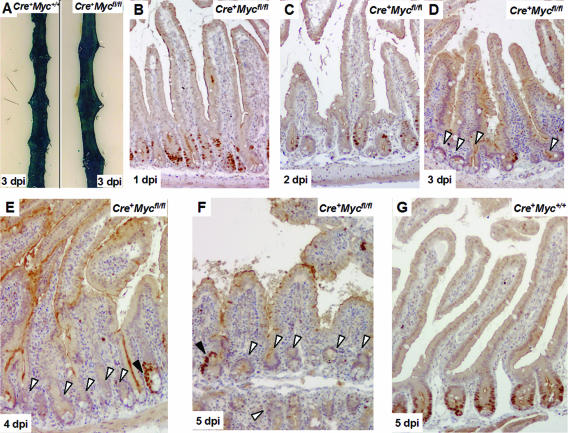
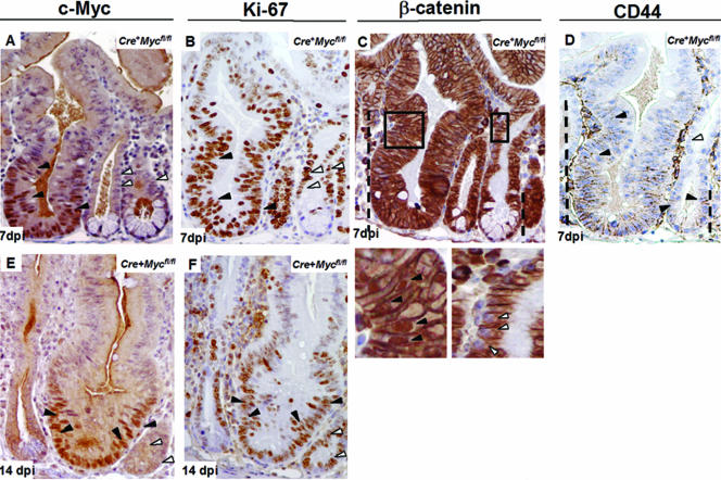
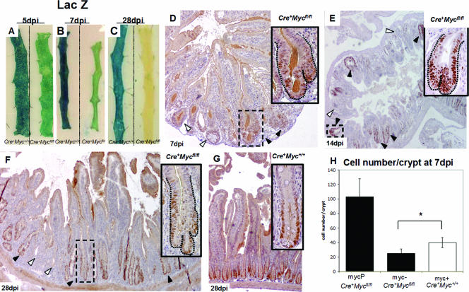
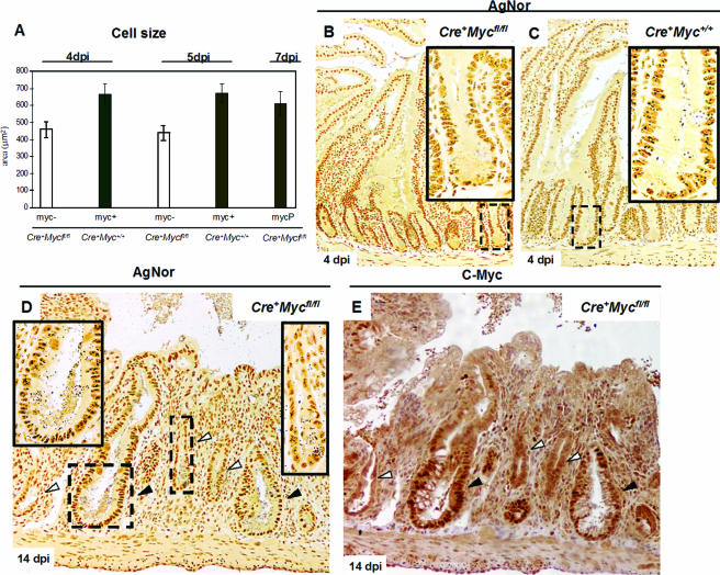
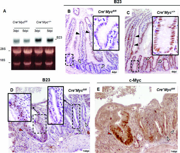
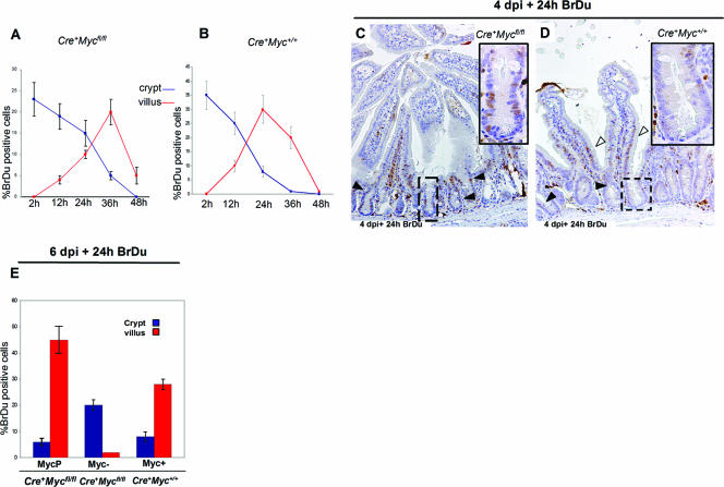
Inhibition of the mutationally activated Wnt cascade in colorectal cancer cell lines induces a rapid G1 arrest and subsequent differentiation. This arrest can be overcome by maintaining expression of a single Tcf4 target gene, the proto-oncogene c-Myc. Since colorectal cancer cells share many molecular characteristics with proliferative crypt progenitors, we have assessed the physiological role of c-Myc in adult crypts by conditional gene deletion. c-Myc-deficient crypts are lost within weeks and replaced by c-Myc-proficient crypts through a fission process of crypts that have escaped gene deletion. Although c-Myc(-/-) crypt cells remain in the cell cycle, they are on average much smaller than wild-type cells, cycle slower, and divide at a smaller cell size. c-Myc appears essential for crypt progenitor cells to provide the necessary biosynthetic capacity to successfully progress through the cell cycle.


Figures

FIG. 1.
FIG. 1.
Assessment of c-Myc deletion in β-napthoflavone-induced Cre+ Mycfl/fl intestines over a 5-day time course. (A) β-Galactosidase staining on intestinal whole mounts 3 days p.i. [dpi], indicating near-100% recombination at the Rosa26 reporter allele. Left, Cre+ Myc+/+; right, Cre+ Mycfl/fl. (B and C) c-Myc staining at days 1 and 2 p.i., showing reduced levels of c-Myc protein but still significant numbers of c-Myc-positive cells decorating the crypts. (D to F) Large areas of c-Myc-deficient epithelium at later time points (white arrows). Black arrows in panels E and F depict occasional c-Myc-proficient cells. Note that at later time points c-Myc-proficient cells clustered. (G) Representative section of Cre+ Myc+/+ control mice, where c-Myc protein was uniformly expressed at the lower half of the crypt region.
FIG. 2.
FIG. 2.
c-Myc-proficient cells in Cre+ Mycfl/fl mice are dominant over c-Myc-deficient cells and form enlarged crypts. (A and E) Representative sections from Cre+ Mycfl/fl mice at 7 and 14 days p.i. [dpi], respectively, stained with anti c-Myc antibody. Enlarged crypts are composed exclusively of c-Myc-proficient cells (black arrows), while the significantly smaller crypts contain cells expressing no c-Myc protein (white arrows). (B and F) Ki-67 staining showed cell proliferation in both c-Myc-proficient and -deficient crypts in Cre+ Mycfl/fl mice (black arrows versus white arrows). Note that c-Myc staining and Ki-67 staining were performed on consecutive sections. (C) Consecutive paraffin section of Cre+ Mycfl/fl mice at 7 days p.i. stained with antibody to β-catenin. The dashed lines depict the position until nuclear β-catenin translocation could be observed. A much higher cell position with nuclear β-catenin from a c-Myc-proficient crypt is also presented as a magnification of the squared area (left lower panel). In contrast, similar cell positions in a Myc-deficient crypt have no nuclear β-catenin (right lower panel). (D) Serial paraffin sections of Cre+ Mycfl/fl mice at 7 days postdeletion stained with antibody to CD44. The CD44 staining pattern follows that of nuclear β-catenin (dashed lines). Expanded CD44 expression in c-Myc-proficient crypts is marked with black arrows. Cells at equivalent positions in c-Myc-deficient crypts have no CD44 expression (white arrows).
FIG. 3.
FIG. 3.
Mycfl/fl crypts are lost in Cre+ Mycfl/fl mice after 4 weeks of induction and are replaced by Myc+/+ crypts through crypt fission. (A to C) LacZ-stained whole-mount preparations from Cre+ Mycfl/fl and control mice at day 5 (A), day 7 (B), and day 28 (C) p.i., indicating preferential repopulation with unrecombined cells in the induced Cre+ Mycfl/fl mice (right intestinal strip in each panel) which was almost complete at 28 days p.i. (D, E, and F) c-Myc staining at 7, 14, and 28 days p.i., respectively, in sections from Cre+ Mycfl/fl mice, indicating repopulation from c-Myc-proficient, hyperplastic crypts through crypt fission (black arrows). (G) Cre+ Myc+/+ control mice. (H) Graph showing reduced cell numbers per crypt in c-Myc-deficient crypts (myc-) from Cre+ Mycfl/fl mice compared to c-Myc-expressing crypts (myc+) of control Cre+ Myc+/+ mice at 7 days p.i. (Cre+ Mycfl/fl, 25 ± 6 [n = 2]; Cre+ Myc+/+, 40 ± 7 [n = 3]; *, P = 0.025 by Mann-Whitney U test). Data are means ± standard deviations; n is the number of mice analyzed. The first bar in the graph represents numbers of cells in c-Myc-proficient crypts (MycP) in Cre+ Mycfl/fl mice (103 ± 24; n = 2). A total of 100 crypts was analyzed per mouse.
FIG. 4.
FIG. 4.
Size and biosynthetic capacity are reduced in c-Myc-deficient cells. (A) Graph summarizing cell size reduction in c-Myc-deficient cells (myc-). Data were analyzed at two time points after induction, and similar results were obtained (Cre+Mycfl/fl, 459 ± 46 μm2, 4 days p.i. [dpi]; Cre+ Myc+/+, 662 ± 61 μm2, 4 days p.i.; Cre+ Mycfl/fl, 437 ± 43 μm2, 5 days p.i.; Cre+ Myc+/+, 672 ± 53 μm2, 5 days p.i., n = 3). The last bar represents the cell size of c-Myc-proficient cells (MycP) of Cre+ Mycfl/fl mice at 7 days p.i. (611 ± 70 μm2; n = 2). Data are means ± standard deviations, and n is the number of mice analyzed per time point. (B) Underrepresentation of AgNor dots, regions of ribosomal gene synthesis, in c-Myc-deficient cells and crypts of Cre+ Mycfl/fl mice at 4 days p.i. (C) Corresponding control Cre+ Myc+/+ mice with multiple large AgNor dots per nucleus. Insets are magnifications of representative squared areas. (D) Sections from Cre+Mycfl/fl mice at 7 days p.i. and incubated with AgNor. Note that at this stage, smaller c-Myc-deficient crypts (white arrows, right-hand magnification inset) clearly have fewer and smaller AgNors compared in the same section with c-Myc-proficient crypts (black arrows, left-hand magnification inset). (E) Consecutive paraffin section stained with c-Myc Ab, showing smaller crypts with fewer AgNors being c-Myc negative (white arrows).
FIG. 5.
FIG. 5.
Nucleophosmin (B23) protein, involved in ribosomal gene synthesis and a component of AgNor dots, is downregulated in Cre+ Mycfl/fl mice. (A) Northern blot showing downregulation of B23 RNA in Cre+ Mycfl/fl mice (two left lanes, upper panel) compared with Cre+ Myc+/+ mice (two right lanes, upper panel). Lower panel: ethidium bromide-stained 1% denatured agarose gel. (B and C) IHC for B23 protein in mouse small intestine samples. Crypt cells from Cre+ Mycfl/fl mice express almost undetectable levels of B23 protein (white arrows in panel B), and expression in the villus is reduced (black arrows in panel B). Cre+ Myc+/+ control intestine samples show uniform B23 expression throughout crypt villus units (white arrows in panel C). (D) B23 IHC at 14 days p.i. of intestine from Cre+ Mycfl/fl mice, clearly underscoring the difference in B23 levels between c-Myc-deficient (black arrows, left inset) and -proficient crypts (white arrows, right inset). (E) IHC with c-Myc Ab on a consecutive paraffin section, confirming that B23-negative crypts are also c-Myc negative. dpi, days p.i.
FIG. 6.
FIG. 6.
c-Myc-deficient crypt cells from Cre+ Mycfl/fl mice cycle profoundly slower than c-myc-expressing cells from Cre+ Myc+/+ control mice. (A and B) BrdU time course. Mice at 4 days p.i. (dpi) were injected with BrdU and harvested 2, 12, 24, 36, and 48 h later. BrdU-positive cells were scored as a percentage where the number of positive cells was normalized with the total number of cells per crypt (blue lines) or villus (red lines). Villus BrdU+ cells reached maximum numbers at 36 h, while the maximum number for control Cre+Myc+/+ mice was already reached at 24 h. (C and D) IHC to BrdU antibody 24 h postinjection. Significant numbers of BrdU+ cells did not exit the crypt in Cre+ Mycfl/fl mice (C, black arrows), while only a few cells decorated the crypts in Cre+ Myc+/+ mice (D, black arrows), with the majority of cells present in the villus (D, white arrows). (E) Graph summarizing the percentage of BrdU-labeled cells in the crypt and villus in Cre+ Mycfl/fl and Cre+ Myc+/+ mice induced for 6 days and pulsed for 24 h. Data are means ± standard deviations, and a minimum of three mice were analyzed per genotype per BrdU time point.

References

    1. Arabi, A., S. Wu, K. Ridderstrale, H. Bierhoff, C. Shine, K. Fatyoul, S. Fahlen, P. Hydbring, O. Soderberg, I. Grummt, L. G. Larson, and A. P. Write. 2005. c-Myc associates with ribosomal DNA and activates RNA polymerase I transcription. Nat. Cell Biol. 7:303-310. - PubMed
    1. Baena, E., A. Gandarillas, M. Vallespinos, J. Zanet, O. Bachs, C. Redondo, I. Fabregat, C. Martinez-A, and I. M. de Alboran. 2005. c-Myc regulates cell size and ploidy but is not essential for postnatal proliferation in liver. Proc. Natl. Acad. Sci. USA 102:7286-7291. - PMC - PubMed
    1. Bettess, M. D., N. Dubois, M. J. Murphy, C. Dubey, C. Roger, S. Robine, and A. Trumpp. 2005. c-Myc is required for the formation of intestinal crypts but dispensable for homeostasis of the adult intestinal epithelium. Mol. Cell. Biol. 25:7868-7878. - PMC - PubMed
    1. Booth, C., and C. S. Potten. 2000. Gut instincts: thoughts on intestinal epithelial stem cells. J. Investig. Dermatol. 114:667-673. - PMC - PubMed
    1. Booth, D., and C. S. Potten. 2001. Protection against mucosal injury by growth factors and cytokines. J. Natl. Cancer Inst. Monogr. 29:16-20. - PubMed

Publication types