. 2006 Nov;26(21):8022-31.
doi: 10.1128/MCB.01091-06. Epub 2006 Sep 5.

Notch1 contributes to mouse T-cell leukemia by directly inducing the expression of c-myc

Affiliations

Notch1 contributes to mouse T-cell leukemia by directly inducing the expression of c-myc

Vishva Mitra Sharma et al. Mol Cell Biol. 2006 Nov.

Abstract

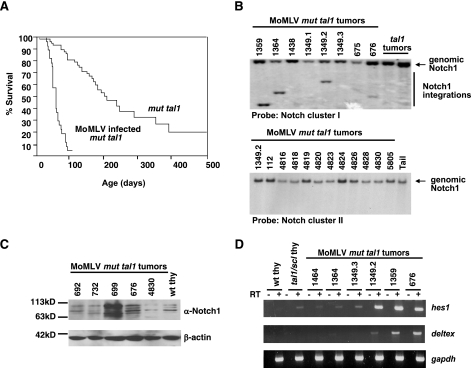
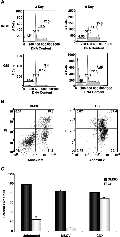
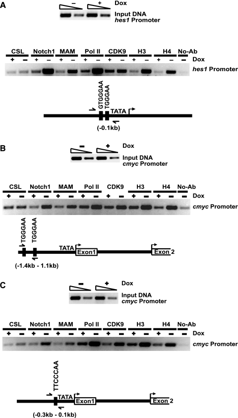
Recent work with mouse models and human leukemic samples has shown that gain-of-function mutation(s) in Notch1 is a common genetic event in T-cell acute lymphoblastic leukemia (T-ALL). The Notch1 receptor signals through a gamma-secretase-dependent process that releases intracellular Notch1 from the membrane to the nucleus, where it forms part of a transcriptional activator complex. To identify Notch1 target genes in leukemia, we developed mouse T-cell leukemic lines that express intracellular Notch1 in a doxycycline-dependent manner. Using gene expression profiling and chromatin immunoprecipitation, we identified c-myc as a novel, direct, and critical Notch1 target gene in T-cell leukemia. c-myc mRNA levels are increased in primary mouse T-cell tumors that harbor Notch1 mutations, and Notch1 inhibition decreases c-myc mRNA levels and inhibits leukemic cell growth. Retroviral expression of c-myc, like intracellular Notch1, rescues the growth arrest and apoptosis associated with gamma-secretase inhibitor treatment or Notch1 inhibition. Consistent with these findings, retroviral insertional mutagenesis screening of our T-cell leukemia mouse model revealed common insertions in either notch1 or c-myc genes. These studies define the Notch1 molecular signature in mouse T-ALL and importantly provide mechanistic insight as to how Notch1 contributes to human T-ALL.


Figures

FIG. 1.
FIG. 1.
Notch1 activation in MoMLV-infected mut tal1 tumors. (A) Disease acceleration in MoMLV-infected mut tal1 transgenic mice. Shown is a Kaplan-Meier survival plot of MoMLV-infected tal1/scl transgenic mice. The cohort of tal1/scl transgenic mice consisted of 62 mice, and the cohort of infected mice consisted of 27 mice. (B) The notch1 locus is a common integration site in infected tumors. Genomic DNA from MoMLV-infected tumors (1359, 1364, 1438, 1349.1, 1349.2, 1349.3, 675, and 676) and from two uninfected tal1 tumors was digested with EcoRV and hybridized with a probe to cluster region I of Notch1 (13). (C) Notch1 activation in MoMLV-infected tumors. Lysates from tumors with MoMLV integrations in notch1 (699, 676, and 4830) were probed with an anti-Notch1 (α-Notch1) antibody and compared to lysates from infected tal1 tumors with no detectable Notch1 integrations (692 and 732) or from wild-type thymus (wt thy). (D) Hes1 and Deltex are expressed in tumors with MoMLV insertions in Notch1. hes1 and deltex expression was examined in wild-type and preleukemic tal1 thymocytes and MoMLV-infected tumors with insertions in Notch1 using RT-PCR. Glyceraldehyde-3-phosphate dehydrogenase (gapdh) was used as an internal control.
FIG. 2.
FIG. 2.
GSI treatment induces G0/G1 arrest followed by apoptosis, and leukemic growth is rescued by expression of intracellular Notch1. (A) G0/G1 arrest precedes the accumulation of sub-G1 cells. Mouse T-ALL cell line 130 with Notch1 PEST truncation was treated with DMSO or 1 μM N-[N-(3,5-difluorophenacetyl)-l-alanyl]-S-phenylglycine t-butyl ester (DAPT) for 3 or 6 days, and cells were assayed for DNA content by staining with PI. (B). GSI treatment of primary tal1 tumor tissue induces apoptosis. Primary thymic masses isolated directly from the animal were left untreated or were treated with 1 μM GSI. Three days later, cells were stained with FITC-Annexin V/PI and analyzed by flow cytometry. (C) GSI-induced effects in mouse T-ALL cells are rescued by human intracellular NOTCH1 (ICNX) expression. Mouse T-ALL line 720 was left uninfected or was infected with vector only or with a retrovirus that expresses human intracellular NOTCH1 (ICNX) and then was left untreated or was treated with 1 μM GSI (MRK-003) for 3 days, and the percentage of viable cells was determined by trypan blue exclusion. The rescue assay was repeated a minimum of three times. A representative experiment is shown.
FIG. 3.
FIG. 3.
A conditional Notch1IC mouse T-ALL cell line. (A) Dox administration represses Notch1IC expression. Fifty micrograms of protein isolated from wild-type thymocytes and doxycycline-regulated T-ALL cell line 3404 was left untreated or was treated with 2 μg/ml Dox. Cell lysates were separated on a 7. 5% SDS-PAGE gel and then probed with anti-Notch1 (α-Notch1) antibody. (B) Decreased hes1, deltex, and pre-Tα expression upon the addition of Dox. Expression of hes1, deltex, and pre-Tα in the Dox-regulated NotchIC T-ALL line was examined by RT-PCR following mock treatment or doxycycline (2 μg/ml) treatment for the time periods indicated. Glyceraldehyde-3-phosphate dehydrogenase (gapdh) was used as an internal control. (C) Dox treatment or Notch1IC inhibition induces G0/G1 arrest followed by an increase in sub-G1 cells. Following 48 or 72 h of doxycycline treatment, cells were assayed for DNA content by staining with PI. Similar to GSI treatment of mouse T-ALL lines, Dox treatment of 3404 cells induces G0/G1 arrest followed by an increase in sub-G1 cells.
FIG. 4.
FIG. 4.
Notch1-regulated genes in mouse T-ALL. Shown is a heat map of clustered samples in columns and clustered genes in rows of expression data of mouse leukemic cell line 3404 cultured in the presence or absence of doxycycline or Notch1IC. The pseudocolor representation of gene expression ratios is shown with the scale below. The hierarchical clustering was performed for genes having a change of more than twofold (P value of 0.005). NotchIC target genes are indicated by formula image
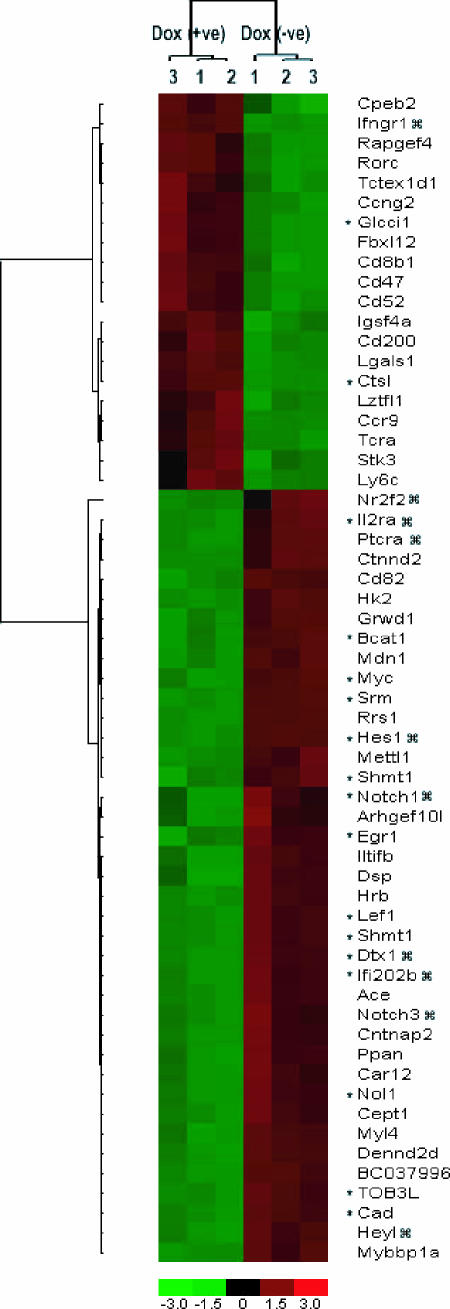
; c-myc target genes are indicated by an asterisk.
FIG. 5.
FIG. 5.
Leukemic growth is Notch1 and c-myc dependent. (A) c-myc expression is increased in primary tal1 tumors that contain mutations in Notch1. c-myc RNA levels were examined using real-time PCR in three primary tal1 tumors with insertions/deletions in the PEST region of Notch1. (B) GSI treatment results in downregulation of c-myc mRNA levels, whereas c-myc levels are maintained in leukemic cells infected with an intracellular Notch1 retrovirus. c-Myc RNA levels were measured using real-time PCR. RNA was harvested from the leukemic cell line 720 treated with DMSO or 1 μM N-[N-(3,5-difluorophenacetyl)-l-alanyl]-S-phenylglycine t-butyl ester (DAPT) and from 720 cells that were infected with MSCV or with a human ICNX retrovirus treated with DMSO or 1 μM DAPT. c-Myc levels were quantified using a real-time PCR assay. (C) Leukemic growth is rescued by c-Myc or intracellular Notch1 expression. Multiple mouse leukemic cell lines were left uninfected or were infected with MSCV or with retroviruses expressing either human intracellular NOTCH1 (ICNX) or mouse c-myc. Cultures were treated with either DMSO or DAPT (1 μm) for 6 days, and viable cells were determined by trypan blue staining. The experiment was repeated at least three times, and one representative experiment is shown.
FIG. 6.
FIG. 6.
Dox-dependent recruitment of Notch1IC and MAM to the hes1 and c-myc promoters. (A) ChIP assay for CSL, Notch1IC, MAM, RNA polymerase II (Pol II), CDK9, and acetylated histones H3 and H4. Briefly, the Dox-dependent 3404 leukemic cell line was grown in RPMI medium with 10% fetal bovine serum at 37°C and treated with doxycycline (2 μg/ml) for 48 h (+Dox) or left untreated (−Dox). Immunoprecipitations (IPs) were carried out using the respective monoclonal or polyclonal antibodies or without antibody (No-ab) as a control. Immunoprecipitated and input DNAs were amplified by PCR using primers specific for the mouse hes1 promoter (A) or the mouse c-myc promoter (B and C). PCR-amplified products were analyzed on 2% agarose gels and stained with ethidium bromide.

References

    1. Adler, S. H., E. Chiffoleau, L. Xu, N. M. Dalton, J. M. Burg, A. D. Wells, M. S. Wolfe, L. A. Turka, and W. S. Pear. 2003. Notch signaling augments T cell responsiveness by enhancing CD25 expression. J. Immunol. 171:2896-2903. - PubMed
    1. Bain, G., E. C. Maandag, D. J. Izon, D. Amsen, A. M. Kruisbeek, B. C. Weintraub, I. Krop, M. S. Schlissel, A. J. Feeney, and M. van Roon. 1994. E2A proteins are required for proper B cell development and initiation of immunoglobulin gene rearrangements. Cell 79:885-892. - PubMed
    1. Beverly, L. J., and A. J. Capobianco. 2003. Perturbation of Ikaros isoform selection by MLV integration is a cooperative event in Notch(IC)-induced T cell leukemogenesis. Cancer Cell 3:551-564. - PubMed
    1. Beverly, L. J., D. W. Felsher, and A. J. Capobianco. 2005. Suppression of p53 by Notch in lymphomagenesis: implications for initiation and regression. Cancer Res. 65:7159-7168. - PubMed
    1. Chervinsky, D. S., X.-F. Zhao, D. H. Lam, M. Ellsworth, K. W. Gross, and P. D. Aplan. 1999. Disordered T-cell development and T-cell malignancies in SCL LMO1 double-transgenic mice: parallels with E2A-deficient mice. Mol. Cell. Biol. 19:5025-5035. - PMC - PubMed

Publication types

MeSH terms

Substances

LinkOut - more resources