. 2006 Oct;26(20):7601-15.
doi: 10.1128/MCB.00836-06. Epub 2006 Sep 5.

A gradient of epidermal growth factor receptor signaling determines the sensitivity of rbf1 mutant cells to E2F-dependent apoptosis

Affiliations

A gradient of epidermal growth factor receptor signaling determines the sensitivity of rbf1 mutant cells to E2F-dependent apoptosis

Nam-Sung Moon et al. Mol Cell Biol. 2006 Oct.

Abstract

The inactivation of retinoblastoma (Rb) family members sensitizes cells to apoptosis. This cell death affects the development of mutant animals and also provides a critical constraint to the malignant potential of Rb mutant tumor cells. The extent of apoptosis caused by the inactivation of Rb is highly cell type and tissue specific, but the underlying reasons for this variation are poorly understood. Here, we characterize a specific time and place during Drosophila melanogaster development where rbf1 mutant cells are exquisitely sensitive to apoptosis. During the third larval instar, many rbf1 mutant cells undergo E2F-dependent cell death in the morphogenetic furrow. Surprisingly, this pattern of apoptosis is not caused by inappropriate cell cycle progression but instead involves the action of Argos, a secreted protein that negatively regulates Drosophila epidermal growth factor receptor (EGFR [DER]) activity. Apoptosis of rbf1 mutant cells is suppressed by the activation of DER, ras, or raf or by the inactivation of argos, sprouty, or gap1, and inhibition of DER strongly enhances apoptosis in rbf1 mutant discs. We show that RBF1 and a DER/ras/raf signaling pathway cooperate in vivo to suppress E2F-dependent apoptosis and that the loss of RBF1 alters a normal program of cell death that is controlled by Argos and DER. These results demonstrate that a gradient of DER/ras/raf signaling that occurs naturally during development provides the contextual signals that determine when and where the inactivation of rbf1 results in dE2F1-dependent apoptosis.


Figures

FIG. 1.
FIG. 1.
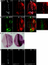
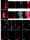
rbf1 mutant cells are prone to apoptosis at the anterior edge of the MF. (A) Third instar rbf1120a eye discs contain a distinctive pattern of apoptosis. Discs were treated with BrdU and immunostained with antibodies to BrdU and C3 to visualize S-phase cells and dying cells, respectively. Note that RBF1 is expressed throughout the disc, but its absence results in a stripe of apoptosis. ELAV stains cells that have initiated neuronal differentiation. (B) The apoptosis triggered by the mutation of rbf1 is cell autonomous. Mosaic clones of an rbf1-null allele, rbf114, were generated in eye discs using ey-FLP and FRT rbf114. rbf114 mutant clones are marked by the lack of green fluorescent protein (GFP) signal. Apoptosis occurs in both rbf1120a discs and rbf114 mutant clones as rbf1 mutant cells enter the MF. The MF passes through the discs shown from left to right.
FIG. 2.
FIG. 2.
Mutation of rbf1 activates dE2F1-dependent transcription, resulting in dE2F1-dependent cell death. Panels A and B show the relative position of apoptotic cells and the distribution of dE2F1 in rbf1120a eye discs using the pattern of cyclin B (Cyc B) expression as a reference point. Arrows indicate the position of the MF. The apoptotic cells, detected by anti-C3, are located in the anterior half of the gap between the stripes of cyclin B expression, while dE2F1 expression is elevated in a broad swath that spans the region between the areas of cyclin B expression. (C) In situ hybridization for the E2F target gene, rnr2, was used to compare the patterns of dE2F1-dependent transcription in wild-type and rbf1120a eye discs. Note that the expression of rnr2 is elevated and expanded in rbf1120a eye discs in a pattern that mirrors the distribution of dE2F1 protein. (D) Apoptosis in rbf1120a discs was completely suppressed by mutation of de2f1. Eye discs were stained with anti-C3 antibody to visualize the pattern of apoptosis.
FIG. 3.
FIG. 3.
Suppression of cell death in rbf1120a eye discs does not result in additional S-phase cells. Cell death in the MF of rbf1120a eye discs, as assayed by C3 staining or TUNEL (data not shown), was completely inhibited in mutant clones of dronc. The distribution of S-phase cells and apoptotic cells in the third instar eye discs was visualized with antibodies to BrdU and C3 as described before; dronc mutant clones are marked by the lack of GFP signal. Note the lack of additional S-phase cells in dronc mutant clones despite the inhibition of apoptosis. GFP, green fluorescent protein.
FIG. 4.
FIG. 4.
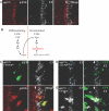
Activation of EGFR suppresses cell death in rbf1120a mutant eye discs. (A) In rbf1120a eye discs, apoptotic cells (stained with anti-C3) are located adjacent to cells that stain with antibodies to p-Erk. (B) A model showing how fluctuations in EGFR activity might regulate cell death in rbf1120a eye discs. The high EGFR activity that is present in differentiating cells and marked by p-Erk may provide a survival signal that protects them from apoptosis. At same time, these cells also express Argos, which inhibits EGFR signaling in the neighboring, uncommitted cells. (C) To test this model, clones of cells that overexpress the activated form of type II EGFR (DERact) were generated in rbf1120a eye discs using a heat shock flip-out technique. Seventy-two hours after the heat shock, eye discs were dissected and stained with anti-p-Erk and anti-C3 antibodies. The overexpressing clones are marked by green fluorescent protein (GFP). Strong p-Erk staining in DERact-expressing cells and its neighboring cells was observed in the furrow. Cells that activate p-Erk as a consequence of DERact expression are protected from apoptosis. (D and E) Clones that overexpress activated forms of Drosophila Raf, Phlgof, or wild-type AKT1 were generated using a heat shock flip-out technique. Clones were marked by GFP signal. Dying cells in the eye discs were visualized by C3 staining.
FIG. 5.
FIG. 5.
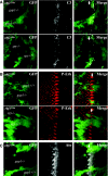
gap1 or sprouty inactivation is sufficient to suppress cell death in rbf1120a eye discs. gap1 or sprouty mutant clones were generated in rbf1120a eye using ey-FLP/FRT. Eye discs were dissected and stained with anti-C3 (A), anti-p-Erk (B), and anti-Atonal (C) antibodies. Mutant clones are marked by absence of green fluorescent protein (GFP) signals. Note the reduced levels of C3-positive cells in gap1 or sprouty mutant clones and the relatively normal pattern of p-Erk staining in the mutant clones.
FIG. 6.
FIG. 6.
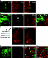
Argos is an important regulator of cell death in rbf1120a eye discs. (A) Eye discs from wild-type control third instar larval were dissected and stained with anti-p-Erk and anti-Argos (Aos) antibodies. The p-Erk staining pattern and the boundary of Argos staining follow the MF and moves across the discs shown from left to right. Note that cells on the right of the panel shown will be exposed to Argos before the stripe of p-Erk staining reaches them (B) argos mutant clones were generated in rbf1120a eye using the ey-FLP/FRT technique. Eye discs were dissected and stained with anti-p-Erk and anti-C3 antibodies. In argos mutant clones (marked by the lack of green fluorescent protein [GFP]), C3 staining is greatly reduced. Note that p-Erk staining is increased but has a relatively normal pattern in argos mutant clones. (C) The level of Argos expression is unaltered in rbf1 mutant eye discs. Twenty-five pairs of control and rbf1120a eye discs were dissected, and protein levels were compared on Western blots. (D) Wild-type and rbf1120a eye discs have a similar pattern of Argos expression. (E) Pupal eye discs containing rbf114 mutant clones (marked by the lack of GFP) were dissected, and dying cells were visualized using anti-C3 antibody. Elevated cell death was observed in rbf114 mutant clones at various time points of pupation (illustrated here 24 h after pupal formation). (F) Pupal eye discs at 42 h after pupal formation, which contain rbf114 mutant clones (marked by the lack of GFP), were dissected, and cell boundaries were visualized using anti-disc large antibody (Dlg). The yellow arrows indicate the places where secondary pigment cells or cone cells are missing. (G) Cell death was inhibited by expression of baculoviral protein p35 in pupal eye discs described in panel F, restoring the missing cells to rbf114 mutant clones and revealing the extent of ectopic proliferation.
FIG. 7.
FIG. 7.
RBF1 and EGFR cooperate to protect cells from apoptosis. This figure shows the effects of expressing a dominant-negative mutant of DER (DERdn) in wild-type (A) and rbf1120a (B) eye discs. Anti-cyclin B (Cyc B) and anti-C3 antibodies were used to visualize the patterns of cell death. The enlarged regions are indicated by the dashed boxes. (C) Eye discs from the indicated genotypes were coimmunostained with anti-Atonal (Ato) and anti-p-Erk antibodies. Expression of DERdn inhibits p-Erk staining patterns in both control and rbf1120a eye discs. The widespread cell death caused by DERdn in rbf1120a eye discs causes the almost complete loss of Atonal-expressing cells.
FIG. 8.
FIG. 8.
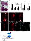
hid is up regulated in rbf1 mutant eye discs and is required for apoptosis. (A) In situ hybridization with a riboprobe of hid antisense sequences shows that hid mRNA is deregulated in rbf1120a eye discs. (B) Real-time quantitative PCR was used to measure the change in hid mRNA levels in rbf1120a eye discs (left panel) or whole larvae (right panel). Two known RBF1/dE2F1 targets, rnr2 and cyclin E, served as positive controls. Expression levels were measured in comparison to the rp49 housekeeping gene and the changes (fold) in rbf1120a samples are shown relative to wild-type controls. Averages were calculated using triplicate samples. (C) Heterozygosity of hid suppresses cell death in rbf1120a mutant eye discs. Apoptotic cells were visualized with anti-C3 in third instar eye discs of the indicated genotypes. Df(3L)x14 removes hid, while Df(3L)x38 removes sickle and reaper. Note the almost complete absence of cell death in rbf1120a eye discs heterozygous for Df(3L)x14. (D) Cells in the MF are highly sensitive to a low level of hid expression. Short heat-shock pulses were used to induce expression from hs-hid or hs-reaper transgenes. Larvae were placed at 37C for 10 min and allowed to recover at 25°C for 1 h. The patterns of cell death before and after heat shock were visualized with anti-C3. Note the similar patterns of cell death in rbf1120a and hid-expressing eye discs. (E) A model illustrating the convergence of RBF1 and DER/Ras on the regulation of HID-induced cell death (see text for details).

References

    1. Abrams, J. M. 1999. An emerging blueprint for apoptosis in Drosophila. Trends Cell Biol. 9:435-440. - PubMed
    1. Asano, M., J. R. Nevins, and R. P. Wharton. 1996. Ectopic E2F expression induces S phase and apoptosis in Drosophila imaginal discs. Genes Dev. 10:1422-1432. - PubMed
    1. Attwooll, C., E. L. Denchi, and K. Helin. 2004. The E2F family: specific functions and overlapping interests. EMBO J. 23:4709-4716. - PMC - PubMed
    1. Baker, N. E., and S. Y. Yu. 2001. The EGF receptor defines domains of cell cycle progression and survival to regulate cell number in the developing Drosophila eye. Cell 104:699-708. - PubMed
    1. Baonza, A., T. Casci, and M. Freeman. 2001. A primary role for the epidermal growth factor receptor in ommatidial spacing in the Drosophila eye. Curr. Biol. 11:396-404. - PubMed

Publication types

MeSH terms

LinkOut - more resources