. 2006 Nov;74(11):6387-97.
doi: 10.1128/IAI.01088-06. Epub 2006 Sep 5.

Cytosolic entry controls CD8+-T-cell potency during bacterial infection

Affiliations

Cytosolic entry controls CD8+-T-cell potency during bacterial infection

Keith S Bahjat et al. Infect Immun. 2006 Nov.

Abstract

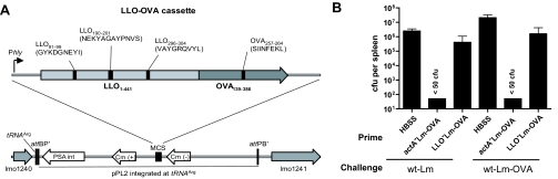
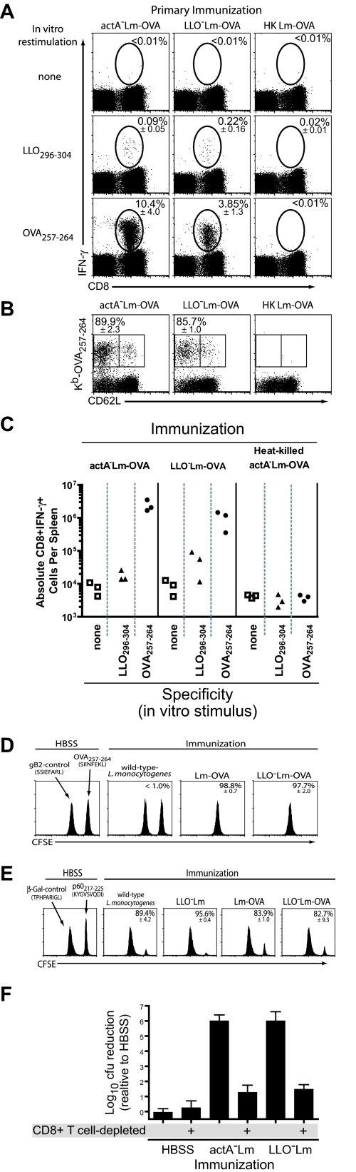
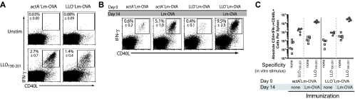
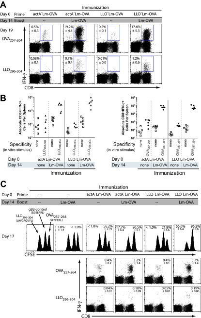
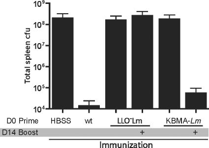
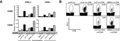
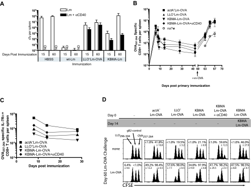
Interaction with host immunoreceptors during microbial infection directly impacts the magnitude of the ensuing innate immune response. How these signals affect the quality of the adaptive T-cell response remains poorly understood. Utilizing an engineered strain of the intracellular pathogen Listeria monocytogenes that infects cells but fails to escape from the phagosome, we demonstrate the induction of long-lived memory T cells that are capable of secondary expansion and effector function but are incapable of providing protective immunity. We demonstrate that microbial invasion of the cytosol is required for dendritic cell activation and integration of CD40 signaling, ultimately determining the ability of the elicited CD8+-T-cell pool to protect against lethal wild-type L. monocytogenes challenge. These results reveal a crucial role for phagosomal escape, not for delivery of antigen to the class I major histocompatibility complex pathway but for establishing the appropriate cellular context during CD8+-T-cell priming.


Figures

FIG. 1.
FIG. 1.
LLO-deleted L. monocytogenes expressing OVA does not elicit protective immunity. (A) Construction of L. monocytogenes strains expressing OVA linked to nonfunctional LLO using the pPL2 site-specific integration vector. This permits the measurement of MHC class I- and II-restricted T-cell responses without the expression of functional LLO. (B) C57BL/6 mice immunized with either HBSS, actALm-OVA (1 × 10⁷ CFU), or LLOLm-OVA (3 × 10⁸ CFU) were challenged 25 days later with 1 × 10⁵ CFU of either wild-type L. monocytogenes (wt-Lm) or wild-type Lm-OVA (wt-Lm-OVA). Spleens were harvested three days after challenge and plated for CFU. Data represent one experiment of two.
FIG. 2.
FIG. 2.
LLOLm elicits primary CD8+ T cells that mediate protection against lethal L. monocytogenes challenge. (A) In C57BL/6 mice at 7 days postimmunization with actALm-OVA (1 × 10⁷ CFU), LLOLm-OVA (3 × 10⁸ CFU), or heat-killed actALm-OVA (1 × 10⁹ particles), the percentage of LLO296-304- or OVA257-264-specific CD8 T cells was determined using intracellular cytokine staining. Numbers represent the percentages of IFN-γ-positive cells within the CD8+-T-cell population, and they are the means of three mice per group. (B) Staining of CD8+ T cells with anti-CD62L and Kb-OVA257-264 multimers. Data shown are gated on MHC class II-negative, CD4-negative events. Numbers represent the percentages of CD62L-negative cells within the CD8+ Kb-OVA257-264+ T-cell population, and they are the means of three mice per group. (C) Absolute antigen-specific T cells per spleen at 7 days postimmunization. Antigen-specific T cells were quantified by staining for intracellular IFN-γ following restimulation with the indicated peptide (x axis). Each symbol represents an individual animal. Data are from a single representative experiment of at least five replicates. (D) C57BL/6 mice were immunized with HBSS (left panel), wild-type L. monocytogenes (5 × 10³ CFU), Lm-OVA (5 × 10³ CFU), or LLOLm-OVA (3 × 10⁸ CFU). In vivo cytolytic activity was determined 7 days later by challenging mice with gB2 (control)- or OVA257-264-loaded targets. (E) BALB/c mice immunized with HBSS (left panel), wild-type L. monocytogenes (5 × 10³ CFU), LLOLm (3 × 10⁸ CFU), Lm-OVA (5 × 10³ CFU), or LLOLm-OVA (3 × 10⁸ CFU) were challenged 7 days later with β-Gal (control)- or p60217-225-loaded splenocytes. Numbers represent the percent killing of targets, and they represent the means of three mice per group. Standard deviations are indicated below. All data represent a single experiment of at least three replicates. (F) C57BL/6 mice were immunized with HBSS, actALm (1 × 10⁷ CFU), or LLOLm (3 × 10⁸ CFU). Five days later, half of the mice were depleted of CD8+ T cells. On day 7 postimmunization, mice were challenged with wild-type L. monocytogenes (1 × 10⁵ CFU), and CFU in the spleen were determined 3 days later. Data represent logs of protection relative to HBSS controls, graphed as the mean and standard deviations of five mice per group. One representative experiment of two is shown.
FIG. 3.
FIG. 3.
LLOLm-OVA elicits functional primary and secondary CD4+ T cells. (A) C57BL/6 mice immunized with actALm-OVA (1 × 10⁷ CFU) or LLOLm-OVA (3 × 10⁸ CFU) were examined seven days later for primary LLO-specific CD4 T-cell responses by intracellular cytokine staining. The upper left corner indicates the percentage of CD4 T cells producing IFN-γ and CD40L in response to the class II-restricted LLO190-201 peptide. Samples are gated on CD4+ CD8 events. The percentages represent the means of three mice per group and represent a single experiment of three. (B) C57BL/6 mice were immunized with the indicated strain, boosted on day 14 with Lm-OVA (1 × 10⁵ CFU), then examined five days later for LLO190-201-specific CD4+ T cells by intracellular cytokine staining (19 days after primary immunization). Samples were restimulated, stained, and gated as above. The percentages represent the means of five to six mice per group and represent a single experiment of three. (C) Absolute numbers of LLO190-201-specific CD4+ T cells five days after challenge with Lm-OVA (1 × 10⁵ CFU). Each symbol represents an individual animal. Data represent a single experiment of three.
FIG. 4.
FIG. 4.
LLOLm-OVA-elicited CD8+ T cells undergo secondary expansion. (A) C57BL/6 mice immunized with actALm-OVA (1 × 10⁷ CFU) or LLOLm-OVA (3 × 10⁸ CFU) were boosted with 1 × 10⁵ CFU Lm-OVA 14 days later. Five days postboost, spleens were harvested and the frequency of antigen-specific CD8+ T cells was determined by IFN-γ intracellular cytokine staining. The peptide used for restimulation is indicated on the far left. The number in each plot represents the percentage of the CD8+-T-cell population, followed by the standard deviation. The percentage is the mean of five to six mice per group from a single representative experiment of four. (B) Absolute numbers of LLO296-304-specific (left) and OVA257-264-specific (right) CD8+ T cells 5 days after challenge with Lm-OVA. (C) The in vivo cytolytic function of the recalled CD8+-T-cell population was assessed using in vivo cytotoxicity assay. Three days following boost immunization with Lm-OVA, mice received an equivalent mixture of differentially CFSE-labeled targets loaded with the indicated peptides. Upper panels show percent killing of the indicated target population relative to the HSV-gB2 control. The numbers in the upper left corners indicate the percentages of LLO296-304 targets killed, while the numbers in the upper right corners indicate the percentages of OVA257-264 targets killed. Lower panels depict the frequency of epitope-specific CD8+ T cells determined by intracellular cytokine staining the day of target cell transfer. Percentages represent the means of three mice per group from a single representative experiment of two replicates.
FIG. 5.
FIG. 5.
Cytosolic entry promotes protective immunity after two immunizations. Boost immunization with cytosolic, nonreplicating L. monocytogenes generates long-lived protective immunity. C57BL/6 mice were immunized with wild-type (wt) L. monocytogenes OVA (5 × 10³ CFU), LLOLm-OVA (3 × 10⁸ CFU), or KBMA-Lm (3 × 10⁸ particles). The indicated groups were boosted 14 days later with the same dose of KBMA-Lm or LLOLm-OVA. Sixty days later, mice were challenged with 1 × 10⁵ CFU of wild-type L. monocytogenes. Spleens were harvested three days postchallenge, and CFU were determined by serial dilution. Data represent the means of five mice per group. Data are from a single experiment of three.
FIG. 6.
FIG. 6.
Cytosolic entry promotes dendritic cell activation and T-cell priming in vivo. (A) C57BL/6 spleens harvested 24 h postimmunization with the indicated L. monocytogenes strain with or without anti-CD40 were labeled with antibodies to class II, CD11c, and CD8α and the antibody indicated above each graph. Median fluorescence intensity of the indicated marker was determined after gating on class II-high and CD11c-high cells and the indicated CD8α-positive or -negative fraction. Data represent means and standard deviations of three mice per group. One representative experiment of three is shown. (B) C57BL/6 mice were immunized with or without the addition of anti-CD40, and then OVA257-264-specific CD8+-T-cell responses were determined in the spleen by intracellular cytokine staining. The percentages represent the frequency of IFN-γ+ cells within the CD8+-T-cell population.
FIG. 7.
FIG. 7.
Cytosolic entry synergizes with anti-CD40 to maintain protective immunity. (A) Mice immunized with wild-type L. monocytogenes (wt-Lm) (5 × 10³ CFU), LLOLm-OVA (3 × 10⁸ CFU), or KBMA-Lm (3 × 10⁸ particles), with or without 100 μg of anti-CD40, were challenged 15 or 60 days later with 1 × 10⁵ CFU of wild-type L. monocytogenes. Three days later, CFU were determined in the spleen. Data represent the means and standard deviations of five mice per group. Data represent one experiment of three. ND, not determined. (B) Mice were immunized with actALm (1 × 10⁷ CFU), LLOLm-OVA (3 × 10⁸ CFU), or KBMA-Lm (3 × 10⁸ particles), with or without 100 μg of anti-CD40. At the indicated time points, spleens were harvested and the frequency of OVA257-264-specific CD8+ T cells were determined by intracellular cytokine staining. On day 60, all groups and an age-matched naive group were infected intraperitoneally with 1 × 10⁶ PFU of vaccinia virus OVA (vv-OVA). (C) Proportion of OVA257-264-specific CD8+ T cells expressing CD127 (IL-7Rα) during contraction of the primary response, determined using Kb-OVA257-264 multimers. Each point represents the average and standard deviation of three mice per group. (D) C57BL/6 mice were immunized with the indicated L. monocytogenes strains (same doses as above). Anti-CD40 was administered to the indicated groups. Boost immunization of KBMA-Lm-OVA was administered 14 days following primary immunization. Sixty days after primary immunization, the indicated groups were challenged with Lm-OVA. Three days later, mice were given CFSE-labeled targets loaded with the indicated peptides. Percentages in the left and right corners correspond, respectively, to LLO296-304- and OVA257-264-specific killing. Data represent a single experiment of two.

References

    1. Albert, M. L., B. Sauter, and N. Bhardwaj. 1998. Dendritic cells acquire antigen from apoptotic cells and induce class I-restricted CTLs. Nature 392:86-89. - PubMed
    1. Auerbuch, V., D. G. Brockstedt, N. Meyer-Morse, M. O'Riordan, and D. A. Portnoy. 2004. Mice lacking the type I interferon receptor are resistant to Listeria monocytogenes. J. Exp. Med. 200:527-533. - PMC - PubMed
    1. Barry, R. A., H. G. Bouwer, D. A. Portnoy, and D. J. Hinrichs. 1992. Pathogenicity and immunogenicity of Listeria monocytogenes small-plaque mutants defective for intracellular growth and cell-to-cell spread. Infect. Immun. 60:1625-1632. - PMC - PubMed
    1. Belz, G. T., K. Shortman, M. J. Bevan, and W. R. Heath. 2005. CD8alpha+ dendritic cells selectively present MHC class I-restricted noncytolytic viral and intracellular bacterial antigens in vivo. J. Immunol. 175:196-200. - PMC - PubMed
    1. Belz, G. T., C. M. Smith, D. Eichner, K. Shortman, G. Karupiah, F. R. Carbone, and W. R. Heath. 2004. Cutting edge: conventional CD8 alpha+ dendritic cells are generally involved in priming CTL immunity to viruses. J. Immunol. 172:1996-2000. - PubMed

MeSH terms