Comparative Study
. 2006 Nov;74(11):6075-84.
doi: 10.1128/IAI.00721-06. Epub 2006 Sep 5.

Evidence that tight junctions are disrupted due to intimate bacterial contact and not inflammation during attaching and effacing pathogen infection in vivo

Affiliations
Comparative Study

Evidence that tight junctions are disrupted due to intimate bacterial contact and not inflammation during attaching and effacing pathogen infection in vivo

Julian A Guttman et al. Infect Immun. 2006 Nov.

Abstract

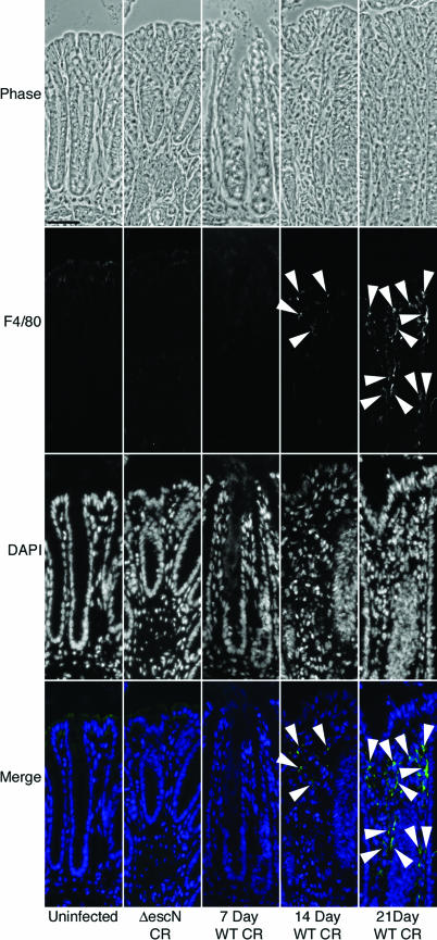
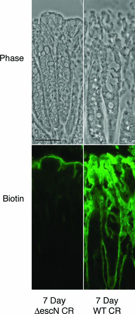
It is widely accepted that tight junctions are altered during infections by attaching and effacing (A/E) pathogens. These disruptions have been demonstrated both in vitro and more recently in vivo. For in vivo experiments, the murine model of A/E infection with Citrobacter rodentium is the animal model of choice. In addition to effects on tight junctions, these bacteria also colonize the colon at high levels, efface colonocyte microvilli, and cause hyperplasia and inflammation. Although we have recently demonstrated that tight junctions are disrupted by C. rodentium, the issue of direct effects of bacteria on epithelial cell junctions versus the indirect effects of inflammation still remains to be clarified. Here, we demonstrate that during the C. rodentium infections, inflammation plays no discernible role in the alteration of tight junctions. The distribution of the tight junction proteins, claudin-1, -3, and -5, are unaffected in inflamed colon, and junctions appear morphologically unaltered when viewed by electron microscopy. Additionally, tracer molecules are not capable of penetrating the inflamed colonic epithelium of infected mice that have cleared the bacteria. Finally, infected colonocytes from mice exposed to C. rodentium for 14 days, which have high levels of bacterial attachment to colonocytes as well as inflammation, have characteristic, altered claudin localization whereas cells adjacent to infected colonocytes retain their normal claudin distribution. We conclude that inflammation plays no discernible role in tight junction alteration during A/E pathogenesis and that tight junction disruption in vivo appears dependent only on the direct intimate attachment of the pathogenic bacteria to the cells.


Figures

FIG. 1.
FIG. 1.
Claudin-1, -3, and -5 and DAPI (DNA) localization on 21-day wild-type C. rodentium-infected mouse sections paired with 21-day ΔescN C. rodentium-infected tissue or uninfected tissue. Claudin staining of 7-day wild-type C. rodentium-infected tissue is also presented. Arrowheads indicate areas of staining at the lateral boundaries of colonocytes. The asterisks in the merged images indicate regions with high levels of cellular infiltrate in the lamina propria of 21-day wild-type C. rodentium-infected inflamed epithelium. WT, wild type; CR. C. rodentium. Scale bars, 50 μm (21-day micrographs) and 25 μm (7-day micrographs).
FIG. 2.
FIG. 2.
Neutrophil-labeled micrographs counterstained with hematoxylin. Neutrophil labeling on uninfected, 7-day ΔescN C. rodentiumescN CR), or 7-day, 14-day or 21-day wild-type C. rodentium (WT CR) infections. Arrowheads indicate some of the neutrophils (pink staining). Increased numbers of neutrophils are present in the lamina propria of 14- and 21-day wild-type C. rodentium-infected colon sections compared to the uninfected, 7-day ΔescN C. rodentium-infected or 7-day wild-type C. rodentium-infected tissue. Scale bar, 50 μm.
FIG. 3.
FIG. 3.
Paired phase, F4/80 (macrophage marker), and DAPI micrographs of uninfected, ΔescN C. rodentiumescN CR)-infected, and 7-, 14-, and 21-day wild-type C. rodentium (WT CR)-infected tissue. Positively labeled cells are not apparent in the lamina propria of uninfected, ΔescN C. rodentium-infected, or 7-day wild-type C. rodentium-infected colon sections. Positively labeled cells are present in the lamina propria of 14- and 21-day wild-type C. rodentium-infected tissue sections (arrowheads). Minimal nonspecific staining is present in the colonocytes and attached C. rodentium cells. Scale bar, 25 μm.
FIG. 4.
FIG. 4.
Histologic and ultrastructure micrographs of infected tissue. The 21-day wild-type C. rodentium (WT CR)-infected tissue (B and B") is paired with 7-day ΔescN C. rodentiumescN CR)-infected mouse tissue (A and A"). Toluidine blue (A and B) and electron micrographs (A′ and B′) of ΔescN and wild-type C. rodentium-infected tissue are shown. The A" and B" images are higher magnifications of the apical junction complexes in the A′ and B′ micrographs. The asterisk indicates a location of cell infiltration in the inflamed epithelium. TJ, tight junction; AJ, adherens junction; DS, desmosome. Scale bars, 50 μm (A), 1.0 μm (A′), and 0.2 μm (A").
FIG. 5.
FIG. 5.
Barrier permeability micrographs of 7-day C. rodentium infections. Phase images of 7-day ΔescN C. rodentiumescN CR)-infected and 7-day wild-type C. rodentium (WT CR)-infected tissue pretreated with a biotin tracer to assess barrier permeability. Biotin is held to the luminal border in ΔescN C. rodentium-infected tissue but permeates the epithelium into the lamina propria in 7-day wild-type C. rodentium-infected tissue. Scale bar, 50 μm.
FIG. 6.
FIG. 6.
Barrier permeability micrographs. (A) Phase images of low magnification of 21-day ΔescN C. rodentiumescN CR)-infected and wild-type C. rodentium (WT CR)-infected tissue pretreated with a biotin tracer to assess barrier permeability. Biotin is held to the luminal border in both ΔescN C. rodentium-infected and wild-type C. rodentium-infected tissue. (B) Higher magnification of biotin- and DAPI-treated murine tissue that was infected with ΔescN C. rodentium or wild-type C. rodentium for 21 days. Double-headed arrows demonstrate the difference in crypt depth between ΔescN C. rodentium-infected and wild-type C. rodentium-infected tissue to indicate that the disease phenotype has not resolved at this time point. The asterisks in the DAPI and merged images indicate some of the regions with high levels of cellular infiltrate in the 21-day wild-type C. rodentium-infected inflamed epithelium. Arrowheads point to regions with normal cellular load in the lamina propria. Scale bars, 100 μm (low magnification) and 50 μm (high magnification).
FIG. 7.
FIG. 7.
(A) Paired phase, C. rodentium Tir, and DAPI micrographs of infected tissue. Tir and DAPI colocalization demonstrates that bacteria labeled by DAPI in infected tissue sections is C. rodentium. Scale bar, 50 μm. (B) Paired phase, claudin-3, and DAPI micrographs of uninfected and 14-day wild-type C. rodentium (WT CR)-infected tissue. Arrows indicate regions of the epithelium without bacterial attachment that label the lateral boundaries of colonocytes. Arrowheads and white asterisks identify regions with bacterial colonization and direct attachment to colonocytes. All infected regions have altered localization of claudin-3. The black asterisks in the DAPI and merged images indicate areas with high levels of cellular infiltrate. Scale bar, 50 μm.

References

    1. Barmeyer, C., M. Harren, H. Schmitz, U. Heinzel-Pleines, J. Mankertz, U. Seidler, I. Horak, B. Wiedenmann, M. Fromm, and J. D. Schulzke. 2004. Mechanisms of diarrhea in the interleukin-2-deficient mouse model of colonic inflammation. Am. J. Physiol. Gastrointest. Liver Physiol. 286:G244-G252. - PubMed
    1. Basuroy, S., A. Seth, B. Elias, A. P. Naren, and R. Rao. 2006. MAPK interacts with occludin and mediates EGF-induced prevention of tight junction disruption by hydrogen peroxide. Biochem. J. 393:69-77. - PMC - PubMed
    1. Canil, C., I. Rosenshine, S. Ruschkowski, M. S. Donnenberg, J. B. Kaper, and B. B. Finlay. 1993. Enteropathogenic Escherichia coli decreases the transepithelial electrical resistance of polarized epithelial monolayers. Infect. Immun. 61:2755-2762. - PMC - PubMed
    1. Chakravortty, D., and K. S. Kumar. 1999. Interaction of lipopolysaccharide with human small intestinal lamina propria fibroblasts favors neutrophil migration and peripheral blood mononuclear cell adhesion by the production of proinflammatory mediators and adhesion molecules. Biochim. Biophys. Acta 1453:261-272. - PubMed
    1. Chang, L., and M. Karin. 2001. Mammalian MAP kinase signalling cascades. Nature 410:37-40. - PubMed

Publication types

MeSH terms

LinkOut - more resources