. 2006 Nov;74(11):6356-64.
doi: 10.1128/IAI.00460-06. Epub 2006 Sep 5.

A newly identified leptospiral adhesin mediates attachment to laminin

Affiliations

A newly identified leptospiral adhesin mediates attachment to laminin

Angela S Barbosa et al. Infect Immun. 2006 Nov.

Abstract

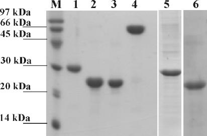
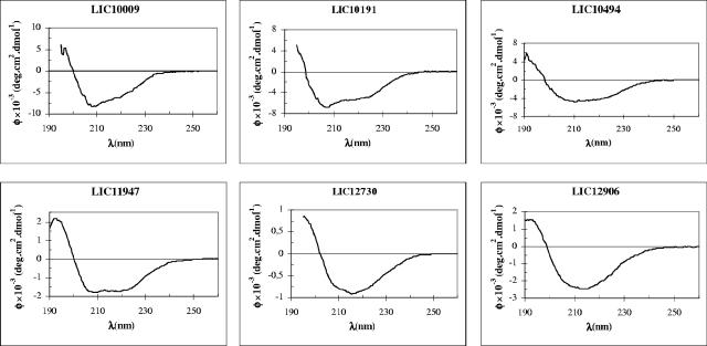
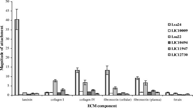
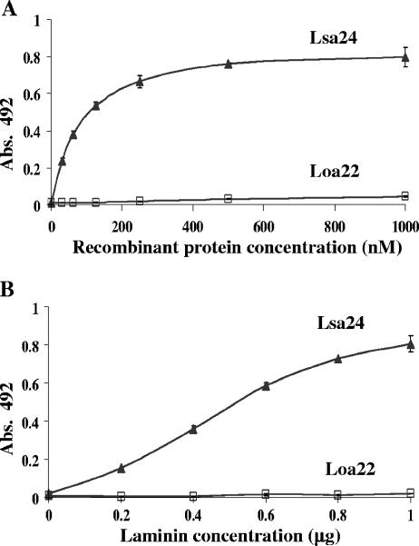
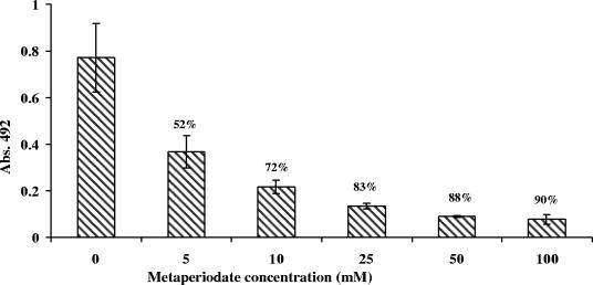
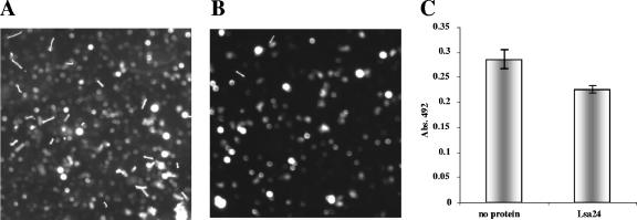
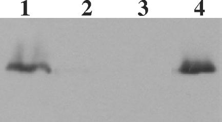
Pathogenic leptospires have the ability to survive and disseminate to multiple organs after penetrating the host. Several pathogens, including spirochetes, have been shown to express surface proteins that interact with the extracellular matrix (ECM). This adhesin-mediated binding process seems to be a crucial step in the colonization of host tissues. This study examined the interaction of putative leptospiral outer membrane proteins with laminin, collagen type I, collagen type IV, cellular fibronectin, and plasma fibronectin. Six predicted coding sequences selected from the Leptospira interrogans serovar Copenhageni genome were cloned, and proteins were expressed, purified by metal affinity chromatography, and characterized by circular dichroism spectroscopy. Their capacity to mediate attachment to ECM components was evaluated by binding assays. We have identified a leptospiral protein encoded by LIC12906, named Lsa24 (leptospiral surface adhesin; 24 kDa) that binds strongly to laminin. Attachment of Lsa24 to laminin was specific, dose dependent, and saturable. Laminin oxidation by sodium metaperiodate reduced the protein-laminin interaction in a concentration-dependent manner, indicating that laminin sugar moieties are crucial for this interaction. Triton X-114-solubilized extract of L. interrogans and phase partitioning showed that Lsa24 was exclusively in the detergent phase, indicating that it is a component of the leptospiral membrane. Moreover, Lsa24 partially inhibited leptospiral adherence to immobilized laminin. This newly identified membrane protein may play a role in mediating adhesion of L. interrogans to the host. To our knowledge, this is the first leptospiral adhesin with laminin-binding properties reported to date.


Figures

FIG. 1.
FIG. 1.
Purification of recombinant proteins: SDS-PAGE (15%) of purified recombinant proteins obtained by metal affinity chromatography. Proteins encoded by LIC10009 (lane 1), LIC10191, known as protein Loa22 (lane 2), LIC11947 (lane 3), LIC12730 (lane 4), LIC10494 (lane 5), LIC12906, named Lsa24 (lane 6) are shown. Lane M, molecular mass standard.
FIG. 2.
FIG. 2.
Circular dichroism spectra of the recombinant proteins. CD spectra of proteins encoded by LIC10009, LIC10191 (Loa22), LIC11947 (predominant α-helical secondary structure), LIC10494 (both α-helical and β-strand), and LIC12730 and LIC12906 (Lsa24) (predominant signal of β-strand) are shown. Far-UV CD spectra are presented as an average of five scans recorded from 190 to 260 nm. φ, molar ellipticity.
FIG. 3.
FIG. 3.
Binding of recombinant proteins to ECM components. Wells were coated with 1 μg of laminin, collagen type I, collagen type IV, cellular fibronectin, plasma fibronectin, and the control proteins BSA and fetuin. Recombinant protein attachment to those ECM macromolecules was assessed by an ELISA-based assay. One microgram of recombinant protein was added per well. Optical densities were taken at 492 nm. Specific profiles of adhesion to the various coating macromolecules are shown as magnitude of attachment (fold) over the negative control protein BSA, arbitrarily set equal to 1. Data represent the mean ± standard error of three independent experiments.
FIG. 4.
FIG. 4.
Binding of Lsa24 to laminin as a function of protein and laminin concentration. (A) Binding of laminin (1 μg) with recombinant protein (concentration ranging from 0 to 1,000 nM); (B) recombinant protein (1 μg) with laminin (amount ranging from 0 to 1 μg). Each point represents the mean absorbance value at 492 nm ± the standard deviation of three independent experiments. Loa22 was included as a negative control since it showed no specific attachment to laminin.
FIG. 5.
FIG. 5.
Sugar moiety contribution to the laminin-Lsa24 interaction. Immobilized laminin was treated with sodium metaperiodate (5 to 100 mM) for 15 min at 4°C in the dark. Mean absorbance values at 492 nm (± the standard deviations of three independent experiments) were compared to those obtained with untreated laminin (0 mM). The percent reduction in protein attachment to metaperiodate-treated laminin as a function of periodate concentration is indicated above each bar.
FIG. 6.
FIG. 6.
L. interrogans attachment to ECM macromolecules. Glass slides coated with laminin (A), collagen I (B), collagen IV (C), cellular fibronectin (D), plasma fibronectin (E), or BSA (F) were incubated with 5 × 10⁷ leptospires (L. interrogans serovar Pomona). Attachment was visualized using dark-field microscopy.
FIG. 7.
FIG. 7.
Inhibition of L. interrogans attachment to laminin by Lsa24. Chamber slides were coated with 4 μg of laminin for 16 h. The slides were blocked with BSA, and 5 × 10⁷ leptospires were added to each chamber. (A) No protein added to the blocking solution; (B) 20 μg of Lsa24 was added to the blocking solution to test for its ability to competitively inhibit attachment of L. interrogans to laminin. Adherence was visualized by dark-field microscopy. (C) Leptospira attachment to the substrate was quantified by ELISA. Laminin-coated microtiter wells were incubated with 0.5 μg of Lsa24 for 1 h 30 min prior to the addition of 1 × 10⁷ leptospires. Wells were probed with anti-LipL32 serum. Data are expressed as the mean A492 ± the standard error of three independent experiments, each performed in triplicate. Significance was assessed by comparison with the no-protein wells by Student's two-tailed t test (*, P < 0.05).
FIG. 8.
FIG. 8.
Cellular localization of Lsa24. Triton X-114 fractions of L. interrogans serovar Copenhageni organisms were separated by SDS-PAGE, transferred to a nitrocellulose membrane, and probed with Lsa24 antiserum. Fractions analyzed were the whole organism (lane 1), Triton X-114-insoluble pellet (lane 2), aqueous phase (lane 3), and detergent phase (lane 4).

Similar articles

Cited by

References

    1. Abreu, P. A., P. A. Miyasato, M. M. Vilar, W. O. Dias, P. L. Ho, M. Tendler, and A. L. T. O. Nascimento. 2004. Sm14 of Schistosoma mansoni in fusion with tetanus toxin fragment C induces immunoprotection against tetanus and schistosomiasis in mice. Infect. Immun. 72:5931-5937. - PMC - PubMed
    1. Areas, A. P.,M. L. Oliveira, C. R. Ramos, M. E. Sbrogio-Almeida, I. Raw, and P. L. Ho. 2002. Synthesis of cholera toxin B subunit gene: cloning and expression of a functional 6XHis-tagged protein in Escherichia coli. Protein Expr. Purif. 25:481-487. - PubMed
    1. Bendtsen, J. D., H. Nielsen, G. von Heijne, and S. Brunak. 2004. Improved prediction of signal peptides: SignalP 3.0. J. Mol. Biol. 340:783-795. - PubMed
    1. Brown, E. L., B. P. Guo, P. O'Neal, and M. Hook. 1999. Adherence of Borrelia burgdorferi. Identification of critical lysine residues in DbpA required for decorin binding. J. Biol. Chem. 274:26272-26278. - PubMed
    1. Cameron, C. E. 2003. Identification of a Treponema pallidum laminin-binding protein. Infect. Immun. 71:2525-2533. - PMC - PubMed

Publication types

MeSH terms

LinkOut - more resources