. 2007 May;27(5):950-62.
doi: 10.1038/sj.jcbfm.9600395. Epub 2006 Sep 6.

Protein SUMOylation is massively increased in hibernation torpor and is critical for the cytoprotection provided by ischemic preconditioning and hypothermia in SHSY5Y cells

Affiliations

Protein SUMOylation is massively increased in hibernation torpor and is critical for the cytoprotection provided by ischemic preconditioning and hypothermia in SHSY5Y cells

Yang-ja Lee et al. J Cereb Blood Flow Metab. 2007 May.

Abstract

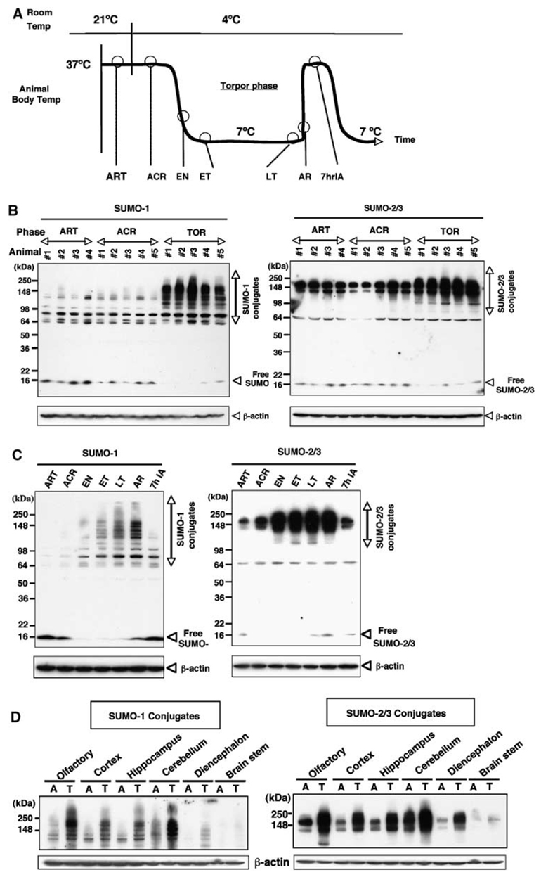
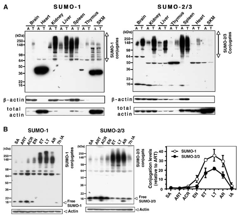
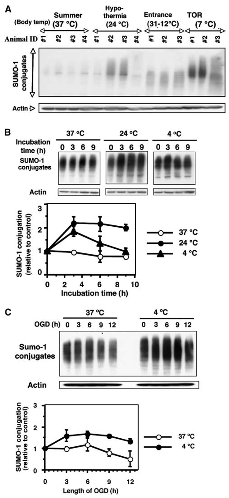
Hibernation torpor provides an excellent natural model of tolerance to profound reductions in blood flow to the brain and other organs. Here, we report that during torpor of 13-lined ground squirrels, massive SUMOylation occurs in the brain, liver, and kidney. The level of small ubiquitin-related modifier (SUMO) conjugation coincides with the expression level of Ubc9, the SUMO specific E2-conjugating enzyme. Hypothermia alone also increased SUMO conjugation, but not as markedly as hibernation torpor. Increased SUMO conjugation (induced by Ubc9 overexpression, ischemic preconditioning (PC)+/-hypothermia) was necessary and sufficient for tolerance of SHSY5Y neuroblastoma cells to oxygen/glucose deprivation (OGD) ('in vitro ischemia'); decreased SUMO conjugation (induced by a dominant-negative Ubc9) severely reduced tolerance to OGD in these cells. These data indicate that post-translational modification of proteins by SUMOylation is a prominent feature of hibernation torpor and is critical for cytoprotection by ischemic PC+/-hypothermia in SHSY5Y cells subjected to OGD.


Figures

Figure 1
Figure 1
Massive SUMO-1 and SUMO-2/3 conjugation occurs during hibernation in the brain of 13-lined ground squirrels. (A) Hibernation bout (cycle) in the 13-lined ground squirrel. ART: active animals at room temperature (Tb=34°C to 37°C); ACR: active animals in cold room (Tb=34°C to 37°C); EN: animals entering torpor (Tb=31°C to 12°C); ET: animals in the early stage of torpor (1 day after entrance into torpor, Tb=5°C to 8°C); LT: animals in the late stage of torpor phase (more than 5 days in torpor, Tb=5°C to 8°C); AR: animals arousing from torpor (Tb=9°C to 12°C); 7hIA: interbout aroused animals that have been in the active state for 7 h (Tb=34°C to 37°C). (B) Immunoblots of SUMO-1 or SUMO-2/3 (free and conjugated forms) in the brain of ART, ACR, and animals in the torpor phase of hibernation bouts (TOR) (n=4 to 5 in each group). (C) Immunoblots of conjugated and free SUMO-1 or SUMO-2/3 in the brain during the phases of the hibernation bout. β-Actin served as a loading control. Each gel is representative of three independent experiments. (D) Immunoblots of protein extracts from different regions of brain (as indicated in the figure) of active (A) and torpor (T) phase animals. β-Actin served as a loading control.
Figure 2
Figure 2
Distribution of SUMO-1 and SUMO-2/3 conjugates in various brain regions and neuronal cells during active and torpor phases of the hibernation bout. (A) Immunohistochemical analysis of the distribution of SUMO-1 in cerebral cortex and cerebellum of ACR and TOR at three different magnifications. Scale bars show 500, 50 and 10 µm (from lower to higher magnification), respectively. Arrows in the photomicrographs of the cerebellum show Purkinje cells. (B) Immunohistochemical analysis of SUMO-1 in cortical neurons and Purkinje cells with hematoxylin counterstaining. Red color labels SUMO-1 and blue labels nuclei. Note the nuclear and perinuclear clustering in TOR samples in contrast to the diffuse cytoplasmic staining in ACR samples. Scale bars show 10 µm. (C) Confocal microscopy showing the subcellular distribution of SUMO-1 in cortical neurons of ACR and TOR. Red: SUMO-1, Blue: nuclei. Scale bars show 10 µm. (D) Confocal microscopy showing the subcellular distribution of PML nuclear bodies in brain sections from ACR and TOR. The far right panels show magnified images of cropped areas in the left panels. Green: PML, Blue: nuclei. Scale bars show 10 µm.
Figure 3
Figure 3
Distribution of SUMO-1 and SUMO-2/3 conjugates in various tissues from active animals and animals in the torpor phase of the hibernation bout. (A) SUMO-1 or SUMO-2/3 immunoblots of protein extracts from different tissues (as indicated in the figure) from active (A) and torpor (T) phase animals. Actin (both β and total) served as loading controls. Each gel is representative of three independent experiments. SKM: skeletal muscle. (B) Two immunoblots, one of conjugated and free SUMO-1 and one of conjugated and free SUMO-2/3, in the kidney during the phases of the hibernation bout. β-Actin served as a loading control. SA: summer active animals. Densities of bands (conjugates) were normalized by the corresponding actin levels and expressed as relative density (–fold) compared with ART samples (right panel). Data represent the mean±s.d. of three independent experiments.
Figure 4
Figure 4
Expression levels of Ubc9 and SENP1 in the brain and kidney of ground squirrels at various stages of the hibernation bout. Immunoblots of total brain extracts (left panel) or kidney extracts (right panel) from animals in the different phases of the hibernation bout. β-Actin served as a loading control. Densities of bands corresponding to Ubc9 or SENP1 were normalized by actin levels and expressed relative (-fold) to levels of samples from ART (bottom). Data represent the mean±s.d. of three independent experiments.
Figure 5
Figure 5
Effect of temperature (hypothermia) and/or OGD on SUMO-1 conjugation level in SHSY5Y cells. (A) Immunoblots of SUMO-1 conjugated protein in ground squirrel brain under various conditions. Body temperatures are shown in parentheses. Summer animal (37°C), hypothermia (24°C), Entrance (31°C to 12°C) and TOR (7°C). Actin served as a loading control. (B) Immunoblots of SUMO-1 conjugated proteins in total cell lysates of SHSY5Y cells grown at 37°C, 24°C, or 4°C for 0, 3, 6, or 9 h (upper panel). Relative density of SUMO-1 conjugates normalized to actin levels and expressed as the ratio to time zero control cells that were incubated at 37°C (lower panel). (C) Immunoblot of SUMO-1 conjugated protein in total cell lysates of SHSY5Y cells exposed to OGD at 37°C or 4°C for 3, 6, 9, or 12 h (upper panel). Relative density of SUMO-1 conjugates shown as the ratio to time zero cells that were incubated at 37°C (lower panel). Data represent the mean±s.d. of three independent experiments.
Figure 6
Figure 6
In SHSY5Y cells, preconditioning (6 h of OGD) and hypothermia increase SUMOylation; increased SUMOylation greatly protects against OGD and decreased SUMOylation eliminates protection from OGD. (A) Preconditioning SHSY5Y cells at either 37°C or 4°C reduces the percentage of cell death caused by 12 h of OGD exposure. Data represent the mean±s.d. of five independent experiments. (B) Upper panel: Preconditioning at 37°C or 4°C increases SUMO-1 conjugation in SHSY5Y cells; OGD depresses SUMO-1 conjugation in nonpreconditioned cells. Lower panel: Densities of the SUMO-1 conjugated proteins were normalized to actin levels at each time point and expressed as ratios to SUMO-1 conjugated protein level of control cells (0 time point of nonpreconditioned cells). (C) Ubc9 expression by T-Rex-SHSY5Y cell lines that stably expressed Myc-tagged Ubc9 (WT) or Myc-tagged Ubc9 (DN) was tightly regulated by tetracycline (tet). Parent cells are T-Rex-SHSY5Y cells that lack a second plasmid. Actin levels served as a loading control. (D) SUMO-1 conjugation levels in T-Rex-SHSY5Y stable transfectant cells were increased by overexpressed Ubc9 (WT) and were decreased by overexpressed Ubc9 (DN) (Top panel). Small ubiquitin-related modifier-1 conjugation band densities were normalized by the corresponding actin band density and expressed relative to the normalized band density of the parent cells. (E) Effect of overexpression of WT or DN Ubc9 on OGD-induced cell death. T-Rex-SHSY5Y cells stably transfected with Ubc9 (WT) or Ubc9 (DN) along with parent cells were grown in the presence of tetracycline (tet) (for 24 h before PC) and subjected to severe OGD (12 h) without or with preconditioning at either 37°C or 4°C. The cell death was assessed by nuclear staining with Hoechst 33342 and PI followed by flourescence-activated cell sorting analysis. The percentage of dead cells was plotted for each treatment. The data are shown as the mean±s.d. of three independent experiments.

References

    1. Azuma Y, Arnaoutov A, Dasso M. SUMO-2/3 regulates topoisomerase II in mitosis. J Cell Biol. 2003;163:477–487. - PMC - PubMed
    1. Carey HV, Andrews MT, Martin SL. Mammalian hibernation: cellular and molecular responses to depressed metabolism and low temperature. Physiol Rev. 2003;83:1153–1181. - PubMed
    1. Chen Y, Matsushita M, Nairn AC, Damuni Z, Cai D, Frerichs KU, Hallenbeck JM. Mechanisms for increased levels of phosphorylation of elongation factor-2 during hibernation in ground squirrels. Biochemistry. 2001;40:11565–11570. - PubMed
    1. De Georgia MA, Krieger DW, Abou-Chebl A, Devlin TG, Jauss M, Davis SM, Koroshetz WJ, Rordorf G, Warach S. Cooling for Acute Ischemic Brain Damage (COOL AID): a feasibility trial of endovascular cooling. Neurology. 2004;63:312–317. - PubMed
    1. Dirnagl U, Simon RP, Hallenbeck JM. Ischemic tolerance and endogenous neuroprotection. Trends Neurosci. 2003;26:248–254. - PubMed

Publication types

MeSH terms