. 2006 Sep;116(9):2333-43.
doi: 10.1172/JCI27154.

Endothelial cell-restricted disruption of FoxM1 impairs endothelial repair following LPS-induced vascular injury

Affiliations

Endothelial cell-restricted disruption of FoxM1 impairs endothelial repair following LPS-induced vascular injury

You-Yang Zhao et al. J Clin Invest. 2006 Sep.

Abstract

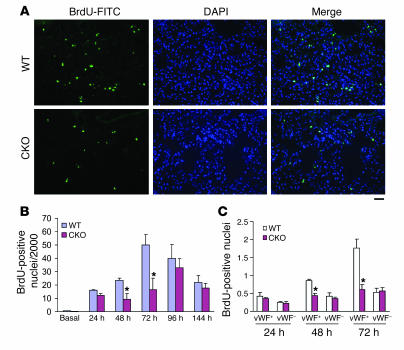
Recovery of endothelial integrity after vascular injury is vital for endothelial barrier function and vascular homeostasis. However, little is known about the molecular mechanisms of endothelial barrier repair following injury. To investigate the functional role of forkhead box M1 (FoxM1) in the mechanism of endothelial repair, we generated endothelial cell-restricted FoxM1-deficient mice (FoxM1 CKO mice). These mutant mice were viable and exhibited no overt phenotype. However, in response to the inflammatory mediator LPS, FoxM1 CKO mice displayed significantly protracted increase in lung vascular permeability and markedly increased mortality. Following LPS-induced vascular injury, FoxM1 CKO lungs demonstrated impaired cell proliferation in association with sustained expression of p27(Kip1) and decreased expression of cyclin B1 and Cdc25C. Endothelial cells isolated from FoxM1 CKO lungs failed to proliferate, and siRNA-mediated suppression of FoxM1 expression in human endothelial cells resulted in defective cell cycle progression. Deletion of FoxM1 in endothelial cells induced decreased expression of cyclins, Cdc2, and Cdc25C, increased p27(Kip1) expression, and decreased Cdk activities. Thus, FoxM1 plays a critical role in the mechanism of the restoration of endothelial barrier function following vascular injury. These data suggest that impairment in FoxM1 activation may be an important determinant of the persistent vascular barrier leakiness and edema formation associated with inflammatory diseases.


Figures

Figure 1
Figure 1. Mouse model of endothelial cell–restricted deletion of FoxM1.
(A) Southern blot demonstrating recombination of the FoxM1 floxed allele in endothelial cell–enriched tissues. Mouse genomic DNA (15 μg /lane) was digested with Bgl II and XbaI. The blots were probed with 3′ probe as described (13). Tie2 promoter/enhancer-driven Cre expression created a predicted 7.6-kb band in the aorta and lungs. The pseudogene band is indicated by an asterisk. (13). fl, FoxM1 floxed allele; AA, aorta; L, lung. (B) Quantitative analysis of mRNA expression of FoxM1 and FoxO1 by QRT-PCR. RNA was extracted from lungs of either FoxM1 WT or FoxM1 CKO mice. mRNA expression of FoxM1 and FoxO1 was normalized to mouse cyclophilin mRNA. There were no differences in FoxM1 mRNA levels in mice with the 3 genotypes FoxM1+/+, FoxM1fl/+, and FoxM1fl/fl; therefore, all these mice were referred to as WT mice. Data are expressed as mean ± SD, n = 3. **P < 0.05. CKO, FoxM1 CKO. (C) Quantitative analysis of FoxM1 expression in population of cells either enriched or depleted of endothelial cells. RNA was extracted from an endothelial cell–enriched primary culture (EC, passage 0) isolated from mouse lungs or from nonendothelial cell primary cultures (non-EC, passage 4), fibroblasts, and epithelial cells. Approximately 70% reduction of FoxM1 mRNA levels was detected in FoxM1 CKO EC and not in FoxM1 CKO non-EC (n = 3), indicating endothelial cell–specific knockdown of FoxM1. #P < 0.01. n = 3.
Figure 2
Figure 2. Impaired lung vascular endothelial barrier reannealing following LPS-induced microvascular injury in FoxM1 CKO mice.
(A) Time course of increase in lung microvessel permeability as measured by Kf,c. Basal, 0.2 ml PBS; LPS, 5 mg/kg BW. Data are expressed as mean ± SD, n = 3–5 per group. *P < 0.001 versus basal; **P < 0.001, CKO versus WT; #P < 0.05, CKO versus WT. (B) Representative graphs of mouse lung wet weights continuously recorded during measurement of microvessel permeability. 48h-LPS, 48 hours after LPS challenge. (C and D) Time course of loss of isogravimetric state of lungs (edema formation) obtained from mouse lungs at indicated times after LPS challenge. WT (C) and FoxM1 CKO mice (D) are represented. Points indicate mean ± 1 SD, n = 3–5 mice per group. #P < 0.05 WT or CKO 6 hours after LPS challenge (6h-LPS) versus basal; ##P < 0.05 CKO 48 hours after LPS challenge (48h-LPS) versus CKO basal or WT 48 hours after LPS challenge; ΧP < 0.05 CKO 96 hours after LPS challenge (96h-LPS) versus CKO basal or WT 96 hours after LPS challenge. (E) Representative micrographs of H&E staining show perivascular leukocyte infiltration in FoxM1 CKO lungs 96 hours after LPS challenge (5 mg/kg). Arrows indicate leukocyte infiltration. Br, bronchia; S, small vessels (<150 μm in diameter); L, large vessels (>150 μm in diameter). Scale bar: 50 mm. (F) Quantitative analysis of infiltrating leukocytes in lungs at 96 hours after LPS challenge. Bar graphs show infiltrating leukocytes in vessels of different diameters. Percentages of vessels of different diameters exhibiting leukocyte infiltration are shown. Data are expressed as mean ± SD, n = 3–4. #P < 0.05 versus WT (infiltrating leukocytes per vessel); ##P < 0.05 versus WT (% of vessels).
Figure 3
Figure 3. Time course of FoxM1 expression following LPS-induced lung microvascular injury.
(A) Quantitative analysis of FoxM1 mRNA levels in mouse lungs by QRT-PCR. Total RNA was extracted from lungs collected from either WT or FoxM1 CKO mice after LPS exposure (5 mg/kg BW) at indicated times. FoxM1 mRNA levels were normalized to cyclophilin. Data are expressed as mean ± SD, n = 3–4. *P < 0.05 WT versus CKO; **P < 0.05 versus basal WT. (B) FoxM1 mRNA expression in endothelial cell–enriched primary cultures isolated from WT mouse lungs. Total RNA was extracted from endothelial cell–enriched primary cultures (passage 2) treated with either PBS (basal), 1,000 U/ml of recombinant mouse TNF-α, or 10 μM of H2O2 for 22 hours. Data are expressed as mean ± SD, n = 3. *P < 0.05.
Figure 4
Figure 4. FoxM1 CKO mice exhibit endothelial apoptosis and lung neutrophil sequestration similar to that of WT mice in the early period following LPS challenge.
(A) Representative micrographs of TUNEL staining. Cryosections of lungs (3- to 5-μm thick) collected 24 hours after LPS challenge were stained with FITC-conjugated TUNEL to identify apoptotic cells and anti-vWF antibody to identify endothelial cells; nuclei were counterstained with DAPI. Arrows, TUNEL+vWF+ cells. Scale bar: 25 mm. (B) Graphic representation of apoptotic endothelial cells and nonendothelial cells in FoxM1 CKO and WT lungs 24 hours after LPS challenge (5 mg/kg BW). The number of TUNEL-positive nuclei and vWF-positive cells from 3 to 4 consecutive cryosections from each mouse lung were averaged. Data are expressed as mean ± SD, n = 3–4 mice per group. There is no difference between WT and FoxM1 CKO. (C) Lung tissue myeloperoxidase (MPO) activity during basal period and 6 hours after LPS challenge (5 mg/kg BW). Data are expressed as mean ± SD, n = 4 mice per group. *P > 0.1 versus WT. Although there is a significant increase in MPO activity in mouse lungs 6 hours following LPS challenge, both WT and FoxM1 CKO lungs exhibit similar MPO activities, indicating a similar extent of polymorphonuclear leukocyte sequestration.
Figure 5
Figure 5. Increased mortality in FoxM1 CKO mice following LPS challenge.
(A) Survival rate following LPS challenge. Mice were challenged i.p. with 12 mg/kg BW of LPS and housed under normal conditions. Approximately 70% of the FoxM1 CKO mice died from day 2 to day 4 after LPS challenge whereas only about 10% of WT mice died in this same period. Differences in the survival rates between the WT and CKO groups after LPS challenge were significant by Peto-Peto-Wilcoxon test. n = 11 mice per group. *P < 0.001. (B) Graphic representation of development of lung edema in FoxM1 CKO mice following LPS challenge (12 mg/kg). Lungs were collected either at 48 hours after LPS challenge (WT and CKO) or in some mice at day 2 just prior to their deaths (CKO-D). Data are expressed as mean ± SD. n = 3–5, #P < 0.05 versus WT; ##P < 0.01 versus WT.
Figure 6
Figure 6. Failure of WT bone marrow cells to rescue the vascular repair defect in FoxM1 CKO mice following LPS challenge.
(A) FACS analysis of GFP-positive bone marrow lymphocytes (lymph) and total white blood cells. At 3 weeks following transplantation of WT bone marrow cells isolated from GFP-transgenic mice, bone marrow cells were isolated from reconstituted FoxM1 CKO and red blood cells were lysed prior to FACS analysis. Bone marrow white blood cells isolated from C57B6 mice and GFP-transgenic mice were used as negative and positive controls, respectively. Approximately 80% of bone marrow reconstitution was achieved. BMT, WT bone marrow cell–transplanted FoxM1 CKO. (B) Sustained increase in lung microvessel permeability in WT bone marrow cell–transplanted FoxM1 CKO following LPS challenge. At 5 weeks following WT bone marrow transplantation, lungs from WT or FoxM1 CKO mice were isolated for Kf,c measurement. Data are expressed as mean ± SD, n = 3–5 per group. *P < 0.001 versus basal; **P < 0.001, CKO versus WT. Basal, 0.2 ml PBS. CKO, FoxM1 CKO mice reconstituted with WT bone marrow cells. (C) Quantitative analysis of FoxM1 mRNA expression in bone marrow cells. Bone marrow white blood cells were isolated from either WT or FoxM1 CKO at the indicated time points following LPS challenge, and RNA was isolated for QRT-PCR analysis. FoxM1 mRNA levels were normalized to cyclophilin. Data are expressed as mean ± SD, n = 3 per group. In contrast to endothelial cells isolated from FoxM1 CKO lungs, the white blood cells from FoxM1 CKO bone marrow expressed FoxM1 at the same level as WT.
Figure 7
Figure 7. Decreased cell proliferation in FoxM1 CKO lungs following LPS-induced lung microvascular injury.
(A) Representative micrographs of BrdU immunostaining. Cryosections of lungs (3- to 5-μm thick), collected 72 hours after LPS challenge, were stained with FITC-conjugated anti-BrdU antibody to identify proliferating cells; nuclei were counterstained with DAPI. Scale bar: 25 mm. (B) Graphic representation of decreased BrdU incorporation in FoxM1 CKO lungs and WT lungs at the indicated times following LPS challenge (5 mg/kg BW). From 3 to 4 consecutive cryosections from each mouse lung were examined, and the average number of BrdU-positive nuclei per 2,000 nuclei was used as the value for the mouse. Data are expressed as mean ± SD, n = 3–4 mice per time point. *P < 0.05 versus WT at 48 and 72 hours after LPS. There are few BrdU-positive nuclei (<1 BrdU positive nuclei per 2,000 nuclei) in both WT and FoxM1 CKO lungs in the absence of LPS challenge. (C) Graphic representation of decreased proliferating endothelial cells (double-positive staining of vWF and BrdU) in FoxM1 CKO lungs. Data are expressed as mean ± SD, percentage of total nuclei, n = 3–4 mice per time point per group. *P < 0.05 versus WT.
Figure 8
Figure 8. Increased nuclear staining and expression of p27Kip1 and decreased expression of Cdc25C and cyclin B1 in FoxM1 CKO lungs.
(A) Representative micrographs of immunostaining of p27Kip1 and vWF. Cryosections of lungs collected 24 hours after LPS challenge (5 mg/kg) were stained with antibodies against p27Kip1 and vWF (marker for endothelial cells); nuclei were counterstained with DAPI. Arrows indicate p27Kip1+vWF+ cells. Scale bar: 25 μm. (B) Graphic representation of p27Kip1-positive endothelial cells and nonendothelial cells. Data are expressed as mean ± SD, n = 3–4 mice per group. *P < 0.05 versus WT. (C) Sustained expression of p27Kip1 in FoxM1 CKO lungs following LPS challenge (5 mg/kg). Protein (50 μg) from mouse lungs was loaded for each lane, the gel was electrophoresed, and proteins were transferred to PVDF and probed with monoclonal antibody against p27Kip1. The same membrane was reprobed with a rabbit polyclonal antibody against β-actin as a loading control. The experiment was repeated 3 times with similar results. (D and E) Time course of expression of Cdc25C (D) and cyclin B1 (E) in lungs following LPS challenge (5 mg/kg). Total RNA was extracted from lungs collected from either WT mice or FoxM1 CKO mice after LPS exposure (5 mg/kg BW) at the indicated times. Quantitative analysis of mRNA levels in mouse lungs was performed with QRT-PCR. Cdc25C and cyclin B1 mRNA levels were normalized to cyclophilin. Data are expressed as mean ± SD, n = 3–4. *P < 0.05 versus WT.
Figure 9
Figure 9. Defective cell cycle progression in FoxM1-deficient endothelial cells.
(A) Representative micrographs of primary cultures of lung endothelial cells isolated from either 2-week-old WT or FoxM1 CKO mice. Phase contrast pictures were taken at day 6 following isolation and culture. Scale bars: 50 μm. Growth of FoxM1 CKO endothelial cells was severely reduced, and cell death was noted after 3–5 days in culture compared with WT cultures. (B) QRT-PCR demonstrated an approximately 80% decrease in levels of FoxM1 mRNA in HMEC-1 transfected with FoxM1 siRNA. Data are expressed as mean ± SD, n = 3. HMEC-1 were transfected with either 100 nM of siRNA (siRNA) against human FoxM1 or its cognate mutant siRNA (miRNA) or PBS (basal). At 65 hours after transfection, cells were collected for RNA isolation. (C) Cell-cycle profiles of HMEC-1. At 72 hours after transfection, cells were fixed and stained with propidium iodide. Representative FACS analyses of cell cycle profiles in asynchronous HMEC-1 are shown. (D) Graphic representation of accumulation of 4 N DNA content and increase of polyploid cells (8 N) in FoxM1-deficient endothelial cells. Data are expressed as mean ± SD, n = 4. *P < 0.01 siRNA versus either basal or miRNA; **P < 0.05 siRNA versus either basal or miRNA; ΧP < 0.05 siRNA versus either basal or miRNA.
Figure 10
Figure 10. FoxM1 regulates expression of a network of genes essential for cell cycle progression in endothelial cells.
(A) Increased expression of the cell cycle inhibitor p27Kip1 seen in endothelial cells following siRNA knockdown of FoxM1. HMEC-1 were transfected with either 100 nM siRNA against human FoxM1 (siRNA) or its cognate mutant siRNA (miRNA) or PBS (basal) for 72 hours, and protein lysates (50 μg/lane) were used for Western blotting. The experiment was repeated twice with similar results. (B) Representative micrographs of immunostaining of p27Kip1 in human microvascular endothelial cells. FoxM1-deficient endothelial cells exhibited extensive nuclear staining of p27Kip1. Scale bar: 25 μm. (C) Graphic representation of decreased activities of Cdk1 and Cdk2 in FoxM1-deficient human microvascular endothelial cells. At 65 hours after transfection, cells were lysed for the Cdk activity assay. Data are expressed as mean ± SD, n = 3. *P < 0.05 versus miRNA. (D) QRT-PCR demonstrated decreased expression of cyclins, Cdc2, and Cdc25C in FoxM1-deficient human microvascular endothelial cells. At 65 hours after transfection, total RNA was isolated for QRT-PCR analysis. Cyclophilin mRNA levels were used for normalization. Data are expressed as mean ± SD, n = 3. *P < 0.05 versus WT. Cyc, cyclin.

Comment in

References

    1. Cines D.B., et al. Endothelial cells in physiology and in the pathophysiology of vascular disorders. Blood. 1998;91:3527–3561. - PubMed
    1. Ross R. Atherosclerosis–an inflammatory disease. N. Engl. J. Med. 1999;340:115–126. - PubMed
    1. Libby P., Ridker P.M., Maseri A. Inflammation and atherosclerosis. Circulation. 2002;105:1135–1143. - PubMed
    1. Austin G.E., Ratliff N.B., Hollman J., Tabei S., Phillips D.F. Intimal proliferation of smooth muscle cells as an explanation for recurrent coronary artery stenosis after percutaneous transluminal coronary angioplasty. J. Am. Coll. Cardiol. 1985;6:369–375. - PubMed
    1. Schwartz R.S., Holmes D.R., Topol E.J. The restenosis paradigm revisited: an alternative proposal for cellular mechanisms. J. Am. Coll. Cardiol. 1992;20:1284–1293. - PubMed

Publication types

MeSH terms