. 2006 Sep;116(9):2356-65.
doi: 10.1172/JCI28988.

Severe facial clefting in Insig-deficient mouse embryos caused by sterol accumulation and reversed by lovastatin

Affiliations

Severe facial clefting in Insig-deficient mouse embryos caused by sterol accumulation and reversed by lovastatin

Luke J Engelking et al. J Clin Invest. 2006 Sep.

Abstract

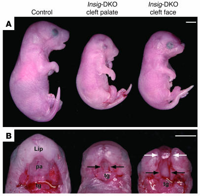
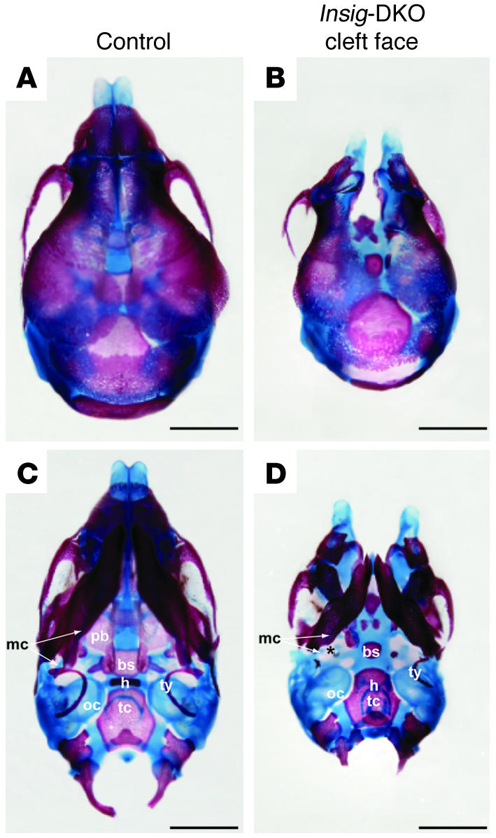
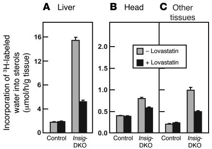
Insig-1 and Insig-2 are regulatory proteins that restrict the cholesterol biosynthetic pathway by preventing proteolytic activation of SREBPs and by enhancing degradation of HMG-CoA reductase. Here, we created Insig-double-knockout (Insig-DKO) mice that are homozygous for null mutations in Insig-1 and Insig-2. After 18.5 days of development, 96% of Insig-DKO embryos had defects in midline facial development, ranging from cleft palate (52%) to complete cleft face (44%). Middle and inner ear structures were abnormal, but teeth and skeletons were normal. The animals were lethargic and runted; they died within 1 day of birth. The livers and heads of Insig-DKO embryos overproduced sterols, causing a marked buildup of sterol intermediates. Treatment of pregnant mice with the HMG-CoA reductase inhibitor lovastatin reduced sterol synthesis in Insig-DKO embryos and reduced the pre-cholesterol intermediates. This treatment ameliorated the clefting syndrome so that 54% of Insig-DKO mice had normal faces, and only 7% had cleft faces. We conclude that buildup of pre-cholesterol sterol intermediates interferes with midline fusion of facial structures in mice. These findings have implications for the pathogenesis of the cleft palate component of Smith-Lemli-Opitz syndrome and other human malformation syndromes in which mutations in enzymes catalyzing steps in cholesterol biosynthesis produce a buildup of sterol intermediates.


Figures

Figure 1
Figure 1. Cholesterol biosynthetic pathway (A) and its relationship to 5 human diseases that result from deficiencies of enzymes in the conversion of lanosterol to cholesterol (B).
For simplicity, the early steps from acetyl-CoA to HMG-CoA are not shown. The clinical features of the genetic diseases are reviewed in articles by Herman (4), Moebius et al. (47), and Porter (3). CHILD syndrome, congenital hemidysplasia with ichthyosiform erythroderma and limb defects; CDPX2 syndrome, X-linked dominant chondrodysplasia punctata type 2.
Figure 2
Figure 2. Craniofacial abnormalities of Insig-DKO embryo.
Male and female Insig-1+/–Insig-2–/– mice were mated, and the resultant embryos were collected at 18.5 dpc, photographed, and genotyped. Lateral (A) and palatal views (B) of control (Insig-1+/+Insig-2–/–littermate) and Insig-DKO embryos with cleft palate or cleft face. The lower jaw was resected to reveal the oral cavity. Black arrows indicate cleft palate; white arrows indicate cleft face. pa, palate; tg, tongue. Scale bars: 2.5 mm.
Figure 3
Figure 3. Histology of control (A, D, G, andJ) andInsig-DKO embryos with cleft palate (B, E, H, andK) or cleft face (C, F, I, and L) at 18.
dpc. (AF) Palate phenotype. The secondary palate was intact in control (A and D) but absent in Insig-DKO embryos (B, C, E, and F). The lateral margins of the palatal shelves are indicated by arrows. In Insig-DKO embryos with cleft face (C), the nasal septum was split, and the brain was displaced rostrally. (GL) Middle and inner ear phenotype. Meckel cartilage (mc) and malleus (m) were normal in the cleft palate Insig-DKO embryo (H) but rudimentary in the cleft face embryo (I). Stapes (s) and the stapedial artery (sa) were absent or vestigial in Insig-DKO embryos (K and L). Compared with that of control embryos (J), the pars canalicularis (pca) of the otic capsule in the Insig-DKO embryos migrated rostral-ventrally (K and L). pa, palate; pco, pars cochlearis; tg, tongue. Scale bars: 0.5 mm.
Figure 4
Figure 4. Developmental study of palate formation in control andInsig -DKO embryos.
Insig-1+/–Insig-2–/– mice were mated, and the resultant embryos were collected at 12.5, 13.5, 14.5, and 15.5 dpc. The Insig-DKO embryos did not have cleft face. The embryos were fixed, sectioned, and stained with H&E. pa, palate; ps, palatal shelves; tg, tongue. Scale bars: 0.5 mm.
Figure 5
Figure 5. Alizarin red– and Alcian blue–stained skull preparations of control embryo andInsig -DKO embryo with cleft face.
(A and B) Dorsal view. (C and D) Ventral view. Compared with that in the control embryo (A and C), the median facial cleft in Insig-DKO embryo was clearly visible (B and D). The palatal bone (pb), basisphenoid (bs), hyoid (h), Meckel cartilage (mc), and tympanic ring (ty) all showed various degrees of abnormality in the Insig-DKO embryo (C and D). oc, otic capsule; tc, thyroid cartilage. The asterisk indicates a breakpoint in Meckel cartilage. Scale bars: 1 mm.
Figure 6
Figure 6. In vivo synthesis rates of sterols in tissues ofInsig -DKO embryos from pregnant females fed a chow diet with or without lovastatin.
Male and female Insig-1+/–Insig-2–/– mice were mated, and mated females were identified and fed ad libitum a regular chow diet or a chow diet containing 0.2% (wt/wt) lovastatin from 5.5 to 18.5 dpc. At 18.5 dpc, pregnant female mice were injected intraperitoneally with 3H-labeled water (50 mCi of in 0.25 ml isotonic saline). One hour later, the embryos were collected by caesarean section for measurement of sterol synthesis in liver (A), head (B), and other tissues (i.e., the whole body minus the liver and head; C). Sterol synthesis was calculated as μmoles of 3H-labeled water incorporated into digitonin-precipitable sterols per hour per gram of tissue. Each bar represents mean ± SEM of values from 16 to 18 embryos.
Figure 7
Figure 7. Prevention of craniofacial defects inInsig -DKO embryos after feeding of lovastatin to pregnant females.
Insig-1+/–Insig-2–/– mice were mated with each other, and mated females were identified by the presence of a postcopulatory vaginal plug. Mated females were fed ad libitum a regular chow diet (red bars) or a chow diet containing 0.2% (wt/wt) lovastatin (blue bars) from 5.5 to 18.5 dpc. (A) Embryos from 50 litters in both dietary groups were collected by caesarean section at 18.5 dpc and analyzed for craniofacial morphology by 2 independent observers prior to genotyping by PCR. The 50 litters in the control chow diet group contained a total of 341 embryos, of which 78 had the Insig-DKO genotype (same data as in Table 1). The 50 litters in the lovastatin-fed group contained a total of 361 embryos, of which 76 had the DKO genotype. The embryos from both groups were harvested in 5 different experiments over a 6-month period in which 5–19 pregnant females in both dietary groups were studied concurrently. The mean ± SEM for body weights of lovastatin-treated DKO embryos was 0.69 ± 0.01 g as compared with 1.08 ± 0.01 g for lovastatin-treated control embryos. (BE) Three Insig-DKO embryos from lovastatin-fed females represented in A were processed for histological analysis. One embryo (B and D) showed a cleft palate phenotype (not corrected) with middle and inner ear abnormalities, and 2 embryos showed normal craniofacial phenotype (corrected; 1 of which is shown in C and E). Arrows indicate the lateral margins of the palatal shelves. pa, palate; pca, pars canalicularis; pco, pars cochlearis; s, stapes; sa, stapedial artery. Scale bars: 0.5 mm.

Comment in

References

    1. Kelley R.I., Herman G.E. Inborn errors of sterol biosynthesis. Annu. Rev. Genomics Hum. Genet. 2001;2:299–341. - PubMed
    1. Nwokoro N.A., Wassif C.A., Porter F.D. Genetic disorders of cholesterol biosynthesis in mice and humans. Mol. Genet. Metab. 2001;74:105–119. - PubMed
    1. Porter F.D. Human malformation syndromes due to inborn errors of cholesterol synthesis. . Curr. Opin. Pediatr. 2003;15:607–613. - PubMed
    1. Herman G.E. Disorders of cholesterol biosynthesis: prototypic metabolic malformation syndromes. . Hum. Mol. Genet. 2003;12:R75–R88. - PubMed
    1. Liu X.Y., et al. The gene mutated in bare patches and striated mice encodes a novel 3β-hydroxysteroid dehydrogenase. Nat. Genet. 1999;22:182–187. - PubMed

Publication types