. 2006 Sep 7:5:24.
doi: 10.1186/1476-069X-5-24.

Over-expression of AhR (aryl hydrocarbon receptor) induces neural differentiation of Neuro2a cells: neurotoxicology study

Affiliations

Over-expression of AhR (aryl hydrocarbon receptor) induces neural differentiation of Neuro2a cells: neurotoxicology study

Eiichi Akahoshi et al. Environ Health. .

Abstract

Background: Dioxins and related compounds are suspected of causing neurological disruption in human and experimental animal offspring following perinatal exposure during development and growth. The molecular mechanism(s) of the actions in the brain, however, have not been fully investigated. A major participant in the process of the dioxin-toxicity is the dioxin receptor, namely the aryl hydrocarbon receptor (AhR). AhR regulates the transcription of diverse genes through binding to the xenobiotic-responsive element (XRE). Since the AhR has also been detected in various regions of the brain, the AhR may play a key role in the developmental neurotoxicity of dioxins. This study focused on the effect of AhR activation in the developing neuron.

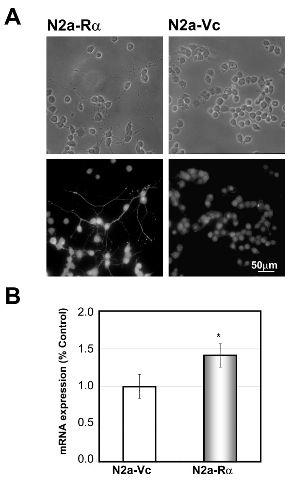
Methods: The influence of the AhR on the developing neuron was assessed using the Neuro2a-AhR transfectant. The undifferentiated murine neuroblastoma Neuro2a cell line (ATCC) was stably transfected with AhR cDNA and the established cell line was named N2a-Ralpha. The activation of exogenous AhR in N2a-Ralpha cells was confirmed using RNAi, with si-AhR suppressing the expression of exogenous AhR. The neurological properties of N2a-Ralpha based on AhR activation were evaluated by immunohistochemical analysis of cytoskeletal molecules and by RT-PCR analysis of mRNA expression of neurotransmitter-production related molecules, such as tyrosine hydroxylase (TH).

Results: N2a-Ralpha cells exhibited constant activation of the exogenous AhR. CYP1A1, a typical XRE-regulated gene, mRNA was induced without the application of ligand to the culture medium. N2a-Ralpha cells exhibited two significant functional features. Morphologically, N2a-Ralpha cells bore spontaneous neurites exhibiting axon-like properties with the localization of NF-H. In addition, cdc42 expression was increased in comparison to the control cell line. The other is the catecholaminergic neuron-like property. N2a-Ralpha cells expressed tyrosine hydroxylase (TH) mRNA as a functional marker of catecholaminergic neurotransmitter production. Thus, exogenous AhR induced catecholaminergic differentiation in N2a-Ralpha cells.

Conclusion: The excessive activation of AhR resulted in neural differentiation of Neuro2a cells. This result revealed that dioxins may affect the nervous system through the AhR-signaling pathway. Activated AhR may disrupt the strictly regulated brain formation with irregular differentiation occurring rather than cell death.


Figures

Figure 1
Figure 1
Observation of N2a-Rα cells by phase contrast microscopy. The parental Neuro2a cells (panel "Neuro2a") were transfected with rat AhR in the mammalian expression vector pcDNA4/V5-His and selected with zeocin. The morphology of the resultant transfectant is shown in panel "N2a-Rα ". The cells transfected with the vector without the insert are shown in panel "N2a-Vc". The N2a-Rα cells exhibited spontaneous neurite outgrowth. Note that no morphological differences in N2a-Vc were observed compared with the parental Neuro2a cells.
Figure 2
Figure 2
Transcriptional activation of exogenous AhR in the N2a-Rα cells. (A) AhR mRNA in N2a-Rα was detected by RT-PCR using primers specific for AhR. In N2a-Vc and Neuro2a, the primers amplified no products. The left panel shows that the primers amplified both mouse and rat AhR. Representative data are presented from triplicate experiments. (B) AhR protein (96kDa) in N2a-Rα was detected by Western blot analysis using a monoclonal antibody specific for AhR. In N2a-Vc and Neuro2a cells, no signals were detected. Representative data are presented from triplicate experiments. (C) The expression of CYP1A1, AhRR and GST mRNAs was detected in N2a-Rα cells. Since these genes were XRE-mediated genes, expression of these mRNAs are functional markers of AhR activation. Representative data are presented from triplicate experiments.
Figure 3
Figure 3
Suppression of CYP1A1 mRNA expression by si-AhR. These graphs demonstrate the repression of AhR and CYP1A1 mRNA expression by si-AhR transfection. N2a-Rα cells were transfected with AhR-targeted siRNA (si-AhR) or scramble siRNA. RNA was isolated from the cells transfected with siRNA and the expression levels were quantified by real-time quantitative PCR. AhR mRNA expression was effectively repressed by si-AhR (panel "AhR"). CYP1A1 mRNA expression was also decreased in cells transfected with si-AhR (panel "CYP1A1"). Scramble siRNA did not exhibit any effect on the mRNA expressions of AhR and CYP1A1 compared with Mock. The results are normalized to the expression level of cyclophilin mRNA and expressed relative to the value in the Mock. Values are presented as the mean of ± S. D. of four replicate determinations; ** p < 0.01 and *p < 0.05.
Figure 4
Figure 4
Properties of neurites elongated from N2a-Rα cells. (A) Immunochemical staining for phosphorylated NF-H was used as an axonal marker in N2a-Rα and N2a-Vc cells. Cells cultured for two-days were stained using the anti-phosphorylated neurofilament 200kDa (NF-H) monoclonal antibody and the FITC-conjugated goat anti-mouse IgG antibody (upper two panels). The lower panels are bright-field images of the upper panels. It should be noted that the outgrowths on the N2a-Rα cells were clearly stained with the anti NF-H antibody (panel "N2a-Rα "). (B) The quantity of cdc42 mRNA was determined in N2a-Rα and N2a-Vc cells with real-time PCR using primers specific for cdc42. A high level of expression of cdc42 mRNA was observed in N2a-Rα compared with the N2a-Vc. The results are normalized to the expression level of cyclophilin mRNA and expressed relative to the value of N2a-Vc. Values are presented as the mean ± S. D. of four replicate determinations; **p < 0.01 and *p < 0.05
Figure 5
Figure 5
Cell proliferation of N2a-Rα cells. 1.0 × 10³ cells were seeded in 96-well plates. After 1, 3 and 7 days of culture in vitro, cell proliferation in each well was measured by Alamar Blue dye reduction assay. Values are presented as the mean ± S. D. of four replicate determinations; *p < 0.05
Figure 6
Figure 6
Expression of catecholaminergic neuronal markers in N2a-Rα cells. The expression of marker of catecholaminergic neuron, namely TH and Nurr is examined. The expression of TH mRNA and Nurr mRNA in the N2a-Rα cells and N2a-Vc cells were measured by RT-PCR with specific primers for TH and Nurr. Approximately twice the amount of TH mRNA was detected in the N2a-Rα cells compared with the N2a-Vc cells. Nurr mRNA was also detected significantly. Data were normalized to the expression level of cyclophilin mRNA and presented relative to the value of N2a-Vc. Values are presented as the mean ± S. D. of four replicate determinations; **p < 0.01 and *p < 0.05

References

    1. Burbach KM, Poland A, Bradfield CA. Cloning of the Ah-receptor cDNA reveals a distinctive ligand-activated transcription factor. Proc Natl Acad Sci USA. 1992;89:8185–8189. doi: 10.1073/pnas.89.17.8185. - DOI - PMC - PubMed
    1. Ema M, Sogawa K, Watanabe N, Chujoh Y, Matsushita N, Gotoh O, Funae Y, Fujii-Kuriyama Y. cDNA cloning and structure of mouse putative Ah receptor. Biochem Biophys Res Commun. 1992;184:246–253. doi: 10.1016/0006-291X(92)91185-S. - DOI - PubMed
    1. Nebert DW, Roe AL, Dieter MZ, Solis WA, Yang Y, Dalton TP. Role of the aromatic hydrocarbon receptor and [Ah] gene battery in the oxidative stress response, cell cycle control, and apoptosis. Biochem Pharmacol. 2000;59:65–85. doi: 10.1016/S0006-2952(99)00310-X. - DOI - PubMed
    1. Machala M, Vondracek J, Blaha L, Ciganek M, Neca JV. Aryl hydrocarbon receptor-mediated activity of mutagenic polycyclic aromatic hydrocarbons determined using in vitro reporter gene assay. Mutat Res. 2001;497:49–62. - PubMed
    1. Hankinson O. The aryl hydrocarbon receptor complex. Annu Rev Pharmacol Toxicol. 1995;35:307–340. doi: 10.1146/annurev.pa.35.040195.001515. - DOI - PubMed

LinkOut - more resources