

Fundus autofluorescence in exudative age-related macular degeneration
- PMID: 16956913
- PMCID: PMC1994758
- DOI: 10.1136/bjo.2006.095109
Fundus autofluorescence in exudative age-related macular degeneration
Abstract
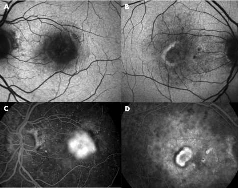
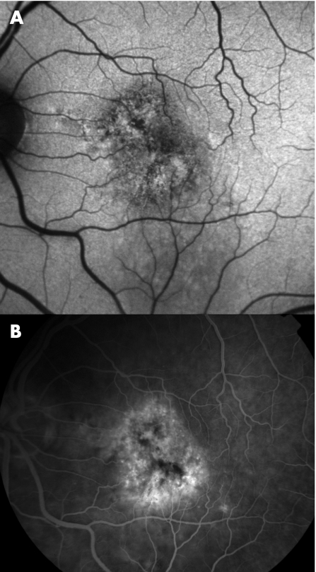
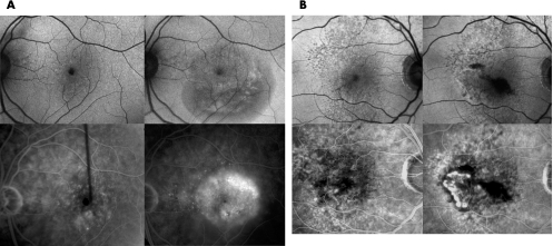
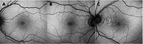
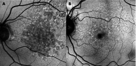
Aim: To evaluate the distribution of fundus autofluorescence in patients with age-related macular degeneration and choroidal neovascularisation (CNV).
Methods: Colour fundus photographs, fundus fluorescein angiograms (FFA) and fundus autofluorescence images were obtained from a group of 40 patients (43 eyes) with age-related macular degeneration and purely classic or occult CNV. Only patients with newly diagnosed CNV and in whom autofluorescence images were obtained within 2 weeks from FFA were included. The distribution of autofluorescence was qualitatively evaluated, and the findings compared with those from colour fundus photographs and FFA.
Results: 29 (67%) eyes had classic CNV and 14 (33%) had occult CNV. In 26 (90%) eyes with classic CNV, a low autofluorescence signal was detected at the site of the CNV; in 7 (50%) eyes with occult CNV, multiple foci of low autofluorescence signal were detected. Outside the area affected by the lesion, homogeneous autofluorescence was observed in most of the cases (n = 33, 77%). Similarly, homogeneous autofluorescence was commonly observed in fellow eyes (62%). A pattern of focal increased autofluorescence was rarely seen in eyes with CNV (n = 4, 9%) or in fellow eyes (n = 4, 15%). In 11 of 43 (25%) eyes, areas of increased autofluorescence, other than a pattern of focal increased autofluorescence, were detected. In four patients, autofluorescence images had been obtained before the development of CNV; in none was any increased autofluorescence detected before the formation of CNV.
Conclusions: Distinct patterns of autofluorescence were observed in eyes with pure classic and occult CNV. Increased autofluorescence was rarely seen in eyes with CNV and in fellow eyes, suggesting that increased autofluorescence, and thus, retinal pigment epithelium lipofuscin, may not play an essential part in the formation of CNV.
Conflict of interest statement
Competing interests: None.
Similar articles
-
Prevalence of reticular pseudodrusen in age-related macular degeneration with newly diagnosed choroidal neovascularisation.Br J Ophthalmol. 2007 Mar;91(3):354-9. doi: 10.1136/bjo.2006.101022. Epub 2006 Sep 14. Br J Ophthalmol. 2007. PMID: 16973663 Free PMC article.
-
Natural course of occult choroidal neovascularization in age-related macular degeneration: development of classic lesions in fluorescein angiography.Acta Ophthalmol Scand. 2005 Apr;83(2):141-7. doi: 10.1111/j.1600-0420.2005.00443.x. Acta Ophthalmol Scand. 2005. PMID: 15799723
-
Autofluorescence imaging in age-related macular degeneration complicated by choroidal neovascularization: a prospective study.Ophthalmology. 2008 Feb;115(2):342-6. doi: 10.1016/j.ophtha.2007.04.023. Epub 2007 Jun 28. Ophthalmology. 2008. PMID: 17599415
-
Fundus autofluorescence and progression of age-related macular degeneration.Surv Ophthalmol. 2009 Jan-Feb;54(1):96-117. doi: 10.1016/j.survophthal.2008.10.004. Surv Ophthalmol. 2009. PMID: 19171212 Review.
-
Fundus autofluorescence in geographic atrophy: a review.Semin Ophthalmol. 2010 Sep-Nov;25(5-6):206-13. doi: 10.3109/08820538.2010.518121. Semin Ophthalmol. 2010. PMID: 21091001 Review.
Cited by
-
Visual cycle modulators versus placebo or observation for the prevention and treatment of geographic atrophy due to age-related macular degeneration.Cochrane Database Syst Rev. 2020 Dec 17;12(12):CD013154. doi: 10.1002/14651858.CD013154.pub2. Cochrane Database Syst Rev. 2020. PMID: 33331670 Free PMC article.
-
[Anti-VEGF therapy of exudative AMD: Prognostic factors for therapy success].Ophthalmologe. 2011 Feb;108(2):124-31. doi: 10.1007/s00347-010-2210-z. Ophthalmologe. 2011. PMID: 20535478 German.
-
Characteristic appearances of fundus autofluorescence in treatment-naive and active polypoidal choroidal vasculopathy: a retrospective study of 170 patients.Graefes Arch Clin Exp Ophthalmol. 2018 Jun;256(6):1101-1110. doi: 10.1007/s00417-018-3980-2. Epub 2018 Apr 14. Graefes Arch Clin Exp Ophthalmol. 2018. PMID: 29656364
-
The pathologic characteristics of pingueculae on autofluorescence images.Korean J Ophthalmol. 2013 Dec;27(6):416-20. doi: 10.3341/kjo.2013.27.6.416. Epub 2013 Nov 15. Korean J Ophthalmol. 2013. PMID: 24311926 Free PMC article.
-
The Minnesota Grading System using fundus autofluorescence of eye bank eyes: a correlation to age-related macular degeneration (an AOS thesis).Trans Am Ophthalmol Soc. 2008;106:383-401. Trans Am Ophthalmol Soc. 2008. PMID: 19277247 Free PMC article.
References
-
- Macular Photocoagulation Study Group Subfoveal neovascular lesions in age‐related macular degeneration. Guidelines for evaluation and treatment in the macular photocoagulation study. Arch Ophthalmol 19911091242–1257. - PubMed
Publication types
MeSH terms
Substances
LinkOut - more resources
Full Text Sources
Medical