. 2006 Nov;80(22):11040-54.
doi: 10.1128/JVI.01056-06. Epub 2006 Sep 6.

Adeno-associated virus type 2 capsids with externalized VP1/VP2 trafficking domains are generated prior to passage through the cytoplasm and are maintained until uncoating occurs in the nucleus

Affiliations

Adeno-associated virus type 2 capsids with externalized VP1/VP2 trafficking domains are generated prior to passage through the cytoplasm and are maintained until uncoating occurs in the nucleus

Florian Sonntag et al. J Virol. 2006 Nov.

Abstract

Common features of parvovirus capsids are open pores at the fivefold symmetry axes that traverse the virion shell. Upon limited heat treatment in vitro, the pores can function as portals to externalize VP1/VP2 protein N-terminal sequences which harbor infection-relevant functional domains, such as a phospholipase A(2) catalytic domain. Here we show that adeno-associated virus type 2 (AAV2) also exposes its VP1/VP2 N termini in vivo during infection, presumably in the endosomal compartment. This conformational change is influenced by treatment with lysosomotropic reagents. While incubation of cells with bafilomycin A1 reduced exposure of VP1/VP2 N termini, incubation with chloroquine stimulated externalization transiently. N-terminally located basic amino acid clusters with nuclear localization activity also become exposed in this process and are accessible on the virus capsid when it enters the cytoplasm. This is an obligatory step in AAV2 infection. However, a direct role of these sequences in nuclear translocation of viral capsids could not be determined by microinjection of wild-type or mutant viruses. This suggests that further modifications of the capsid have to take place in a precytoplasmic entry step that prepares the virus for nuclear entry. Microinjection of several capsid-specific antibodies into the cell nucleus blocked AAV2 infection completely, supporting the conclusion that AAV2 capsids bring the infectious genome into the nucleus.


Figures

FIG. 1.
FIG. 1.
BCs on VP1/VP2 N termini of AAV2. (A) Schematic representation of the localization of BC1, BC2, BC3, and the phospholipase A2 domain on the capsid proteins of AAV2. (B) Sequence alignment of the 11 known AAV serotypes. Arginine and lysine residues in the basic clusters are highlighted in bold letters with gray background. (C) Mutations of the basic clusters and of the PLA2 domain. Single, double, and triple mutation of BC elements were generated by substitution of positively charged amino acids to glutamic acids. The HD/AN PLA2 mutant is characterized by two mutated residues in the catalytic center (21). Mutations are highlighted with gray background. (D) Effect of the analyzed mutations on infectivity. Virus supernatants obtained from 293T cells transfected with wt AAV2 or mutated genomic plasmids and infected with Ad5 (MOI = 10) were assayed for particle/infectivity ratios by calculating the amount of genome-containing particles per infectious particle. Means ± standard deviations from three independent experiments are shown.
FIG. 2.
FIG. 2.
Externalization of VP1/VP2 N termini during infection. (A) Subcellular localization of exposed VP1/VP2 N termini. HeLa cells were incubated with wt AAV2 (25,000 particles/cell) for 30 min at 4°C and subsequently washed to remove unbound virus. After further incubation for the indicated time periods at 37°C, the externalization of VP1/VP2 N termini was examined by immunofluorescence with VP1/VP2-specific antibody A69 and tyramide signal amplification. Fluorescence images were superimposed on the corresponding phase-contrast images. (B) Analysis of cell lysates. HeLa cells were incubated with wt AAV2 (10,000 particles/cell) for 30 min at 4°C and washed. Following incubation at 37°C, the cells were harvested and lysed by freezing-thawing at the indicated time points. Virus-containing supernatant was analyzed by a native immuno-dot blot using VP1-specific antibody A1, VP1/VP2, specific antibody A69, C-terminus-specific antibody B1, or capsid-specific antibody A20. Extracts of noninfected cells were used as a negative control (n). Signal intensities were measured with Image Quant TL software, and the ratios of A1/A20 and A69/A20 signal were determined to quantify exposure of N termini on capsids. Means ± standard deviations from three independent experiments are shown. (C) Externalization of VP1/VP2 N termini and capsid stability of mutants with exchanges of amino acids at the fivefold symmetry axes of the capsid: N335A, L336A, and V221Y. Means ± standard deviations from three independent experiments are shown for the wt and the N335A mutant; values for L336A and V221Y mutants are from a single experiment. (D) Capsid stability calculated by A20 signal intensity (values are from a representative experiment).
FIG. 3.
FIG. 3.
Influence of lysosomotropic drugs on infectivity, capsid stability, and externalization of VP1/VP2 N termini. (A) Influence of chloroquine and bafilomycin A1 (BFLA) on viral infectivity. HeLa cells were infected with AAV2 in the presence or absence of lysosomotropic drugs. Early viral gene expression was analyzed 20 h p.i. by immunofluorescence using the Rep-specific antibody 76.3. Cell nuclei were visualized with DAPI. (B to F) Native immuno-dot blot analysis of cell lysates obtained at the indicated time points. HeLa cells were incubated with wt AAV2 (10,000 particles/cell) for 30 min at 4°C and washed. Following incubation at 37°C in the presence or absence of drugs, the cells were harvested and lysed, and virus-containing supernatant was analyzed by a native immuno-dot blot using VP1-specific antibody A1, VP1/VP2-specific antibody A69, or capsid-specific antibody A20. Signal intensity was measured with Image Quant TL software. (B) Capsid stability was calculated from A20 signal intensity. (C) Externalization of VP1 N termini, determined as the ratio of A1/A20 signal intensity, and (D) externalization of VP1/VP2 N termini, determined as the ratio of A69/A20 signal intensity, in the presence of chloroquine. (E) Externalization of VP1 N termini, determined as the ratio of A1/A20 signal intensity, and (F) externalization of VP1/VP2 N termini, determined as the ratio of A69/A20 signal intensity, in the presence of bafilomycin A1. Results of one representative of two independent experiments are shown.
FIG. 4.
FIG. 4.
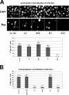
AAV2 enters the cytoplasm with exposed VP1/VP2 N termini. (A) Extracellular neutralization of infection. AAV2 particles were preincubated with 100 μg/ml of VP1-specific antibody A1, VP1/VP2-specific antibody A69, C-terminus-specific antibody B1, or capsid-specific antibody A20. In a control experiment, no antibody (no Ab) was added. The samples were transferred to HeLa cells (2 × 10³ particles/cell), infected with Ad5 (MOI = 4), and incubated for 20 h. Early viral gene expression was analyzed by immunofluorescence with Rep-specific antibody 76.3. Cell nuclei were visualized with DAPI. Means ± standard deviations from two independent experiments are shown. (B) Intracellular neutralization of infection by cytoplasmic microinjection of antibodies. Antibodies were microinjected into the cytoplasm of HeLa cells at a concentration of 5 mg/ml. Cells were subsequently infected with AAV2 (MOI = 20) and superinfected with Ad5 (MOI = 4). After 20 h, early gene expression was analyzed in injected cells by immunofluorescence with a Rep-specific polyclonal antiserum. Means ± standard deviations from at least two independent experiments are shown. Rep pos., Rep positive.
FIG. 5.
FIG. 5.
Nuclear localization activities of BC elements. Different synthetic peptides containing a C-terminal cysteine were cross-linked to fluorescein-labeled BSA and microinjected into the cytoplasm of HeLa cells. After 2 h, the subcellular localization (subcell. loc.) of the peptide-protein conjugates was examined by confocal fluorescence microscopy. BSA, no peptide coupled; SV40-BSA, NLS of SV40 large T antigen coupled to BSA; aa 20-43, amino acids 20 to 43 of the AAV2 VP1 sequence coupled to BSA; BC1-, BC2-, and BC3-BSA, separate basic clusters coupled to BSA; BC1/2- and BC2/3-BSA, combined BC elements coupled to BSA; cyt, cytoplasm; nuc, nucleus.
FIG. 6.
FIG. 6.
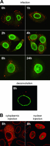
Subcellular localization of AAV2 particles. (A) Time course after infection with AAV2. HeLa cells were incubated with 500,000 particles/cell at 4°C for 30 min and then shifted to 37°C for the designated time periods. After fixation, the localization was examined by confocal immunofluorescence microscopy using an antibody directed against capsids (A20; red) and an antibody directed against the nuclear lamina (Lamin B; green). Deconvolution of signals located above or below the nucleus at 8 h p.i. (B) Subcellular localization of AAV2 particles 8 h after microinjection into the cytoplasm (2 × 10³ particles/cell) or the nucleus (5 × 10² particles/cell) of HeLa cells, followed by superinfection with Ad5 (MOI = 5) in the presence of the proteasome inhibitor MG-132 (30 μM). The green signal corresponds to AAV2 particles detected with antibody A20. The red signal corresponds to goat IgG injection marker and Lamin B1.
FIG. 7.
FIG. 7.
Infectivities of wt and mutant AAV2 particles after infection, cytoplasmic microinjection, or nuclear microinjection. (A) Infection. HeLa cells were infected with full wt or mutant AAV2 (17 genome-containing particles/cell) and superinfected with Ad5 (MOI = 4). Early viral gene expression was analyzed after 20 h by immunofluorescence using Rep-specific MAb 76.3. Cell nuclei were visualized with DAPI, and Rep-positive (Rep pos.) cells were counted. Means ± standard deviations from two independent experiments are shown. (B) Cytoplasmic injection. Full virions (approximately 70 genome-containing particles/cell) and IgG injection marker were injected into the cytoplasm of HeLa cells, followed by superinfection with Ad5 (MOI = 4). Early viral gene expression was analyzed in injected cells after 20 h by immunofluorescence using a Rep-specific polyclonal antiserum. (C) Nuclear injection. Full virions (approximately 18 genome-containing particles/cell) and IgG injection marker were injected into the nucleus of HeLa cells, followed by superinfection with Ad5 (MOI = 4). Early viral gene expression was analyzed in injected cells after 20 h by immunofluorescence using a Rep-specific polyclonal antiserum. Means ± standard deviations from two independent experiments are shown. Heat treatment for panels A, B, and C was performed for 3 min at the indicated temperatures. To block effectively infection by virus that had been released into the culture medium during the injection procedure, the medium was supplemented with the neutralizing antibody C37-B. To compare the infectivities of the different virus constructs after application in the extracellular, cytoplasmic, or nuclear compartment particle/infectivity ratios were determined by calculating the amount of genome-containing particles per infectious particle on the basis of Rep-positive cells.
FIG. 8.
FIG. 8.
Inhibition of AAV2 infection by injection of antibodies into the nucleus. (A) Nuclear injection. Antibodies were microinjected into the nucleus of HeLa cells at a concentration of 5 mg/ml. Cells were subsequently infected with AAV2 (MOI = 20) and superinfected with Ad5 (MOI = 4). After 20 h, early gene expression was analyzed in injected cells by immunofluorescence using a Rep-specific polyclonal antiserum. Means ± standard deviations from at least two independent experiments are shown. Rep pos., Rep positive; not inj., not injected. (B) Cytoplasmic injection of 1:10-diluted antibodies. Antibodies were injected into the cytoplasm of HeLa cells in a concentration of 0.5 mg/ml, corresponding to the estimated maximal leakage during nuclear injection. Samples were processed further as described above. (C) Indicated concentrations of goat IgG were injected into the nucleus or the cytoplasm and stained for immunofluorescence using Alexa 488-labeled secondary antibodies.
FIG. 9.
FIG. 9.
Determination of DNA release. HeLa cells were infected with 5,000 AAV2 particles/cell, harvested after the indicated time periods, and separated into the cytoplasmic (cyt) and nuclear (nuc) fractions. Viral particles were immunoprecipitated by protein A-Sepharose-bound antibody A20. Samples were then digested (+) or not digested (−) with MNase. Coprecipitated genomes were isolated and transferred to a nylon membrane, and genomes were detected with a rep-specific probe.

References

    1. Bantel-Schaal, U., B. Hub, and J. Kartenbeck. 2002. Endocytosis of adeno-associated virus type 5 leads to accumulation of virus particles in the Golgi compartment. J. Virol. 76:2340-2349. - PMC - PubMed
    1. Bartlett, J. S., R. Wilcher, and R. J. Samulski. 2000. Infectious entry pathway of adeno-associated virus and adeno-associated virus vectors. J. Virol. 74:2777-2785. - PMC - PubMed
    1. Bayer, N., D. Schober, E. Prchla, R. F. Murphy, D. Blaas, and R. Fuchs. 1998. Effect of bafilomycin A1 and nocodazole on endocytic transport in HeLa cells: implications for viral uncoating and infection. J. Virol. 72:9645-9655. - PMC - PubMed
    1. Becerra, S. P., F. Koczot, P. Fabisch, and J. A. Rose. 1988. Synthesis of adeno-associated virus structural proteins requires both alternative mRNA splicing and alternative initiations from a single transcript. J. Virol. 62:2745-2754. - PMC - PubMed
    1. Bleker, S., F. Sonntag, and J. A. Kleinschmidt. 2005. Mutational analysis of narrow pores at the fivefold symmetry axes of adeno-associated virus type 2 capsids reveals a dual role in genome packaging and activation of phospholipase A2 activity. J. Virol. 79:2528-2540. - PMC - PubMed

Publication types

LinkOut - more resources