. 2006 Nov;80(21):10836-46.
doi: 10.1128/JVI.00966-06. Epub 2006 Sep 6.

Posttranslational modification and cell type-specific degradation of varicella-zoster virus ORF29p

Affiliations

Posttranslational modification and cell type-specific degradation of varicella-zoster virus ORF29p

Christina L Stallings et al. J Virol. 2006 Nov.

Abstract

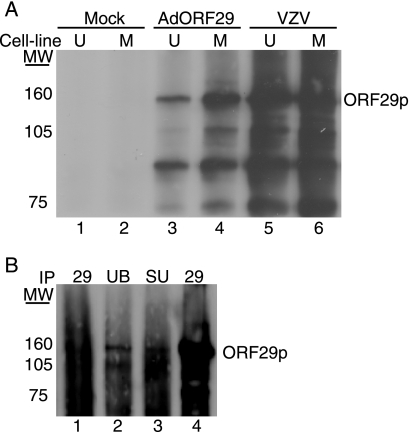
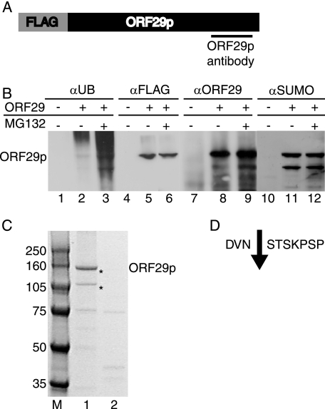
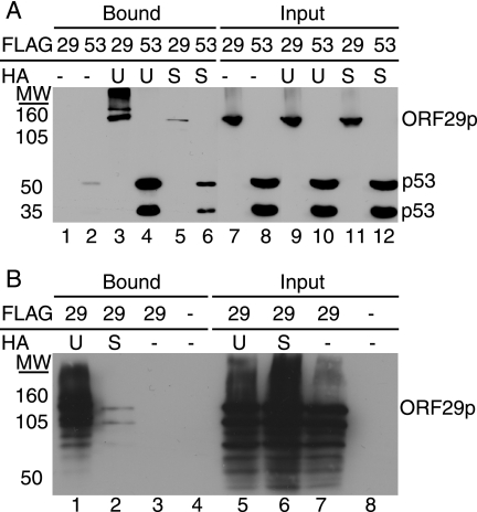
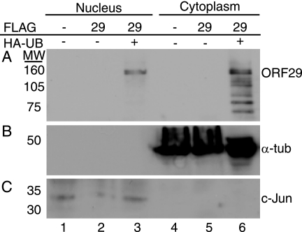
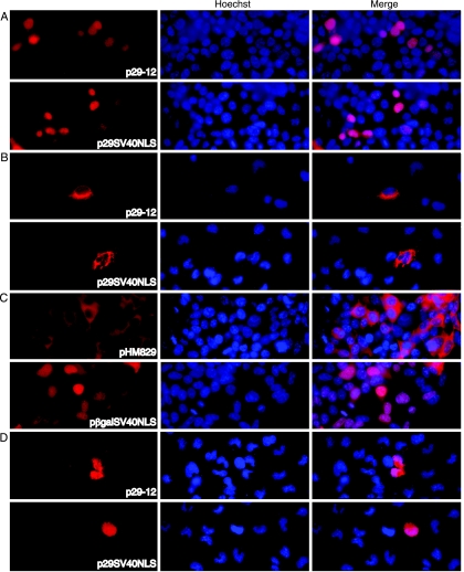
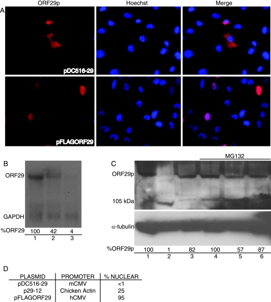
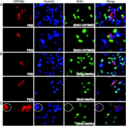
The ORF29 gene of varicella-zoster virus encodes a single-stranded DNA binding protein that is predominantly nuclear during lytic infection but appears to be restricted to the cytoplasm of latently infected neurons. Following reactivation, ORF29p accumulates in the nuclei of neurons, suggesting that its confinement to the cytosol may be critical for maintaining quiescence. When autonomously expressed, ORF29p accumulates in the nuclei of fibroblasts and the cytoplasm of cells (guinea pig enteric neurons) and cell lines (U373MG) of neuronal origin. Inhibition of the 26S proteasome redirects the accumulation of ORF29p to the nucleus in cells of neuronal origin. Here, we show that ORF29p is ubiquitinated and sumoylated in 293T cells and subsequently degraded from the N terminus. Ubiquitinated ORF29p accumulates in both the nuclei and the cytoplasm of fibroblasts, but degradation products are seen primarily in the cytoplasm. Modification and degradation of ORF29p occurs in 293T, U373MG, and MeWo cells. Therefore, these processes are ubiquitous; however, the robustness of the degradation process is cell type specific. The proteasome-mediated mechanism of nuclear exclusion in U373MG cells is an active process that is not specific for the endogenous ORF29p nuclear localization signal but can be saturated by protein stabilization or overexpression, which leads to nuclear accumulation of ORF29p. The evidence for ORF29p ubiquitination and previous data regarding the effect of proteasome inhibitors on the abundance and distribution of ORF29p implicate the 26S proteasome in influencing the protein's cell type-specific localization.


Figures

FIG. 1.
FIG. 1.
Analysis of ORF29p electrophoretic mobility. (A) MeWo (M) (lanes 2, 4, and 6) and U373MG (U) (lanes 1, 3, and 5) cells were either infected with cell-free Jones VZV at an MOI of 1 (lanes 5 and 6), infected with AdORF29 at an MOI of 50 (lanes 3 and 4), or mock infected. At 3 days postinfection, cells were labeled with 500 μCi/ml Tran35S-label for 24 h, harvested, and lysed in RIPA buffer. ORF29p was immunoprecipitated from the labeled cell lysates by using an ORF29p-specific antiserum, and bound proteins were subjected to SDS-PAGE analysis on a 5% gel. Proteins were visualized by autoradiography. (B) MeWo cells were either mock infected (lane 1) or infected with cell-free VZV at an MOI of 1 (lanes 2 to 4). At 5 days postinfection, the cells were harvested, lysed in RIPA buffer, and immunoprecipitated (IP) with ubiquitin-specific antibody (UB) (lane 2), SUMO-specific antibody (SU) (lane 3), or ORF29p-specific antiserum (lanes 1 and 4) (29). The bound proteins were subjected to SDS-PAGE and analyzed by Western blotting with antiserum specific for ORF29p. MW, molecular weights in thousands.
FIG. 2.
FIG. 2.
Analysis of two species of ORF29p. (A) The N-terminal, FLAG-tagged ORF29 expression cassette from pFLAGORF29 is diagrammed (not to scale), with the region recognized by the ORF29p-specific antiserum underlined. 293T cells were transiently transfected with either pFLAGORF29 (B, lanes 2, 3, 5, 6, 8, 9, 11, and 12; C, lane 1) or empty pCF2HN (B, lanes 1, 4, 7, and 10; C, lane 2). At 48 h posttransfection, cells were either treated with DMSO (B, lanes 1, 2, 4, 5, 7, 8, 10, and 11; C) or treated with 20 μM MG132 (B, lanes 3, 6, 9, and 12) for 6 h. Cells were lysed in RIPA buffer and incubated with an anti-FLAG M2 agarose matrix. Bound proteins were eluted with FLAG peptide and resolved by SDS-PAGE. (B) Western blot analysis of the eluates with antiserum specific to ubiquitin (αUB) (lanes 1 to 3), FLAG peptide (αFLAG) (lanes 4 to 6), ORF29p (αORF29) (lanes 7 to 9), or SUMO (αSUMO) (lanes 10 to 12). (C) Coomassie blue staining of bound proteins. Asterisks indicate the bands that were sequenced by MALDI mass spectrophotometry. MW, molecular weights in thousands. (D) Sequence of ORF29p amino acids 301 to 310 and the cleavage site (↓) that produces the 105-kDa ORF29p peptide as determined by MALDI mass spectrophotometry.
FIG. 3.
FIG. 3.
Analysis of ORF29p modifications. 293T cells were transiently transfected with either pFLAGORF29 (A, lanes 1, 3, 5, 7, 9, and 11; B, lanes 1 to 3 and 5 to 7), pCMV-Flag-53 (A, lanes 2, 4, 6, 8, 10, and 12), or empty pCF2HN (B, lanes 4 and 8) either alone (A, lanes 1, 2, 7, and 8; B, lanes 3, 4, 7, and 8) or in conjunction with pCMV-HA-UB (UB) (A, lanes 3, 4, 9, and 10; B, lanes 1 and 5) or pHis6-HA-SUMO (SU) (A, lanes 5, 6, 11, and 12; B, lanes 2 and 6). At 48 h posttransfection, cells were lysed in RIPA buffer and reacted with an anti-HA matrix. Bound proteins were eluted with HA peptide. Bound and input fractions were resolved by SDS-PAGE, and ORF29p and p53 were detected by Western blot analysis with FLAG-specific antiserum (A) or ORF29p-specific antiserum (B). MW, molecular weights in thousands.
FIG. 4.
FIG. 4.
Subcellular distribution of ubiquitinated ORF29p. 293T cells were transiently transfected with either empty pCF2HN (lanes 1 and 4) or pFLAGORF29 (lanes 2, 3, 5, and 6) either alone (lanes 1, 2, 4, and 5) or in conjunction with pCMV-HA-UB (lanes 3 and 6). At 48 h posttransfection, cells were harvested, separated into nuclear and cytoplasmic fractions, and incubated with an anti-HA matrix. Bound proteins were eluted with HA peptide. Eluates (A) and input fractions (B and C) were resolved by SDS-PAGE. ORF29p (ORF29) (A), c-Jun (B), and α-tubulin (α-tub) (C) were detected by Western blotting. MW, molecular weights in thousands.
FIG. 5.
FIG. 5.
Localization of an ORF29p SV40 NLS fusion protein. 293T (A and C) and U373MG (B and D) cells were transfected with p29-12, p29SV40NLS, pHM829, or pβgalSV40NLS. At 48 h posttransfection, cell cultures were treated with either DMSO (A to C) or 20 μM MG132 in DMSO (D) for 6 h prior to fixing them onto glass coverslips. ORF29p, β-galactosidase, and the SV40 NLS fusions of these proteins were detected by indirect immunofluorescence microscopy after reacting them with specific antisera. The nuclei were visualized by counterstaining with Hoechst.
FIG. 6.
FIG. 6.
Overexpression of ORF29p in U373MG cells. U373MG cells were transfected with pDC516-29 (B, lane 3; C lanes 2 and 5), pFLAGORF29 (B, lane 1; C, lanes 1 and 4), or p29-12 (B, lane 2; C, lanes 3 and 6), which express ORF29p from mCMV, hCMV, or chicken actin promoters, respectively. At 48 h posttransfection, the cells were treated with either DMSO (A, B, and C, lanes 1 to 3) or 20 μM MG132 (C, lanes 4 to 6) for 6 h before fixing them onto glass coverslips (A) or harvesting them for Northern (B) and Western (C) blot analyses. (A) ORF29p was detected by indirect immunofluorescence microscopy after it was reacted with specific antiserum. (B) Total RNA was isolated, separated by gel electrophoresis, and transferred to a nylon membrane. The mRNA was probed with radiolabeled probes specific for ORF29 and GAPDH (glyceraldehyde-3-phosphate dehydrogenase). Following hybridization, these RNAs were visualized by exposure to film. The percentages of ORF29 transcript were calculated in comparison to transcript levels from the hCMV promoter (100%) after normalization to GAPDH levels by using the ImageJ program. (C) Infected U373MG cells were lysed in RIPA buffer and subjected to SDS-PAGE. After the transfer of proteins to a nitrocellulose membrane, Western blot analysis was done using ORF29p- and α-tubulin-specific antisera. The percentages of ORF29p were calculated in comparison to protein levels from the hCMV promoter (100%) after normalization to α-tubulin levels by using the ImageJ program. (D) The percentages of ORF29p-expressing cells that exhibited nuclear staining when reacted with antiserum specific for ORF29p during IF experiments, according to the promoter.
FIG. 7.
FIG. 7.
ORF29p localization in U373MG and MeWo cell heterokaryons. MeWo (A) and U373MG (B and C) cells were infected with AdORF29 at an MOI of 50. At 48 hours postinfection, infected cells were mixed with U373MG (A) or MeWo (B and C) cells that were labeled by incubation with BrdU and seeded onto coverslips for 12 h. The mixed cultures were incubated with either PBS or 50% PEG in PBS for 90 s at 37°C, washed four times, and allowed to recover in normal growth media for 12 h at 37°C before they were fixed and analyzed by indirect immunofluorescence with antisera specific for ORF29p and BrdU. For each panel, the cell line containing BrdU is indicated in parentheses. The white circle surrounds the heterokaryon containing a 5:1 MeWo-to-U373MG nucleus ratio.

Similar articles

Cited by

References

    1. Arvin, A. M. 1996. Varicella-zoster virus. Clin. Microbiol. Rev. 9:361-381. - PMC - PubMed
    1. Blank, V., P. Kourilsky, and A. Israel. 1991. Cytoplasmic retention, DNA binding and processing of the NF-kappa B p50 precursor are controlled by a small region in its C-terminus. EMBO J. 10:4159-4167. - PMC - PubMed
    1. Chen, J. J., A. A. Gershon, Z. S. Li, O. Lungu, and M. D. Gershon. 2003. Latent and lytic infection of isolated guinea pig enteric ganglia by varicella zoster virus. J. Med. Virol. 70(Suppl. 1):S71-S78. - PubMed
    1. Cohrs, R. J., D. H. Gilden, P. R. Kinchington, E. Grinfeld, and P. G. Kennedy. 2003. Varicella-zoster virus gene 66 transcription and translation in latently infected human ganglia. J. Virol. 77:6660-6665. - PMC - PubMed
    1. Croen, K. D., and S. E. Straus. 1991. Varicella-zoster virus latency. Annu. Rev. Microbiol. 45:265-282. - PubMed

Publication types

MeSH terms

LinkOut - more resources