. 2006;34(16):4561-71.
doi: 10.1093/nar/gkl376. Epub 2006 Sep 6.

Recognition of T-rich single-stranded DNA by the cold shock protein Bs-CspB in solution

Affiliations

Recognition of T-rich single-stranded DNA by the cold shock protein Bs-CspB in solution

Markus Zeeb et al. Nucleic Acids Res. 2006.

Abstract

Cold shock proteins (CSP) belong to the family of single-stranded nucleic acid binding proteins with OB-fold. CSP are believed to function as 'RNA chaperones' and during anti-termination. We determined the solution structure of Bs-CspB bound to the single-stranded DNA (ssDNA) fragment heptathymidine (dT7) by NMR spectroscopy. Bs-CspB reveals an almost invariant conformation when bound to dT7 with only minor reorientations in loop beta1-beta2 and beta3-beta4 and of few aromatic side chains involved in base stacking. Binding studies of protein variants and mutated ssDNA demonstrated that Bs-CspB associates with ssDNA at almost diffusion controlled rates and low sequence specificity consistent with its biological function. A variation of the ssDNA affinity is accomplished solely by changes of the dissociation rate. 15N NMR relaxation and H/D exchange experiments revealed that binding of dT7 increases the stability of Bs-CspB and reduces the sub-nanosecond dynamics of the entire protein and especially of loop beta3-beta4.


Figures

Figure 1
Figure 1
The 2D 1H/15N HSQC spectrum of free (black) and dT7-bound (red) Bs-CspB in 50 mM Na-cacodylate, 3 mM MgCl2, pH 7.0 at 15°C. The spectra were recorded at a Bs-CspB-concentration of 0.8 mM and a final dT7 concentration of 1.2 mM.
Figure 2
Figure 2
Protection factors of amide protons from H/D exchange of free (hatched bars) and dT7-bound (solid bars) Bs-CspB. Missing bars indicate amides, which got fully deuterated in the dead-time of the experiment (5 min). The solid bars on top represent the five β-strands of Bs-CspB.
Figure 3
Figure 3
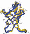
Backbone superposition of the 18 lowest energy NMR structures of free [blue, 1NMF.pdb (19)] and dT7-bound (yellow) Bs-CspB. The five β-strands and their connecting loops are indicated. The r.m.s.d. values of dT7-bound Bs-CspB of all 67 residues are 0.54 Å (backbone) and 0.94 Å (all heavy atoms). Main deviations between apo and holo form of Bs-CspB are located in loop β1–β2 (L12) and loop β3–β4 (L34).
Figure 4
Figure 4
15N relaxation and internal motion parameter of free (48) (open symbols) and dT7-bound (closed symbols) Bs-CspB. (a) heteronuclear NOE hNOE and (c) transversal relaxation rates R2, as well as (b) order parameters S2 and (d) chemical exchange contributions Rex to R2. S2 and Rex were obtained from extended Lipari-Szabo analyses of longitudinal relaxation rates R1 (data not shown), R2 rates and hNOE using the program MODELFREE (49). Solid and gray bars represent the five β-strands of Bs-CspB.
Figure 5
Figure 5
Nucleic acid-binding site of Bs-CspB determined by NMR spectroscopy and site-directed mutagenesis at (a) front view and (b) back view. The following residues experienced substantial chemical shift changes of their NMR resonances upon binding to dT7 and are indicated in blue: 7, 10–17, 20, 25, 26–31, 33, 35, 38–42, 54, 56–59 (backbone) and 8, 25, 34, 59, 62 (side chain). Yellow highlighted side chains illustrate aromatic residues, for which the KD increased above 100 nM after substitution with alanine. For this illustration the NMR structure with the lowest energy was used.
Figure 6
Figure 6
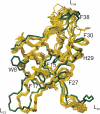
Backbone superposition of the 18 lowest energy NMR structures (yellow) of Bs-CspB/dT7 and the crystal structure (green) of Bs-CspB/dT6 (2ES2.pdb). The side chain conformations of the aromatic residues, which facilitate binding of dT7, are depicted and labeled.

Similar articles

Cited by

References

    1. Graumann P.L., Marahiel M.A. A superfamily of proteins that contain the cold-shock domain. Trends Biochem. Sci. 1998;23:286–290. - PubMed
    1. Jones P.G., van Bogelen R.A., Neidhardt F.C. Induction of proteins in response to low temperature in Escherichia coli. J. Bacteriol. 1987;169:2092–2095. - PMC - PubMed
    1. Jones P.G., Inouye M. RbfA, a 30S ribosomal binding factor, is a cold-shock protein whose absence triggers the cold-shock response. Mol. Microbiol. 1996;21:1207–1218. - PubMed
    1. Phadtare S., Inouye M., Severinov K. The nucleic acid melting activity of Escherichia coli CspE is critical for transcription antitermination and cold acclimation of cells. J. Biol. Chem. 2002;277:7239–7245. - PubMed
    1. Jones P.G., Krah R., Tafuri S.R., Wolffe A.P. DNA gyrase, CS7.4, and the cold shock response in Escherichia coli. J. Bacteriol. 1992;174:5798–5802. - PMC - PubMed

Publication types