. 2006 Sep 19;103(38):13956-61.
doi: 10.1073/pnas.0606381103. Epub 2006 Sep 6.

Crystal structure of human histone lysine-specific demethylase 1 (LSD1)

Affiliations

Crystal structure of human histone lysine-specific demethylase 1 (LSD1)

Yong Chen et al. Proc Natl Acad Sci U S A. .

Abstract

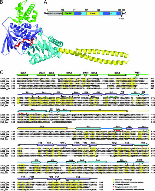
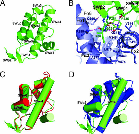
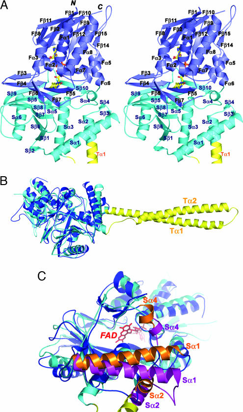
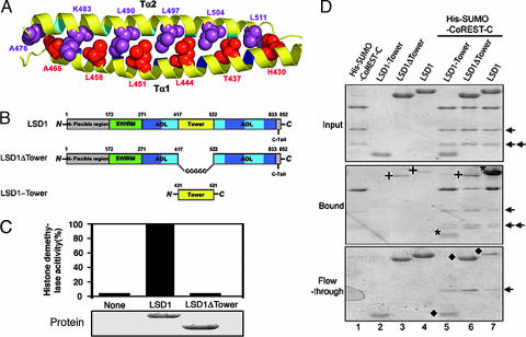
Lysine-specific demethylase 1 (LSD1) was recently identified as the first histone demethylase that specifically demethylates monomethylated and dimethylated histone H3 at K4. It is a component of the CoREST and other corepressor complexes and plays an important role in silencing neuronal-specific genes in nonneuronal cells, but the molecular mechanisms of its action remain unclear. The 2.8-A-resolution crystal structure of the human LSD1 reveals that LSD1 defines a new subfamily of FAD-dependent oxidases. The active center of LSD1 is characterized by a remarkable 1,245-A3 substrate-binding cavity with a highly negative electrostatic potential. Although the protein core of LSD1 resembles other flavoenzymes, its enzymatic activity and functions require two additional structural modules: an N-terminal SWIRM domain important for protein stability and a large insertion in the catalytic domain indispensable both for the demethylase activity and the interaction with CoREST. These results provide a framework for further probing the catalytic mechanism and the functional roles of LSD1.


Conflict of interest statement

The authors declare no conflict of interest.

Figures

Fig. 1.
Fig. 1.
Overview of the LSD1 structure. (A) Domain organization of LSD1. The SWIRM domain is shown in green, the AOL domain is in blue (the FAD-binding subdomain) and cyan (the substrate-binding subdomain), and the Tower domain is in yellow. The N-terminal flexible region and the C-terminal tail that are not included in the structure determination are colored in gray. (B) Ribbon diagram of the LSD1 structure. The molecule is colored as in A. FAD is in ball-and-stick representation and is colored in red. (C) Structural-based sequence alignments of the SWIRM and AOL domains of LSD1 and its homologs. Secondary structure assignments from the LSD1 crystal structure are shown as cylinders (α-helices) and arrows (β-strands) above the aligned sequences and colored as in B.
Fig. 2.
Fig. 2.
The structure of SWIRM domain and its interactions with the AOL domain. (A) Ribbon diagram of the SWIRM domain. (B) A three-amino-acid motif (F264G265I266) of the SWIRM domain mediates the interactions between the SWIRM and AOL domains. The SWIRM and AOL domains are colored in green and blue, respectively. Side chains of residues important for the interactions are shown explicitly. The hydrogen bonds are shown as dotted purple lines. (C and D) Superposition of the LSD1 SWIRM domain (green) with those of Swi3 (red) and Ada2α (blue).
Fig. 3.
Fig. 3.
The structure of the AOL domain. (A) Stereo ribbon diagram of the AOL domain of LSD1. The molecule is colored as in Fig. 1B. The FAD is in ball-and-stick representation with carbon colored yellow, nitrogen colored blue, oxygen colored red, and phosphorus colored orange. (B) Superposition of LSD1 (the AOL and the Tower domains) on the structure of PAO. LSD1 is in cyan, and PAO is in blue. The Tower domain of LSD1 is colored in yellow. (C) Superposition of the AOL domain of LSD1 on the structure of PAO highlighting the differences in the catalytic cavity. LSD1 is in cyan and purple, and PAO is in blue and orange. The Tower domain of LSD1 is colored in yellow. The FAD, in red and ball-and-stick representation, lies in the back of the cavity. The positional change of three α-helices (Sα1, Sα2, and Sα4) of PAO relative to those of LSD1 significantly reduces the volume of the catalytic cavity.
Fig. 4.
Fig. 4.
The catalytic center of LSD1. (A) Stereo ribbon diagram of the catalytic center of LSD1. The LSD1 and FAD are colored as in Fig. 3A. The modeled Cξ atom (green sphere) is located 3.6 Å from the N5 atom of the FAD. The side chain of the catalytically important K661 is shown in ball-and-stick representation. The conserved water molecule, which makes hydrogen-bonding interactions with both K661 and FAD, is shown as a red sphere. The hydrogen-bonding interactions are represented by dotted purple lines. Also highlighted are the side chains of the residues that may be involved in substrate recognition. (B) Electrostatic surface representation of the substrate-binding pocket. The view is the same as in A. The 3D coordination system denotes the orientation relative to that in C. (C) Electrostatic surface representation of LSD1 shows an acidic surface formed by helices Sα1 and Sα3 at the entrance of the catalytic cavity. The 3D coordination system denotes the orientation relative to that in B. (D) Histone demethylase activity assay of LSD1 and its mutants. Approximately equal amounts (2 μg) of wild-type and mutant recombinant LSD1 were incubated with equal amounts of 3H-labeled H3K4 methylated substrates. Released 3H-formaldehyde from each reaction was quantified by scintillation counting (cpm).
Fig. 5.
Fig. 5.
The Tower domain of LSD1 is an antiparallel coiled coil and is indispensable for both the demethylase activity and the interaction between LSD1 and CoREST. (A) Ribbon diagram of the coiled coil of the Tower domain. The amino acids at the d positions of the heptad repeat of the two helices are in space-filling representation and colored in red (Tα1) and purple (Tα2), respectively. (B) Schematic diagrams of LSD1 and the LSD1 deletion mutants. The domains are colored as in Fig. 1A. (C) Histone demethylase activity assay of LSD1 and its deletion mutant LSD1ΔTower. Approximately equal amounts (2 μg) of wild-type and mutant recombinant LSD1 (Lower) were incubated with equal amounts of 3H-labeled H3K4 methylated substrates. Released 3H-formaldehyde from each reaction was quantified by scintillation counting (cpm) (Upper). (D) The Tower domain of LSD1 interacts with CoREST. Comparable wild-type and deletion mutants of LSD1 were incubated with Ni2+ beads in the presence or absence of His-SUMO-CoREST-C. The bound proteins (Middle) and the flow-through proteins (Bottom) were analyzed by SDS/PAGE. The input proteins are shown in Top. The asterisks in Middle indicate the bound LSD1 and LSD1-Tower, and the diamonds in Bottom indicate the unbound LSD1 (lane 7) and LSD1 mutants (lanes 5 and 6) after incubation with His-SUMO-CoREST-C. LSD1-Tower did not bind the Ni2+ beads (no LSD1-Tower band in Middle). The crosses indicate the nonspecifically bound LSD1 and LSD1ΔTower (lanes 3, 4, and 6 in Middle). The arrows indicate two degradation products of the His-SUMO-CoREST-C protein.

Similar articles

Cited by

References

    1. Fischle W, Wang Y, Allis CD. Curr Opin Cell Biol. 2003;15:172–183. - PubMed
    1. Margueron R, Trojer P, Reinberg D. Curr Opin Genet Dev. 2005;15:163–176. - PubMed
    1. Martin C, Zhang Y. Nat Rev Mol Cell Biol. 2005;6:838–849. - PubMed
    1. Shi Y, Lan F, Matson C, Mulligan P, Whetstine JR, Cole PA, Casero RA, Shi Y. Cell. 2004;119:941–953. - PubMed
    1. Tsukada Y, Fang J, Erdjument-Bromage H, Warren ME, Borchers CH, Tempst P, Zhang Y. Nature. 2006;439:811–816. - PubMed

Publication types

Associated data