. 2006 Sep 6;26(36):9130-4.
doi: 10.1523/JNEUROSCI.1628-06.2006.

Contrasting roles of corticosteroid receptors in hippocampal plasticity

Affiliations

Contrasting roles of corticosteroid receptors in hippocampal plasticity

Avi Avital et al. J Neurosci. .

Abstract

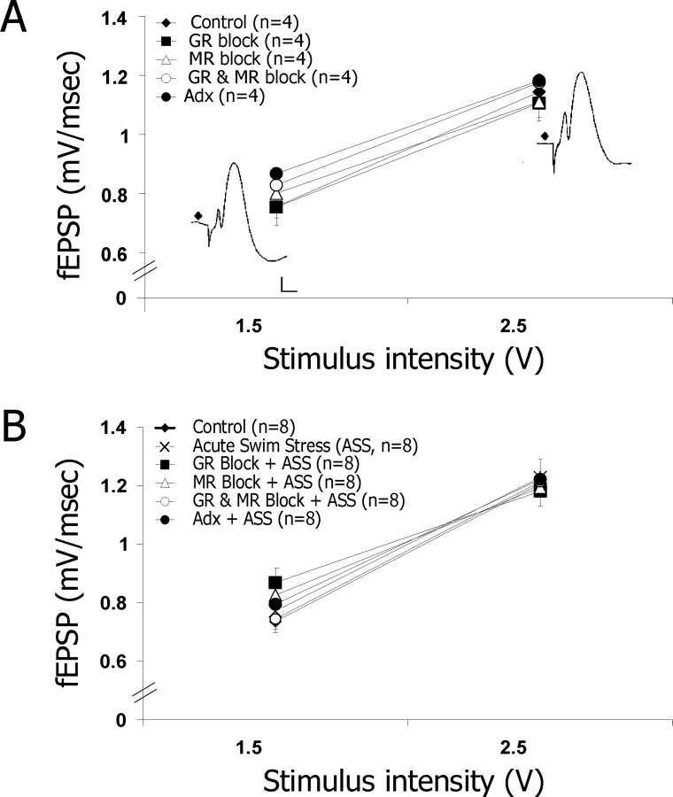
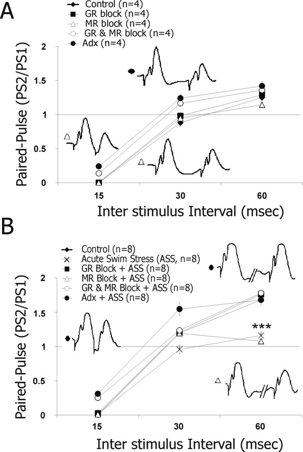
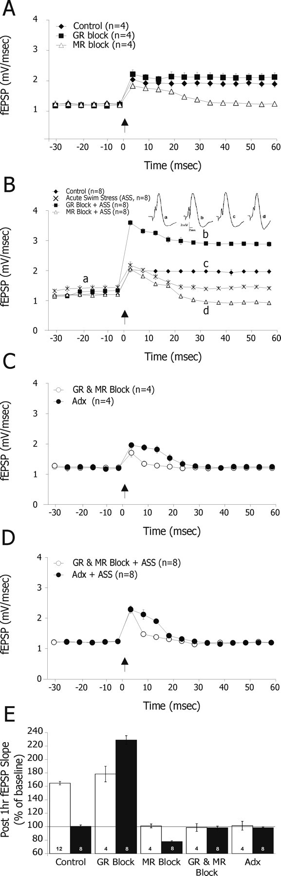
Elevated levels of corticosteroid hormones, presumably occupying both mineralocorticoid receptors (MRs) and glucocorticoid receptors (GRs), have been reported to impair synaptic plasticity in the hippocampus as well as the acquisition of hippocampus-dependent memories. In contrast, recent evidence suggests that activation of MRs enhance cognitive functions. To clarify the roles of different steroid receptors in hippocampal plasticity, young adult rats were injected with the GR antagonist RU38486 (mifepristone) or the MR antagonist Spironolactone before the exposure to an acute swim stress. Hippocampal responses to perforant path stimulation were then recorded in anesthetized rats. Stress combined with RU38486 produced a striking facilitation of LTP. Spironolactone enabled only short-term potentiation that reversed to long-term depression (LTD) in the stressed animals. Finally, the blockade of both MRs and GRs led to impairment of long-term potentiation. These findings indicate that MRs and GRs assume opposite roles in regulation of synaptic plasticity after acute exposure to stressors.


Figures

Figure 1.
Figure 1.
The dynamic range of reactivity to afferent stimulation is similar under control, stress, or drug conditions without (F(4,15) < 1) (A) and with (F(5,42) < 1) (B) stress. This indicates that there was no significant difference among the groups in the excitatory synaptic input to the hippocampus recorded in the DG.
Figure 2.
Figure 2.
DG responses to paired-pulse stimulation applied to the PP: under control conditions, MR or GR blockade did not affect paired-pulse responses (A). B, Strikingly, after stress, there was a decrease in paired-pulse facilitation at 60 ms ISI compared with controls. GR but not MR blockade restored paired-pulse facilitation in the stressed rats (***p < 0.0001). Adrenalectomy restored paired-pulse facilitation as well, presumably because of the lack of GR activation under this condition (traces of MR block and Adx groups are shown under control and stress conditions).
Figure 3.
Figure 3.
Effects of GR and MR antagonists on LTP. After one-half hour of stable baseline recording, an HFS was applied to the PP, and its consequence was recorded in the DG. A, Control rats expressed a sustained and typical, 63% increase in fEPSPs slope. Rats treated with GR antagonist (RU38486; 20 mg/kg) 75 min before HFS expressed a similar level of LTP (84.16 ± 7.64%; n = 4). In contrast, rats that were treated with the MR antagonist (Spironolactone; 20 mg/kg) showed no LTP but only a short-term potentiation that lasted for ∼30 min. B, Under stress, the difference between these two latter groups was accentuated; rats that were injected with the GR antagonist and subjected to behavioral stress (trace b) showed remarkable and long-lasting potentiation (228 ± 6.41%; n = 8) compared with their counterparts controls (trace c). Interestingly, the application of the MR antagonist before stress led to decreased post-HFS potentiation (trace d) to the extent that it reversed within 25 min to depotentiation/long-term depression (22.23 ± 3.42%; n = 8). C, D, Finally, rats that were either pretreated with the combination of GR and MR antagonists or adrenalectomized rats that showed no LTP. E, The fEPSP slope recorded 1 h post-HFS stimulation compared with baseline level was calculated in all groups. Both MR blockade and an exposure to stress blocked LTP. In contrast, GR blockade facilitated LTP, although mainly under stress.

References

    1. Alfarez DN, Wiegert O, Joels M, Krugers HJ. Corticosterone and stress reduce synaptic potentiation in mouse hippocampal slices with mild stimulation. Neuroscience. 2002;115:1119–1126. - PubMed
    1. Avital A, Richter-Levin G. Exposure to juvenile stress exacerbates the behavioural consequences of exposure to stress in the adult rat. Int J Neuropsychopharmacol. 2005;8:163–173. - PubMed
    1. Avital A, Goshen I, Kamsler A, Segal M, Iverfeldt K, Richter-Levin G, Yirmiya R. Impaired interleukin-1 signaling is associated with deficits in hippocampal memory processes and neural plasticity. Hippocampus. 2001;13:826–834. - PubMed
    1. Berger S, Wolfer DP, Selbach O, Alter H, Erdmann G, Reichardt HM, Chepkova AN H, Haas HL, Lipp HP, Schutz G. Loss of the limbic mineralocorticoid receptor impairs behavioral plasticity. Proc Natl Acad Sci USA. 2006;103:195–200. - PMC - PubMed
    1. Cahill L, McGaugh JL. Mechanisms of emotional arousal and lasting declarative memory. Trends Neurosci. 1998;21:294–299. - PubMed

Publication types

Substances