Clinical Trial
. 2006 Sep 6;26(36):9162-72.
doi: 10.1523/JNEUROSCI.2877-06.2006.

Differential encoding mechanisms for subsequent associative recognition and free recall

Affiliations
Clinical Trial

Differential encoding mechanisms for subsequent associative recognition and free recall

Bernhard P Staresina et al. J Neurosci. .

Erratum in

  • J Neurosci. 2006 Sep 20;26(38):9836

Abstract

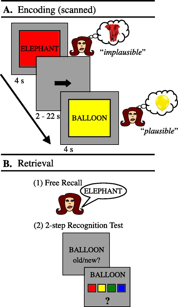
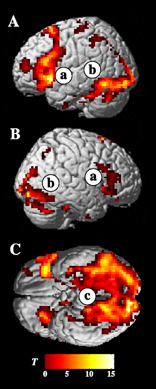
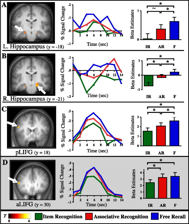
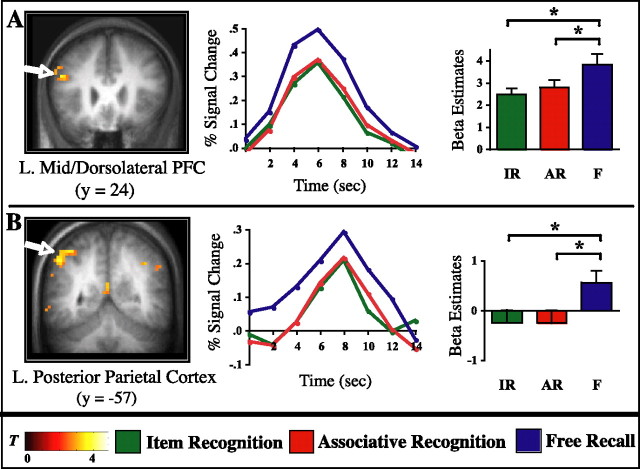
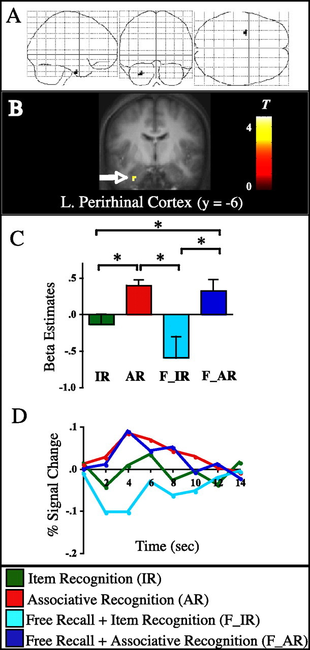
Recent neuroimaging studies have successfully identified encoding mechanisms that support different forms of subsequent episodic recognition memory. In our everyday lives, however, much of our episodic memory retrieval is accomplished by means of free recall, i.e., retrieval without an external recognition cue. In this study, we used functional magnetic resonance imaging to investigate the encoding mechanisms that support later free recall and their relationship to those that support different forms of later recognition memory. First, in agreement with previous work, we found that activation in the left inferior frontal gyrus and hippocampus correlated with later associative/relational recognition. In these regions, activation was further enhanced for items later freely recalled, pointing to shared underlying relational encoding mechanisms whose magnitude of activation differentiates later successful free recall from successful associative recognition. Critically, we also found evidence for free recall-specific encoding mechanisms that did not, in our paradigm, support later associative recognition compared with item recognition. These free recall-specific effects were observed in left mid/dorsolateral prefrontal (DLPFC) and bilateral posterior parietal cortices (PPC). We speculate that the higher-level working memory operations associated with DLPFC and attention to internal mnemonic representations perhaps mediated via PPC may serve to embed an item into a rich associative network during encoding that facilitates later access to the item. Finally, activation in the perirhinal cortex correlated with successful associative binding regardless of the form of later memory, i.e., recognition or free recall, providing novel evidence for the role of the perirhinal cortex in episodic intra-item encoding.


Figures

Figure 1.
Figure 1.
Experimental design. A, Encoding. Two example trials from the scanned encoding task. Subjects were instructed to vividly imagine the referent of the noun in the color presented and to decide whether this combination was plausible or not. B, Three-step subsequent memory test, consisting of a free recall phase (1), followed by assessment of both item and associative recognition (2).
Figure 2.
Figure 2.
Task effects. Statistical parametric maps (T-maps) of activation during encoding task performance (collapsed across all subsequent memory outcomes) compared with the sensorimotor baseline condition, rendered on a canonical brain template. A, Left lateral view. B, Right lateral view. C, Ventral view (cerebellum removed for illustration purposes). Robust activation was found in the prefrontal cortex (a), in ventrolateral temporal lobe regions (b), and in the medial temporal lobes (c).
Figure 3.
Figure 3.
Linearly increasing activation across subsequent memory conditions. Left column, Statistical parametric maps (T-maps) revealing regions that show linearly increasing encoding activation across subsequent IR, AR, and F, superimposed on the coronal slices of the mean anatomical image across subjects. A, Left hippocampus (L. Hippocampus). B, Right hippocampus (R. Hippocampus). C, Posterior left inferior frontal gyrus. Highlighted in gray (D) is the anterior left inferior frontal gyrus, in which the statistical pattern is distinguished from A–C because IR < AR = F. Middle column, Deconvolved BOLD time course data are shown for the peak voxel (A, −24, −15, −18; B, 21, −21, −21; C, −51, 18, 18; D, −51, 30, 9). Percentage signal change is graphed for each memory condition across 14 s (7 time points) after trial onset. Right column, Results from pairwise contrasts across memory conditions. Bar graphs reflect the mean of the β parameter estimates across subjects for each condition, averaged across all voxels in a given cluster. Error bars represent the SEM. * indicates statistically significant at p < 0.05; ∼ indicates trending toward significance at p = 0.085.
Figure 4.
Figure 4.
Free recall-specific effects. Encoding activation is significantly greater in these regions for subsequent F (blue) compared with both subsequent IR (green) and subsequent AR (red), without differing between IR and AR. Left column, Statistical parametric maps (T-maps) are superimposed on the coronal slices of the mean anatomical image across subjects. A, Left mid/dorsolateral prefrontal cortex. B, Left inferior parietal lobule/intraparietal sulcus. Middle column, Time course data are shown for the peak voxel (A, −45, 21, 21; B, −39, −54, 57). Percentage signal change is graphed for each memory condition across 14 s (7 time points) after trial onset. Right column, Results from pairwise contrasts across memory conditions. Bar graphs reflect the mean of the β parameter estimates across subjects for each condition, averaged across all voxels in a given cluster. Error bars represent the SEM. *p < 0.05. Note that bar graphs are mainly shown here for confirmatory and illustrative purposes, because the analysis was specifically designed to reveal regions that show this statistical pattern of activation. L., Left.
Figure 5.
Figure 5.
Successful word/color binding in left perirhinal cortex. A, SPM2 “glass brain” view of regions selectively involved in successful word/color binding regardless of form of memory, thresholded at five contiguous voxels exceeding an uncorrected p < 0.001. B, Statistical parametric map (T-map) superimposed on the coronal slice of the mean anatomical image across subjects. C, Results from pairwise contrasts across all memory conditions. Bar graphs show the mean of the β parameter estimates across subjects for each condition, averaged across all voxels from the perirhinal cortex cluster. Error bars represent the SEM. *p < 0.05. D, Time course data for the peak voxel (−30, −6, −36). Percentage signal change is graphed for each memory condition across 14 s (7 time points) after trial onset.

References

    1. Alkire MT, Haier RJ, Fallon JH, Cahill L. Hippocampal, but not amygdala, activity at encoding correlates with long-term, free recall of nonemotional information. Proc Natl Acad Sci USA. 1998;95:14506–14510. - PMC - PubMed
    1. Anderson JR, Bower GH. Recognition and retrieval processes in free recall. Psychol Rev. 1972;79:97–123.
    1. Awh E, Jonides J, Smith EE, Schumacher EH, Koeppe RA, Katz S. Dissociation of storage and rehearsal in verbal working memory: evidence from positron emission tomography. Psychol Sci. 1996;7:25–31.
    1. Badre D, Poldrack RA, Pare-Blagoev EJ, Insler RZ, Wagner AD. Dissociable controlled retrieval and generalized selection mechanisms in ventrolateral prefrontal cortex. Neuron. 2005;47:907–918. - PubMed
    1. Bahrick HP. Two-phase model for prompted recall. Psychol Rev. 1970;77:215–222.

Publication types

LinkOut - more resources