. 2006 Sep 6;26(36):9293-303.
doi: 10.1523/JNEUROSCI.1740-06.2006.

The B' protein phosphatase 2A regulatory subunit well-rounded regulates synaptic growth and cytoskeletal stability at the Drosophila neuromuscular junction

Affiliations

The B' protein phosphatase 2A regulatory subunit well-rounded regulates synaptic growth and cytoskeletal stability at the Drosophila neuromuscular junction

Natasha M Viquez et al. J Neurosci. .

Abstract

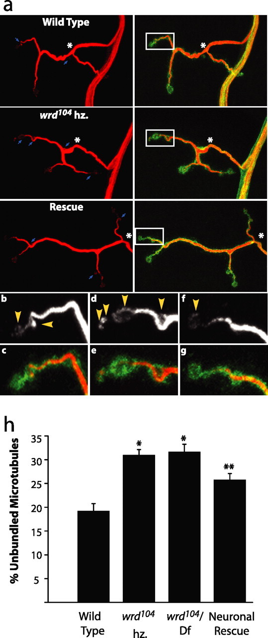
Synaptic growth is essential for the development and plasticity of neural circuits. To identify molecular mechanisms regulating synaptic growth, we performed a gain-of-function screen for synapse morphology mutants at the Drosophila neuromuscular junction (NMJ). We isolated a B' regulatory subunit of protein phosphatase 2A (PP2A) that we have named well-rounded (wrd). Neuronal overexpression of wrd leads to overgrowth of the synaptic terminal. Endogenous Wrd protein is present in the larval nervous system and muscle and is enriched at central and neuromuscular synapses. wrd is required for normal synaptic development; in its absence, there are fewer synaptic boutons and there is a decrease in synaptic strength. wrd functions presynaptically to promote normal synaptic growth and postsynaptically to maintain normal levels of evoked transmitter release. In the absence of wrd, the presynaptic cytoskeleton is abnormal, with an increased proportion of unbundled microtubules. Reducing PP2A enzymatic activity also leads to an increase in unbundled microtubules, an effect enhanced by reducing wrd levels. Hence, wrd promotes the function of PP2A and is required for normal cytoskeletal organization, synaptic growth, and synaptic function at the Drosophila NMJ.


Figures

Figure 1.
Figure 1.
well-rounded promotes synaptic growth. a, Gene structure of well-rounded (CG 7913). well-rounded has five splice forms. Exons are designated by boxes. Blue boxes indicate exons common to all five splice forms, pink boxes designate noncoding exons, green boxes designate a cluster of exons present in three of the wrd splice forms, and lavender boxes designate a second cluster of exons present in two of the wrd splice forms. Triangles indicate P-element insertions obtained in gain-of-function screen (GS). Imprecise excision of P[2535] was used to generate the wrd104 and Df(3R)189 mutants, whose deletions are illustrated by bars at the bottom. wrd104 is missing exons 4 and 5 as well as some exons common to all splice forms, whereas Df(3R)189 removes all of the wrd gene and five adjacent genes. Star marks the location of the antigen for the Well-rounded antibody. b, Representative confocal images of muscle 4 synapses from wild-type third-instar larvae (elav Gal4/+) and wrd overexpressing (O/E) third instars, which express additional wrd via the P[676] insert driven pan-neuronally with elav Gal4 (elav Gal4/P[676]). Synapses are costained with antibodies to DVGLUT (green), which highlights the cytoplasm of the synaptic terminal, and HRP (blue), which highlights neuronal membranes.
Figure 2.
Figure 2.
Well-rounded is present at synapses. a, Confocal images of larval brains stained with polyclonal antibody to Wrd. Left displays a wild-type brain, and right displays a brain from an animal expressing a wrd cDNA driven pan-neuronally under elav Gal4. Wrd labeling is prominent in brain lobes and the synapse-rich region of neuropil (arrow, right). Overexpression leads to increased staining of brain lobes and cell bodies in the cortex of neuropil (arrowhead, left). b, Confocal images of muscle 4 larval NMJs costained with antibody to Wrd (green) and the neuronal marker HRP (red). Top row displays an NMJ from a wild-type animal, and bottom row is from an animal with pan-neuronal expression of Wrd under elav Gal4. Overexpression (O/E) leads to increased Wrd staining at the NMJ. c, Costaining of Wrd at wild-type NMJ with presynaptic and postsynaptic markers. Image demonstrates a single confocal slice. Left, The NMJ is stained for Wrd alone (green). Middle, The same synapse is stained for Wrd and Dlg (red), a postsynaptic marker. Right, The synapse costained with Wrd and HRP (blue), a presynaptic marker. Synaptic Wrd staining colocalizes nearly completely with HRP, as demonstrated by the increased intensity throughout the NMJ in the overlay of Wrd and HRP. Overlay of Wrd and Dlg demonstrates partial localization, because a ring of Dlg staining falls outside the Wrd staining.
Figure 3.
Figure 3.
Generation of wrd loss-of-function mutants. a, Western blot confirming absence of Wrd protein in excision mutants. Genotypes are as follows: wild type, elav Gal4/+; wrd189/Df(3R)DG4, Df(3R)189/Df(3R)DG4; and wrd104 hz, wrd104/wrd104. α-Wrd reveals three bands (arrows) in wild-type animals, consistent with the presence of multiple wrd splice forms. These bands are absent in the lane for wrd189/Df(3R)DG4, a definitive null lacking the wrd gene, showing that the antibody specifically recognizes Wrd protein. The Wrd bands are also absent in the wrd104 homozygote, which is therefore a protein null. Bottom half of blot shows a nonspecific background band, here shown as a loading control. b, Representative neuropil staining for Wrd in wild-type animals, wrd104 mutants, and wrd189 hz [Df(3R)189/Df(3R)189]. Animals were costained for Wrd and Bruchpilot, an active zone marker with strong neuropil staining. Bruchpilot staining is shown in insets. Absence of Wrd staining in wrd104 mutants and wrd189 hz demonstrates specificity of staining at synapses in neuropil. Remaining staining outside of neuropil represents background, because it persists in wrd189 hz, a definitive null. c, Representative synaptic staining for Wrd at the NMJ on muscle 4 of wild-type animals, wrd104 mutants, and wrd189 hz. The NMJ is stained for Wrd as well as HRP to delineate the synaptic terminal. Whereas Wrd staining is present at the NMJ in wild-type animals, it is essentially absent in the wrd104 mutant and the wrd189 hz, demonstrating specificity of Wrd staining at NMJ.
Figure 4.
Figure 4.
Wrd is required for normal synaptic growth and morphology. a–c, Representative NMJs from muscle 4 in wild-type animals (a), wrd104/Df(3R)DG4 mutants (b), and wrd104 homozygotes rescued with neuronal expression of UAS wrd under elav Gal4 (c). Synapses are costained with antibodies to DVGLUT (green) and HRP (red). d, e, Enlarged images of boutons in wild-type animals (d) and wrd104 homozygotes (e) highlighting increased size and rounded contours of boutons in wrd mutant. f, g, Quantification of bouton number and bouton area in segments A2 and A3 of muscle 4 1b innervation of third-instar larvae. Genotypes include wild type (elav Gal4/+), wrd104/Df(3R)DG4 [elav Gal4/+; wrd104/Df(3R)DG4), wrd104 hz (elav Gal4/+; wrd104/wrd104)], and neuronally rescued wrd104 hz (elav Gal4/UAS wrd; wrd104/wrd104). Bouton number and bouton area in wrd mutants are significantly different from wild-type animals; *p < 0.01 for bouton number in 4b; *p < 0.001 for bouton area in 4c. wrd104/Df(3R)DG4 and wrd104 hz are not significantly different for either bouton number or bouton area (p > 0.5). Neuronal expression of wrd cDNA rescues both bouton number and bouton area defects in wrd mutants. **p < 0.01 for bouton number in 4b; **p < 0.001 for bouton area in 4c. For both quantifications, wild type, n = 36; wrd104/Df(3R)DG4, n = 32; wrd104 hz, n = 36; and rescue, n = 16. Error bars represent SEM.
Figure 5.
Figure 5.
wrd is required postsynaptically for synaptic function. a, Representative traces from wild type (elav Gal4/+), wrd104 hz (elav Gal4/+; wrd104/wrd104), and muscle rescue (G7 Gal4/UAS wrd; wrd104/wrd104). Calibration: 10 ms, 10 mV for evoked release; 250 ms, 2 mV for spontaneous release. b–d, The mean mEJP amplitude, EJP amplitude, and quantal content are plotted for wild type (elav Gal4/+), wrd104 hz. (elav Gal4/+; wrd104/wrd104), wrd104/Df(3R)DG4 (elav Gal4/+; wrd104/Df(3R)DG4), neuronal Wrd (elav Gal4/UAS wrd), Wrd neuronal rescue (elav Gal4/UAS wrd; wrd104/wrd104), muscle Wrd (G7 Gal4/UAS wrd), and Wrd muscle rescue (G7 Gal4/UAS wrd; wrd104/wrd104) (n = 8 for wild type and wrd104/Df(3R)DG4; n = 9 for wrd104 hz and neuronal Wrd; n = 6 for Wrd neuronal rescue; n = 12 for muscle Wrd; and n = 10 for Wrd muscle rescue). mEJPs are not significantly different for any of the genotypes compared with wild type (wrd104/Df(3R)DG4, p > 0.3; all others p > 0.9). Mean EJP value and quantal content are decreased by 56 and 51%, respectively, in wrd104 hz mutants, with similar decreases observed in wrd104/Df(3R)DG4 [wrd104 hz and wrd104/Df(3R)DG4 are not significantly different; p > 0.2 (EJP) and p > 0.3 (quantal content)]. Muscle expression of Wrd in the wrd104 hz mutants rescues both mean EJP and quantal content to wild-type levels [wrd104 hz vs muscle rescue, p < 0.05 (EJP) and p < 0.001 (quantal content); muscle rescue vs WT, p > 0.8 (EJP) and p > 0.3 (quantal content)]. *p < 0.01 and **p < 0.001 for mean EJP relative to wild type in c. In d, *p < 0.05, **p < 0.01, and ***p < 0.001 for mean quantal content relative to wild type. Error bars represent SEM. e, Wrd staining of NMJ in Wrd muscle rescue (G7 Gal4/UAS wrd; wrd104/wrd104) demonstrates localization of Wrd at the NMJ when expressed in muscle in the absence of endogenous Wrd. Inset shows similar staining pattern with Dlg, a primarily postsynaptic marker.
Figure 6.
Figure 6.
Loss of wrd disrupts cytoskeletal stability. a, Representative confocal images of muscle 4 synapses from wild type (elav Gal4/+), wrd104 hz (elav Gal4/+; wrd104/wrd104), and rescue (elav Gal4/UAS wrd; wrd104/wrd104). Left column displays synapses stained with antibody to Futsch, a reporter for microtubules, and right column displays corresponding synapses costained with α-Futsch and α-HRP. Futsch staining demonstrates bundled microtubules within the core of the synaptic terminal. Unbundled microtubules are visible at terminal boutons and, in some instances, along the shaft of the terminal, as marked by arrows. The wrd mutant shows an increased occurrence of unbundled microtubules along the shaft of the terminal. Asterisks mark junction between the synaptic terminal and preterminal axon. Insets mark a representative sample of unbundled microtubules at terminal boutons and are enlarged in b–g. b–g, Enlarged insets displaying representative terminal boutons from wild type (b, c), wrd104 hz (d, e), and rescue (f, g). Top row shows staining for Futsch alone, and bottom row shows costaining of Futsch and HRP. Arrowheads in top row indicate instances of unbundled microtubules. Although insets display a similar number of boutons for each genotype, instances of unbundled microtubules are greatest in the wrd104 mutant and take up a greater proportion of the total microtubule area than in the other genotypes. c, Quantification of unbundled microtubules as a percentage of total area of synaptic microtubules. For wild type (elav Gal4/+), wrd104 hz (elav Gal4/+; wrd104/wrd104), wrd104/Df(3R)DG4 [elav Gal4/+; wrd104/Df(3R)DG4], and neuronal rescue (elav Gal4/UAS wrd; wrd104/wrd104), n = 27, 27, 23, and 28, respectively. Both wrd mutants have increased unbundled microtubules relative to WT; *p < 0.01. Neuronal rescue significantly decreases unbundled microtubules relative to both wrd mutants; **p < 0.05. Error bars indicate SEM.
Figure 7.
Figure 7.
PP2A activity is required for cytoskeletal stability of synapse. a, Representative confocal images of muscle 4 synapses from wild type (elav Gal4/+), wrd104 het (elav Gal4/+; wrd104/+), dnMts (elav Gal4/UAS dnMts), and dnMts; wrd104 het (elav Gal4/+; wrd104/UAS dnMts). Left column shows synapses labeled with Futsch, and right column shows corresponding synapses labeled with α-DVGLUT and α-HRP. In the elav dnMts case, arrows mark boutons with dramatic unbundling of microtubules. b, Quantification of percentage unbundled microtubules at NMJ. Unbundled microtubules are measured as a proportion of total area of microtubules at synaptic terminal. n = 19, 14, 27, 16 for WT, wrd104 het, dnMts, and wrd104 het; dnMts, respectively. Reduced PP2A activity leads to significantly increased microtubule unbundling relative to wild type; *p < 0.05. The combined wrd104 het; dnMts mutant shows a significant increase in unbundled microtubules relative to dnMts alone; **p < 0.01. The heterozygous wrd mutant thus enhances the effect of reduced PP2A activity, suggesting that it can positively regulate the activity of PP2A toward the synaptic cytoskeleton. Error bars indicate SEM.

Similar articles

Cited by

References

    1. Abdelilah-Seyfried S, Chan Y-M, Zeng C, Justice NJ, Younger-Shepherd S, Sharp LE, Barbel S, Meadows SA, Jan LY, Jan YN. A gain-of-function screen for genes that affect the development of the Drosophila adult external sensory organ. Genetics. 2000;155:733–752. - PMC - PubMed
    1. Barnes GN, Slevin JT, Vanaman TC. Rat brain protein phosphatase 2A: an enzyme that may regulate autophosphorylated protein kinases. J Neurochem. 1995;64:340–353. - PubMed
    1. Belmeguenai A, Hansel C. A role for protein phosphatases 1, 2A, and 2B in cerebellar long-term potentiation. J Neurosci. 2005;25:10768–10772. - PMC - PubMed
    1. Betz WJ, Henkel AW. Okadaic acid disrupts clusters of synaptic vesicles in frog motor nerve terminals. J Cell Biol. 1994;124:843–854. - PMC - PubMed
    1. Brand AH, Perrimon N. Targeted gene expression as a means of altering cell fates and generating dominant phenotypes. Development. 1993;118:401–415. - PubMed

Publication types

LinkOut - more resources