Comparative Study
. 2006 Dec;54(12):1413-23.
doi: 10.1369/jhc.6A7001.2006. Epub 2006 Sep 6.

Analysis of protein expression in cell microarrays: a tool for antibody-based proteomics

Affiliations
Comparative Study

Analysis of protein expression in cell microarrays: a tool for antibody-based proteomics

Ann-Catrin Andersson et al. J Histochem Cytochem. 2006 Dec.

Abstract

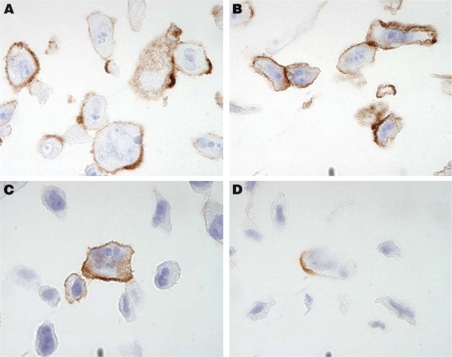
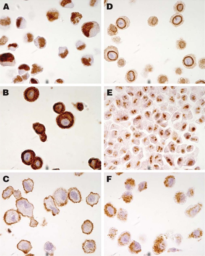
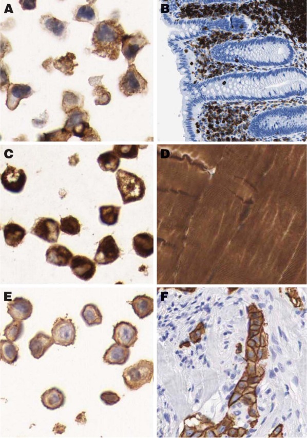
Tissue microarray (TMA) technology provides a possibility to explore protein expression patterns in a multitude of normal and disease tissues in a high-throughput setting. Although TMAs have been used for analysis of tissue samples, robust methods for studying in vitro cultured cell lines and cell aspirates in a TMA format have been lacking. We have adopted a technique to homogeneously distribute cells in an agarose gel matrix, creating an artificial tissue. This enables simultaneous profiling of protein expression in suspension- and adherent-grown cell samples assembled in a microarray. In addition, the present study provides an optimized strategy for the basic laboratory steps to efficiently produce TMAs. Presented modifications resulted in an improved quality of specimens and a higher section yield compared with standard TMA production protocols. Sections from the generated cell TMAs were tested for immunohistochemical staining properties using 20 well-characterized antibodies. Comparison of immunoreactivity in cultured dispersed cells and corresponding cells in tissue samples showed congruent results for all tested antibodies. We conclude that a modified TMA technique, including cell samples, provides a valuable tool for high-throughput analysis of protein expression, and that this technique can be used for global approaches to explore the human proteome.


Figures

Figure 1
Figure 1
Immunohistochemical staining using antibodies directed toward the transmembrane glycoproteins CD44 (A,B) and N-cadherin (C,D) shows a distinct membranous pattern of immunoreactivity suggesting preserved antigenicity in PC-3 cells. No difference was detected between cells detached by trypsinization (A,C) and cells mechanically detached using a rubber policeman (B,D).
Figure 2
Figure 2
Examples of staining patterns using six antibodies toward antigens in specific subcellular compartments. The antibody toward the nuclear phosphoprotein p53 shows distinct positive nuclei in the epidermoid carcinoma cell line A431 (A). A cytoplasmic staining pattern was observed in the breast adenocarcinoma cell line SK-BR-3 using an antibody toward cytokeratin 7, which is an intermediate filament distributed to glandular and transitional epithelia (B). A membranous staining pattern was revealed in the glioblastoma cell line U-138-MG with an antibody recognizing the transmembrane protein N-cadherin (C). In A-549, a lung carcinoma cell line, a clear positive staining pattern around the nuclear membrane could be observed with the antibody against emerin, a protein associated with the nuclear envelope (D). The fibrillarin antibody recognizing a component of the nucleolar small ribonucleoprotein particle shows a nucleolar staining pattern in the hepatocellular carcinoma cell line Hep-G2 (E). The antibody recognizing a protein component of mitochondria showed a granular cytoplasmic staining pattern in cells of the A-549 cell line (F).
Figure 3
Figure 3
Examples of congruent staining patterns in tissue and corresponding cell line. Immunostaining with the anti-LCA (CD45) antibody recognizing the CD45 glycoprotein shows strong positivity in the THP-1 cell line (A), representing an acute monocytic leukemia as well as in lymphoid cells in appendix (B). The antibody recognizing desmin, which is the characteristic intermediate filament of all three types of muscle cells, shows cytoplasmic positivity in the rhabdomyosarcoma cell line RH-30 (C) as well as in striated muscular tissue (D). HER-2 antibody recognizing the c-erbB-2 oncoprotein, which is frequently overexpressed in human carcinomas, shows strong membranous reaction in the breast carcinoma cell line SK-BR-3 (E) and corresponding breast carcinoma (F).

Similar articles

Cited by

References

    1. Agaton C, Galli J, Hoiden Guthenberg I, Janzon L, Hansson M, Asplund A, Brundell E, et al. (2003) Affinity proteomics for systematic protein profiling of chromosome 21 gene products in human tissues. Mol Cell Proteomics 2:405–414 - PubMed
    1. Battifora H. (1986) The multitumor (sausage) tissue block: novel method for immunohistochemical antibody testing. Lab Invest 55:244–248 - PubMed
    1. Battifora H, Mehta P. (1990) The checkerboard tissue block. An improved multitissue control block. Lab Invest 63:722–724 - PubMed
    1. Boukamp P, Petrussevska RT, Breitkreutz D, Hornung J, Markham A, Fusenig NE. (1988) Normal keratinization in a spontaneously immortalized aneuploid human keratinocyte cell line. J Cell Biol 106:761–771 - PMC - PubMed
    1. Butterfield JH, Weiler D, Dewald G, Gleich GJ. (1988) Establishment of an immature mast cell line from a patient with mast cell leukemia. Leuk Res 12:345–355 - PubMed

Publication types