. 2006 Oct 4;25(19):4448-57.
doi: 10.1038/sj.emboj.7601335. Epub 2006 Sep 7.

Distinct roles of doublecortin modulating the microtubule cytoskeleton

Affiliations

Distinct roles of doublecortin modulating the microtubule cytoskeleton

Carolyn A Moores et al. EMBO J. .

Abstract

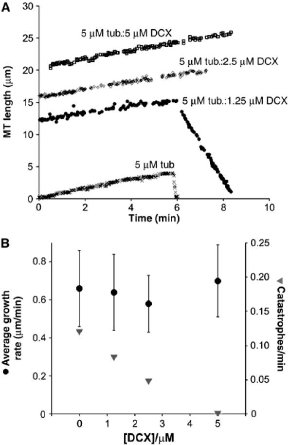
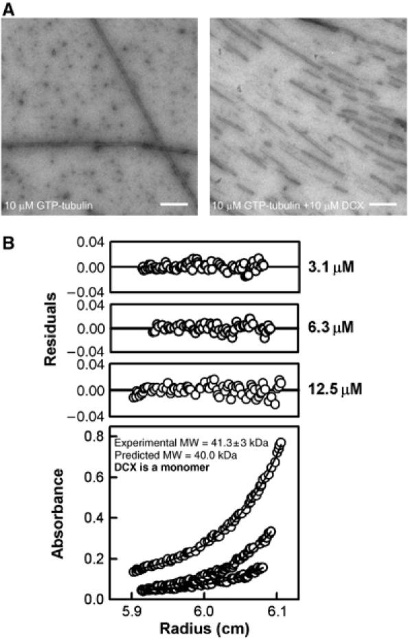
Doublecortin is a neuronal microtubule-stabilising protein, mutations of which cause mental retardation and epilepsy in humans. How doublecortin influences microtubule dynamics, and thereby brain development, is unclear. We show here by video microscopy that purified doublecortin has no effect on the growth rate of microtubules. However, it is a potent anti-catastrophe factor that stabilises microtubules by linking adjacent protofilaments and counteracting their outward bending in depolymerising microtubules. We show that doublecortin-stabilised microtubules are substrates for kinesin translocase motors and for depolymerase kinesins. In addition, doublecortin does not itself oligomerise and does not bind to tubulin heterodimers but does nucleate microtubules. In cells, doublecortin is enriched at the distal ends of neuronal processes and our data raise the possibility that the function of doublecortin in neurons is to drive assembly and stabilisation of non-centrosomal microtubules in these doublecortin-enriched distal zones. These distinct properties combine to give doublecortin a unique function in microtubule regulation, a role that cannot be compensated for by other microtubule-stabilising proteins and nucleating factors.


Figures

Figure 1
Figure 1
DCX is an anti-catastrophe factor but has no effect on MT growth rate. (A) Representative life histories of individual MT plus-ends grown from axonemes and 5 μM tubulin in the presence of 0, 1.25, 2.5 and 5 μM DCX. The parallel positive gradient of each MT trace illustrates that DCX has no effect on MT growth rate, whereas the difference in negative gradient between two of the traces illustrates that when catastrophes do occur in the presence of DCX, they are slower. The individual growth and shrinkage rates are as follows: 5 μM tubulin (x) +0.68 μm/min, −32.77 μm/min, respectively; 5 μM tubulin:1.25 μM DCX (•) +0.53 μm/min, −5.80 μm/min, respectively; 5 μM tubulin:2.5 μM DCX (◊) +0.58 μm/min; 5 μM tubulin:5 μM DCX (□) +0.64 μm/min. (B) DCX has no effect on the average MT plus-end growth rate (•) but reduces the frequency of catastrophes (▾).
Figure 2
Figure 2
DCX MT lattice binding. (A) Cosedimentation assay of DCX with 13pf-enriched MTs. A constant concentration of MTs (2.9 μM polymerised tubulin) was titrated with a range of [DCX], the mixture was centrifuged and the supernatant (s) and pellet (p) fractions were visualised by SDS–PAGE. (B) Binding of DCX to 13pf-enriched MTs is bimodal, one mode of which corresponds to simple decoration with a 1:1 stoichiometry of DCX:tubulin dimer and an apparent affinity of 2 μM (left side of graph), and another in the presence of excess DCX where MT bundles are formed (right side). The curve was fitted to the equation [DCX]bound/[polymerised tubulin]=(stoichiometrymax*[DCX]total)/(Kd+[DCX]total)+[DCX]total/p as described previously (Ackmann et al, 2000). (C) MTs polymerised in the presence of substoichiometric DCX (0.8:1 DCX:tubulin dimer) form individual filaments (i), whereas in the presence of excess DCX (3:2 DCX:tubulin dimer), MT bundles form (ii) and exhibit aggregates on their surface (arrows in iii and iv). Bar=500 Å. (D) GDP-tubulin copolymerised with DCX for 30 min at 37°C forms MTs. Bar=2500 Å. (E) DCX favours polymerisation of 13pf MTs independent of the guanine nucleotide bound to β-tubulin.
Figure 3
Figure 3
DCX-MTs are substrates for transport and depolymerisation by kinesin motors. (A) Series of frames, separated by 2 s, from movies of in vitro ensemble gliding assays showing mammalian neuronal kinesin-1 moving paclitaxel-stabilised (left frames) or DCX-stabilised (right frames) MTs (see also Supplementary Movies). The trailing end of one MT is marked by an arrow at 0 s and tracked by a dot as it moves between consecutive frames. Bar=5 μm. (B) (i) Graph showing the increase of tubulin released from stabilised MTs as a result of increasing concentrations of the ATP-dependent depolymerising motor pkin13; n=4. (ii) Characteristic pkin13-induced tubulin rings (arrows) were observed by negative stain EM at the ends of DCX-MTs in the presence of the non-hydrolysable analogue AMPPNP. This supports the idea that individual DCX-MTs are substrates for depolymerising kinesins. Bar=500 Å.
Figure 4
Figure 4
MT nucleation by monomeric DCX. (A) Electron micrographs of 10 μM tubulin polymerised in the absence (left) and presence (right) of DCX, imaged 5 min after polymerisation was initiated. Bar=5000 Å. (B) Absorbance data, curve fits (bottom panel) and residual of the fits (top panels) from the analytical ultracentrifugation experiment with 3.1, 6.3 and 12.5 μM DCX centrifuged at 20 000 r.p.m. The curve fits show that DCX is predominantly monomeric.
Figure 5
Figure 5
DCX does not bind dimeric tubulin but nucleates MTs. (A) Size-exclusion chromatography trace showing the migration of dimeric tubulin, DCX and dimeric kinesin-1, which were run separately down a Superose-12 column. SDS–PAGE of the relevant fractions is shown above the trace. (B) Size-exclusion chromatography trace and SDS–PAGE analysis of co-complexes of DCX-tubulin, tubulin+kinesin and tubulin+kinesin+DCX, demonstrating that whereas kinesin binds dimeric tubulin and shifts its migration profile, DCX and tubulin do not interact, either as a bi-complex or as a tri-complex in the presence of kinesin. (C) Electron micrograph of 5 μM tubulin polymerised in the presence of 5 μM DCX imaged 5 min after polymerisation was initiated. The asterisk and arrows indicate a fully polymerised MT and small DCX-induced tubulin sheets, respectively. Bar=1000 Å.
Figure 6
Figure 6
A model for the effects of DCX on MT function. DCX does not bind individual tubulin dimers but stabilises the tubulin–tubulin lateral contacts in tubulin oligomers, thereby mediating nucleation in the early stages of MT polymerisation (left). DCX stabilises the fully polymerised MT lattice (viewed from the end) against spontaneous catastrophe by counteracting the outward-directed forces of MT depolymerisation (right) that disrupt inter-pf lateral contacts. It does not, however, greatly inhibit kinesin motors involved in either cargo movement or MT depolymerisation.

References

    1. Ackmann M, Wiech H, Mandelkow E (2000) Nonsaturable binding indicates clustering of tau on the microtubule surface in a paired helical filament-like conformation. J Biol Chem 275: 30335–30343 - PubMed
    1. Al-Bassam J, Ozer RS, Safer D, Halpain S, Milligan RA (2002) MAP2 and tau bind longitudinally along the outer ridges of microtubule protofilaments. J Cell Biol 157: 1187–1196 - PMC - PubMed
    1. Aldaz H, Rice LM, Stearns T, Agard DA (2005) Insights into microtubule nucleation from the crystal structure of human γ-tubulin. Nature 435: 523–527 - PubMed
    1. Andersen SSL (2005) The search and prime hypothesis for growth cone turning. BioEssays 27: 86–90 - PubMed
    1. Arnal I, Heichette C, Diamantopoulos GS, Chretien D (2004) CLIP-170/tubulin-curved oligomers coassemble at microtubule ends and promote rescues. Curr Biol 14: 2086–2095 - PubMed

Publication types

MeSH terms

LinkOut - more resources