. 2006 Sep 20;25(18):4175-86.
doi: 10.1038/sj.emboj.7601317. Epub 2006 Sep 7.

Secretion of L-glutamate from osteoclasts through transcytosis

Affiliations

Secretion of L-glutamate from osteoclasts through transcytosis

Riyo Morimoto et al. EMBO J. .

Abstract

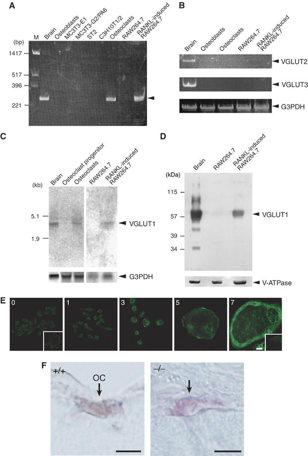
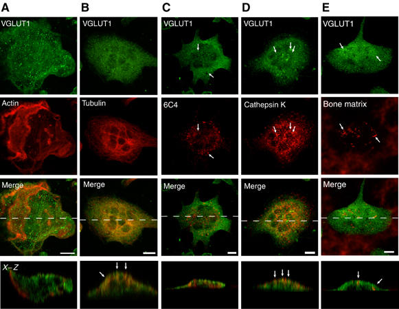
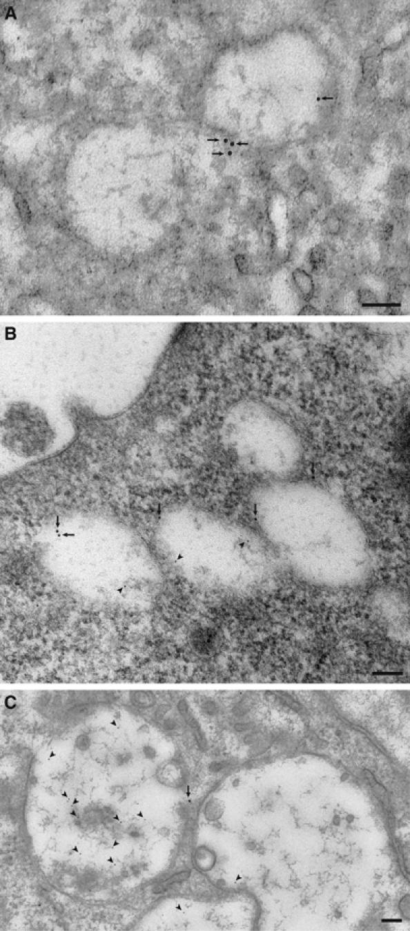
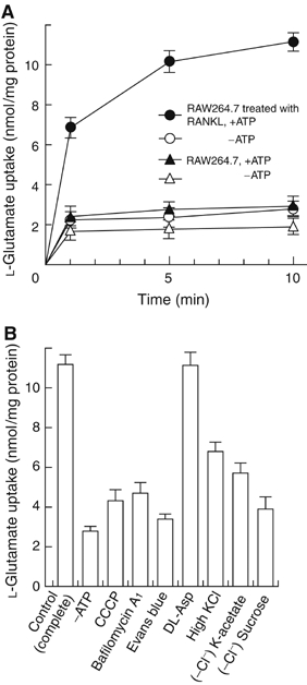
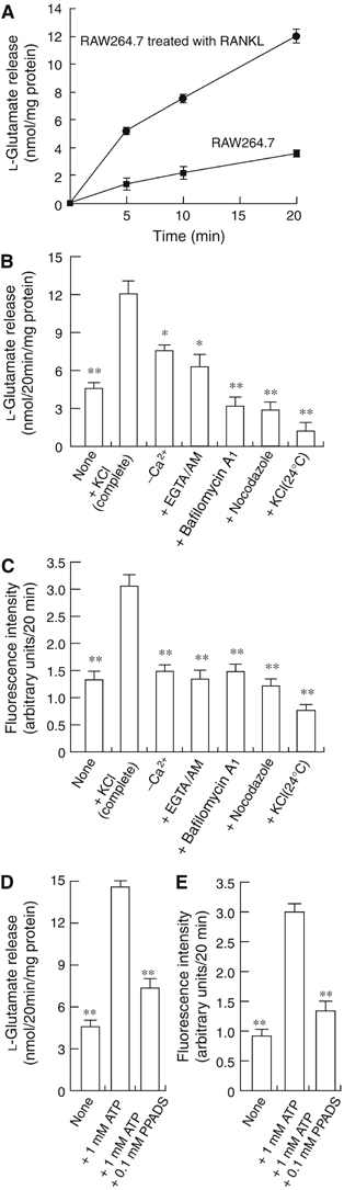
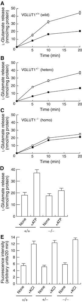
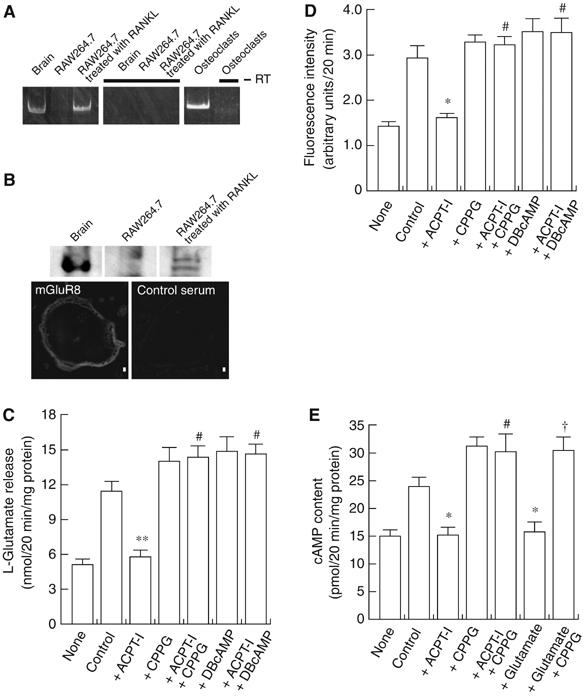
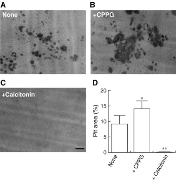
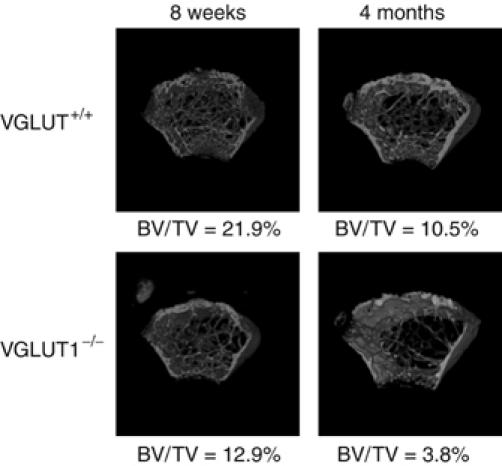
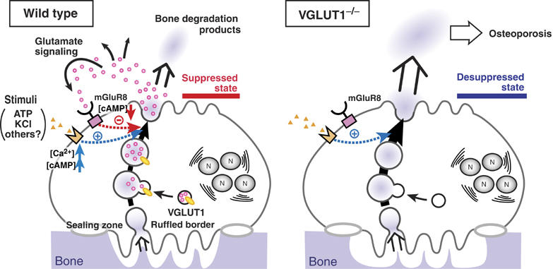
Osteoclasts are involved in the catabolism of the bone matrix and eliminate the resulting degradation products through transcytosis, but the molecular mechanism and regulation of transcytosis remain poorly understood. Upon differentiation, osteoclasts express vesicular glutamate transporter 1 (VGLUT1), which is essential for vesicular storage and subsequent exocytosis of glutamate in neurons. VGLUT1 is localized in transcytotic vesicles and accumulates L-glutamate. Osteoclasts secrete L-glutamate and the bone degradation products upon stimulation with KCl or ATP in a Ca2+-dependent manner. KCl- and ATP-dependent secretion of L-glutamate was absent in osteoclasts prepared from VGLUT1-/- knockout mice. Osteoclasts express mGluR8, a class III metabotropic glutamate receptor. Its stimulation by a specific agonist inhibits secretion of L-glutamate and bone degradation products, whereas its suppression by a specific antagonist stimulates bone resorption. Finally, it was found that VGLUT1-/- mice develop osteoporosis. Thus, in bone-resorbing osteoclasts, L-glutamate and bone degradation products are secreted through transcytosis and the released L-glutamate is involved in autoregulation of transcytosis. Glutamate signaling may play an important role in the bone homeostasis.


Figures

Figure 1
Figure 1
Inducible expression of VGLUT1 in osteoclasts. (A) RT–PCR analysis of brain, osteoblasts, MC3T3-E1 (clonal osteoblasts), MC3T3-G2/PA6 (clonal stroma cell line from calvariae), ST2 (clonal stroma cell line from bone marrow), C3H10T1/2 (clonal fibroblast cell line), osteoclasts, RAW264.7 macrophages and RAW264.7 cells treated with RANKL. The arrowhead indicates the VGLUT1 transcript. (B) VGLUT2 and VGLUT3 genes were not detectable in bone cells. Results of RT–PCR analysis of total cellular RNA are shown. Expression of G3PDH gene is also shown as a control. (C) Northern blotting revealing expression of the VGLUT1 gene in mature osteoclasts and RAW264.7 cells treated with RANKL. The G3PDH transcript, as a loading control, is also shown (lower panel). (D) Western blotting reveals the presence of VGLUT1 in RAW264.7 cells treated with RANKL. The presence of V-ATPase subunit A on the same blot is also shown. (E) RAW264.7 cells were cultured in the presence of RANKL for the indicated incubation periods (days) and the expression of VGLUT1 during osteoclastogenesis was observed by immunohistochemistry. Negative control with control IgG is also shown in insets. Bar=10 μm. (F) Osteoclasts (OC) in the femora of VGLUT1+/+ (wild type) mice visualized by TRAP staining (red) contain VGLUT1, which was visualized by the horseradish peroxidase-diaminobenzidine (HRP-DAB) method (charcoal). No VGLUT1 immunoreactivity was seen in osteoclasts from VGLUT1−/− mice. Bar=10 μm.
Figure 2
Figure 2
Immunohistochemical localization of VGLUT1 in bone-resorbing osteoclasts. Osteoclasts were grown on pieces of bone. Localization of VGLUT1 was examined by double-labeling immunofluorescence microscopy. The staining pairs were as follows: VGLUT1 and actin (A), VGLUT1 and tubulin (B), VGLUT1 and lysobisphosphatidic acid (6C4) (C), VGLUT1 and cathepsin K (D) and VGLUT1 and fluorescent bone degradation products (E). The immunological localization was observed under a confocal microscope. A horizontal view of a merged picture is also shown. Bar=10 μm.
Figure 3
Figure 3
Double gold labeling immunoelectron microscopy of bone-resorbing osteoclasts. Arrows and arrowheads indicate VGLUT1 (10 nm in diameter) and bone degradation product (5 nm in diameter), respectively. (A) VGLUT1-containing small vesicles; (B) vesicles containing both VGLUT1 and bone degradation products localized near the basolateral region; (C) vesicles containing bone degradation products but little VGLUT1 localized near the ruffled border membrane. Bar=100 nm.
Figure 4
Figure 4
VGLUT1-mediated vesicular storage of L-glutamate. (A) Time course of L-glutamate uptake by membrane vesicles isolated from RAW264.7 cells treated with or without RANKL determined in the presence or absence of ATP; n=3. (B) Effects of various compounds on the ATP-dependent L-glutamate uptake by membrane vesicles of RAW264.7 cells treated with RANKL for 1 week. Additions: bafilomycin A1, 1 μM; CCCP, 1 μM; Evans blue, 1 μM; D,L-aspartate, 10 mM; KCl, 100 mM. In some experiments, KCl was substituted with K-acetate or omitted from the assay medium. The results are shown as means±s.e.m., n=4.
Figure 5
Figure 5
Regulated secretion of L-glutamate and fluorescent bone degradation products from RAW264.7 cells treated with RANKL. (A) RAW264.7 cells treated with RANKL or RAW264.7 cells (2.0 × 10⁵ cells/dish) were stimulated with 50 mM KCl. The L-glutamate released was measured. (B) L-Glutamate secretion after 20 min is shown. In some experiments, cells were treated for 2 h with 1 μM bafilomycin A1, 50 μM EGTA-AM or 10 μM nocodazole, and then stimulated with KCl. (C) Fluorescent bone degradation products in the medium under the conditions in panel B were assessed fluorometrically. The results are means±s.e.m., n=4. Asterisks indicate statistically significant numbers (*P<0.01, **P<0.001). ATP stimulates the secretion of L-glutamate (D) and fluorescent bone degradation products (E) from RAW264.7 cells treated with RANKL. The assay was started by the addition of 1 mM ATP. In some experiments, 0.1 mM PPADS was also included. The results are means±s.e.m., n=4. Asterisks indicate statistically significant numbers (*P<0.01, **P<0.001).
Figure 6
Figure 6
Both KCl- and ATP-dependent L-glutamate secretion were impaired in osteoclasts from VGLUT1−/− mice. The time course of KCl-dependent L-glutamate secretion from osteoclasts (2.0 × 10⁵ cells/dish) prepared from bone marrow of wild-type VGLUT1+/+ (A), VGLUT1+/− (B) or VGLUT1−/− (C) was measured as in Figure 5 in the presence (open squares) or absence (closed squares) of 50 mM KCl. (D) In osteoclasts (2.0 × 10⁵ cells/dish) prepared from wild-type and VGLUT1−/− mice, L-glutamate secretion were measured 20 min after stimulation with 1 mM ATP. (E) Secretion of bone degradation products from osteoclasts under the conditions in panels A–C was assessed fluorometrically after 20 min. The results are means±s.e.m., n=3.
Figure 7
Figure 7
mGluR-mediated regulation of the secretion of L-glutamate and fluorescent bone degradation products from RAW264.7 cells treated with RANKL. (A) Expression of mGluR8 gene as revealed by RT–PCR. (B) Expression of mGluR8 as revealed by immunoblotting (upper panel) and immunohistochemistry (lower panel). Bar=10 μm. (C) RAW264.7 cells treated with RANKL (2.0 × 10⁵ cells/dish) were incubated in the presence or absence of agonists or antagonists of glutamate receptors or DBcAMP as indicated and then stimulated with 50 mM KCl. The L-glutamate released after 20 min is shown. The results are means±s.e.m., n=3. Asterisks and marks indicate statistically significant numbers (**P<0.001 compared with control, #P<0.001 compared with +ACPT-I). (D) Fluorescent bone degradation products in the medium under the conditions in panel A were assessed fluorometrically. The results are means±s.e.m., n=3. Asterisks and marks indicate statistically significant numbers (*P<0.01 compared with control, #P<0.001 compared with +ACPT-I). (E) cAMP content in the osteoclast-like cells (2.0 × 10⁵ cells/dish) under the conditions in panel A was measured. The results are means±s.e.m., n=3. Asterisks and marks indicate statistically significant numbers (*P<0.01 compared with control, #P<0.001 compared with +ACPT-I, P<0.001, compared with +L-glutamate). Additions: L-glutamate, 0.5 mM; ACPT-I, 50 μM; CPPG, 100 μM; DBcAMP, 1 mM.
Figure 8
Figure 8
Inhibition of L-glutamate signal stimulated bone resorptive activity. Osteoclasts were cultured on dentine slices in the absence (A) or presence (B) of 100 μM CPPG. In some experiments, 10 nM eel calcitonin was added (C). After 24 h, the resorption pits formed on the dentine slices were stained with Mayer's hematoxylin and quantified using the NIH Image program. The pictures of typical resorption pits formed on the dentine slices under respective conditions are shown. (D) Ratio of resorption area to total area (pit area expressed in %) is shown. *P<0.01, **P<0.001 compared with control. The results are means±s.e.m., n=6. Bar=100 μm.
Figure 9
Figure 9
Microcomputed tomographs of female mouse femora from VGLUT1+/+ and VGLUT1−/− mice. Microcomputed tomography 3D images of femoral metaphyses of VGLUT1+/+ and VGLUT1−/− at the ages of 8 weeks (left) and 4 months (right) were constructed. The values of BV/TV are shown under each picture.
Figure 10
Figure 10
Proposed L-glutamate signaling during bone resorption. (left) Under the normal physiological conditions, a proportion of VGLUT1-containing small clear vesicles may fuse with the transcytotic vesicle. Upon stimulation with KCl or ATP, L-glutamate and bone degradation products are secreted through transcytosis. Then, the released L-glutamate acts as a negative feedback regulator through mGluR8-mediated signaling pathway, keeping osteoclasts in the suppressed state. (right) In osteoclasts of VGLUT1−/− mice, L-glutamate-mediated signaling pathway is impaired. This may induce the desuppressive state of osteoclasts, causing stimulated bone resorption followed by osteoporosis. N, nucleus.

Similar articles

Cited by

References

    1. Acher FC, Tellier FJ, Azerad R, Brabet IN, Fagni L, Pin JP (1997) Synthesis and pharmacological characterization of aminocyclopentanetricarboxylic acids: new tools to discriminate between metabotropic glutamate receptor subtypes. J Med Chem 40: 3119–3129 - PubMed
    1. Baron R, Bartkiewicz M, David P, Hernando-Sobrino N (1994) Acidification and bone resorption: the role and characteristics of V-ATPases in the osteoclast. In Molecular Biology Intelligence Unit. Organellar Proton-ATPases, Nelson N (ed) pp 49–73. Springer-Verlag: RG Landes Company
    1. Bhangu PS, Genever PG, Spencer GJ, Grewal TS, Skerry TM (2001) Evidence for targeted vesicular glutamate exocytosis in osteoblasts. Bone 29: 16–23 - PubMed
    1. Blair HC (1998) How the osteoclast degrades bone. BioEssays 20: 837–846 - PubMed
    1. Bowman EJ, Siebers A, Altendorf K (1988) Bafilomycins: a class of inhibitors of membrane ATPases from microorganisms, animal cells, and plant cells. Proc Natl Acad Sci USA 85: 7972–7976 - PMC - PubMed

Publication types

MeSH terms

Substances