. 2006 Oct 4;25(19):4638-49.
doi: 10.1038/sj.emboj.7601311. Epub 2006 Sep 7.

Silencing by plant Polycomb-group genes requires dispersed trimethylation of histone H3 at lysine 27

Affiliations

Silencing by plant Polycomb-group genes requires dispersed trimethylation of histone H3 at lysine 27

Daniel Schubert et al. EMBO J. .

Abstract

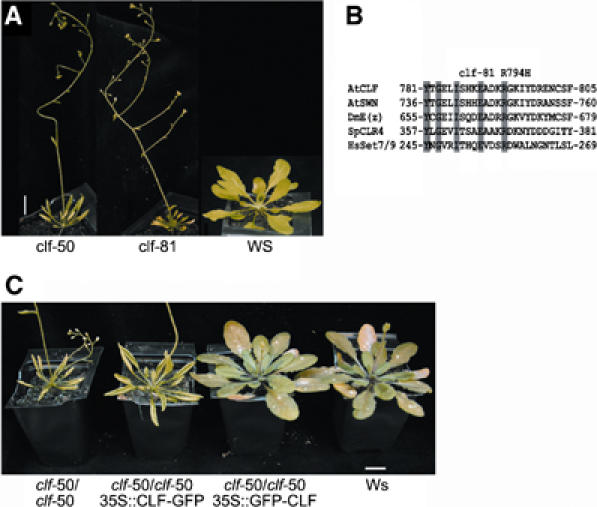
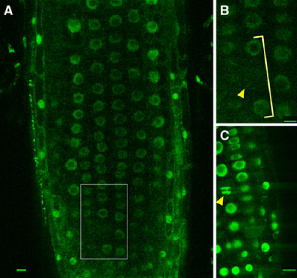
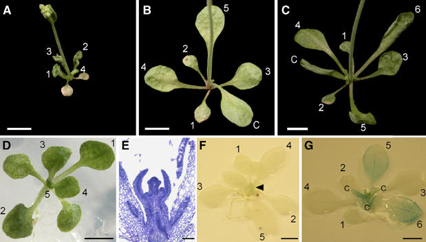
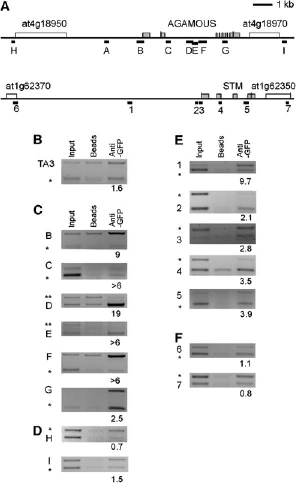
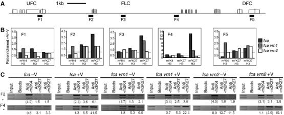
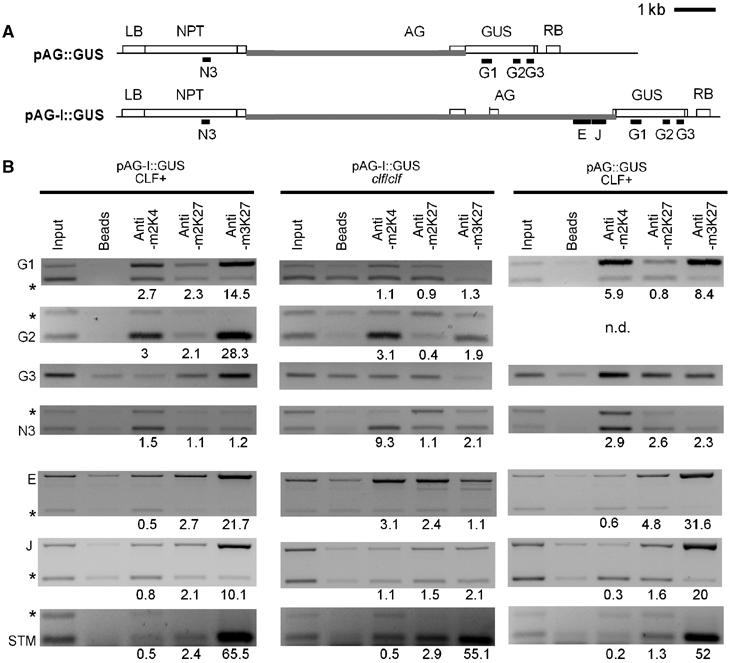
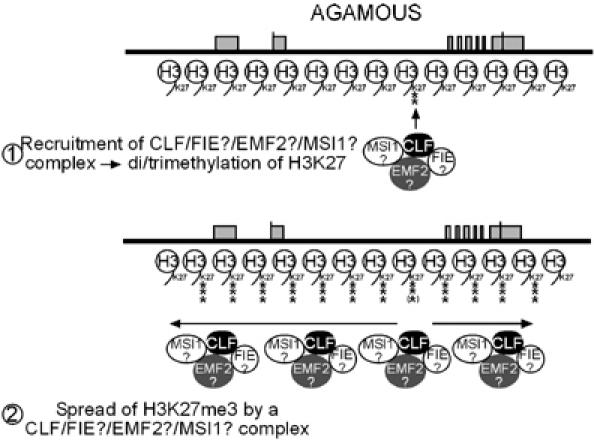
The plant Polycomb-group (Pc-G) protein CURLY LEAF (CLF) is required to repress targets such as AGAMOUS (AG) and SHOOTMERISTEMLESS (STM). Using chromatin immunoprecipitation, we identify AG and STM as direct targets for CLF and show that they carry a characteristic epigenetic signature of dispersed histone H3 lysine 27 trimethylation (H3K27me3) and localised H3K27me2 methylation. H3K27 methylation is present throughout leaf development and consistent with this, CLF is required persistently to silence AG. However, CLF is not itself an epigenetic mark as it is lost during mitosis. We suggest a model in which Pc-G proteins are recruited to localised regions of targets and then mediate dispersed H3K27me3. Analysis of transgenes carrying AG regulatory sequences confirms that H3K27me3 can spread to novel sequences in a CLF-dependent manner and further shows that H3K27me3 methylation is not sufficient for silencing of targets. We suggest that the spread of H3K27me3 contributes to the mitotic heritability of Pc-G silencing, and that the loss of silencing caused by transposon insertions at plant Pc-G targets reflects impaired spreading.


Figures

Figure 1
Figure 1
A severe clf allele carries a point mutation in the SET domain. (A) Alignment of a part of the SET domains of different SET domain proteins; residues conserved in all proteins are shaded, residue R794 that is mutated to H in clf-81 is marked. (B) clf-81 shows leaf curling and early flowering (compare to wild-type Ws (Wassilewskija)), similar to the clf-50 deletion allele. (C) clf-50 is complemented by a 35S∷GFP-CLF transgene but only partially by a 35S∷CLF-GFP transgene. All plants were grown under short day conditions; scale bar is 1 cm. AtCLF, Arabidopsis thaliana CURLY LEAF; AtSWN, A. thaliana SWINGER; DmE(z), Drosophila melanogaster ENHANCER OF ZESTE; SpCLR4, Schizosaccharomyces pombe CLR4; HsSET7/9, Homo sapiens SET7/9.
Figure 2
Figure 2
CLF protein is nuclear localised but is not present in nuclei throughout mitosis. (A) Root of a clf-50/clf-50 35SGFP-CLF plant showing GFP expression in most cells in the nuclei. (B) Close-up of the inset in (A) showing a cell without nuclear GFP expression (arrowhead). Adjacent cells in the same cell file show nuclear GFP (bracket). (C) Details of a VRN1VRN1-GFP root exhibiting a mitotic figure (arrowhead). At least 10 roots per line were analysed by confocal laser microscopy; in no case, GFP-stained mitotic figures were identified in clf/clf 35SGFP-CLF roots, whereas VRN1VRN1-GFP roots showed green-fluorescing mitotic figures in all roots. Scale bars, 10 μm.
Figure 3
Figure 3
CLF is persistently required for repression of AG in leaves. (A–G) All plants are clf/clf and carry the pCLFCLF-GR and the pAG-IGUS transgenes. Plants were initially grown on MS medium under long-day condition with or without dex steroid and transferred at day 10 to soil and either sprayed or not sprayed with dex. (A) A 27-day-old plant grown without dex throughout. (B) A 27-day-old plant grown with dex throughout. (C) A 27-day-old plant grown for 10 days with dex, then transferred from soil and dex withdrawn. Note leaf curling of leaves 4 and 5 and cauline leaf (C). (D) Plant grown on dex for 10 days shows no leaf curling. (E) Longitudinal section through a 10-day-old seedling grown with dex. Note that all leaf primordia have formed and that the plant has started flowering, so that the influorescence bolt is visible. (F, G) GUS staining reflects AG expression. (F) A 27-day-old plant grown on dex throughout only shows GUS staining in the flowers (arrowhead). (G) A 27-day old plant shifted on day 10 from dex to no dex shows strong GUS staining in leaves 5 and 6 and cauline leaves. Numbers indicate leaf number emerging after germination. Scale bars: 5 mm (A–C, F, G), 2 mm (D), 50 μm (E).
Figure 4
Figure 4
CLF is bound to AG and STM. (A) Schematic structure of the AG and STM loci including the 5′- and 3′-flanking genes. For AG and STM, the exon/intron structure (exons are shaded), and for the flanking genes, the transcribed regions are depicted. Black bars with letters (for AG) or numbers (for STM) indicate regions amplified in ChIP PCRs. (B, C) IP on chromatin preparations isolated from 10-day-old seedlings of a 35SGFP-CLF transgenic line was performed without antibody (‘beads') or anti-GFP antibodies and precipitated DNA was amplified by PCR. Semiquantitative ChIP duplex PCRs were used to amplify ACTIN 2/7 (marked with *) or PHOSPHOFRUCTOKINASE (PFK) sequences (marked with **) as internal controls and regions from TA3 retrotransposon (B) as negative control, from AG (C) and AG flanking genes (D), or from STM (E) and STM flanking genes (F). Numbers below the lanes indicate the ratio of the intensity of TA3, AG or STM products, respectively, compared to ACTIN or PFK intensity after IP normalised to the ratio before IP (‘input'). Each experiment was performed at least twice; representative experiment is shown.
Figure 5
Figure 5
Histone methylation patterns of AG and STM in wild-type and Pc-G mutant plants. (A–D) ChIPs were performed with antibodies against H3K4me2, H3K27me2 and H3K27me3 from wild-type (A–D), clf (A, C), emf2 (A, C), clf swn (C) and vrn2 emf2-3 (C) chromatin extracted from 10-day-old seedlings. Regions amplified are shown in Figure 4A and calculation of relative enrichment is described in Figure 4. Results of ChIP PCRs on the AG locus (A), on the AG flanking genes (B), the STM locus and TA3 retrotransposon (latter is H3K27me2control) (C) and on the STM flanking genes (D). (E) RT–PCRs with primers amplifying AG, STM or eIF2 coding regions on cDNAs generated from mRNA isolated from wild-type, clf and clf swn seedlings. EIF2 was used as a loading control.
Figure 6
Figure 6
The entire FLC locus shows strong enrichment with H3K27me3 after vernalisation. (A) Schematic structure of the FLC locus including the flanking genes UFC and DFC. The FLC exons are shaded. Black bars indicate the regions amplified. (B) Summary of ChIP PCRs performed with antibodies against H3K4me2, H3K27me2 and H3K27me3 on chromatin extracted from the vernalisation-requiring background fca, vrn1 fca and vrn2 fca mutants before and after vernalisation. Plants were grown for either 2 days at 4°C (−V) or 40 days at 4°C (+V), then transferred to 22°C and harvested after 15 days. Relative enrichment for ChIPs was determined as described in Figure 4 and the ratio of +V/−V for each genotype and antibody was calculated. Dashed lines indicate relative enrichment of 1. (C) Example of ChIPs performed for the different genotypes, antibodies and growth conditions. The whole data set is available in Supplementary Figure S4.
Figure 7
Figure 7
H3K27me3 spreads over AG transgenic sequences but is not sufficient for silencing. (A) Schematic structure of the pAGGUS and pAG-IGUS transgenes, constructed by Sieburth and Meyerowitz (1997). Black bars indicate regions amplified in ChIP PCRs. Grey bars indicate sequences homologous to AG. The pAGGUS T-DNA contains a NEOMYCIN PHOSPHOTRANSFERASE (NPT) resistance gene close to the left border (LB) of the T-DNA, around 6 kb of AG upstream region, the first AG exon, the β-GLUCURONIDASE (GUS) coding region and the right border (RB) of the T-DNA. The pAG-IGUS T-DNA consists of the same T-DNA backbone and AG upstream region but contains the first two exons and the first two introns of AG. The pAG-IGUS reporter reflects the endogenous AG expression pattern in wild-type and clf leaves, whereas pAGGUS is strongly mis-expressed in wild-type cotyledons and leaves (Sieburth and Meyerowitz, 1997). (B) Results of ChIP PCRs performed on IP with antibodies against H3K4me2, H3K27me2 and H3K27me3 on chromatin samples extracted from 10-day-old wild-type or clf seedlings carrying the pAG-IGUS transgene or wild-type seedlings carrying the pAGGUS transgene. Regions F and J of AG are present both at the endogenous AG locus and on the pAG-IGUS transgene. A region of STM (region 2 in Figure 4) served as an H3K27me3 control. Relative enrichment for ChIPs was determined as described in Figure 4.
Figure 8
Figure 8
Model of the regulation of AG (and other Pc-G targets) by plant Pc-G proteins and H3K27 methylation. EMF2 and MSI1 are likely part of at least one of the putative complexes, as a reduction in their protein level causes phenotypes similar to clf and lines with reduced FIE levels.

References

    1. Annunziato AT (2005) Split decision: what happens to nucleosomes during DNA replication? J Biol Chem 280: 12065–12068 - PubMed
    1. Bannister AJ, Schneider R, Kouzarides T (2002) Histone methylation: dynamic or static? Cell 109: 801–806 - PubMed
    1. Bastow R, Mylne JS, Lister C, Lippman Z, Martienssen RA, Dean C (2004) Vernalization requires epigenetic silencing of FLC by histone methylation. Nature 427: 164–167 - PubMed
    1. Baumbusch LO, Thorstensen T, Krauss V, Fischer A, Naumann K, Assalkhou R, Schulz I, Reuter G, Aalen RB (2001) The Arabidopsis thaliana genome contains at least 29 active genes encoding SET domain proteins that can be assigned to four evolutionarily conserved classes. Nucleic Acids Res 29: 4319–4333 - PMC - PubMed
    1. Bernstein BE, Mikkelsen TS, Xie X, Kamal M, Huebert DJ, Cuff J, Fry B, Meissner A, Wernig M, Plath K, Jaenisch R, Wagschal A, Feil R, Schreiber SL, Lander ES (2006) A bivalent chromatin structure marks key developmental genes in embryonic stem cells. Cell 125: 315–326 - PubMed

Publication types

MeSH terms

LinkOut - more resources