. 2006 Sep 20;25(18):4372-80.
doi: 10.1038/sj.emboj.7601303. Epub 2006 Sep 7.

Hippocampal neurons recycle BDNF for activity-dependent secretion and LTP maintenance

Affiliations

Hippocampal neurons recycle BDNF for activity-dependent secretion and LTP maintenance

Spartaco Santi et al. EMBO J. .

Abstract

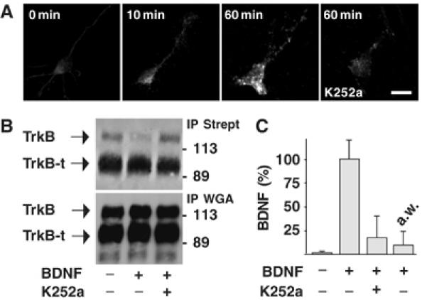
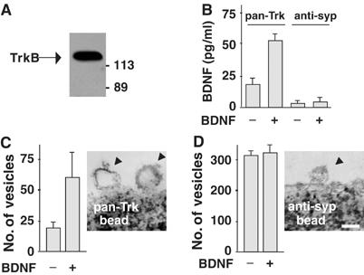
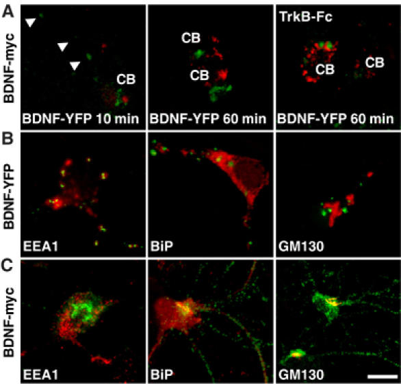
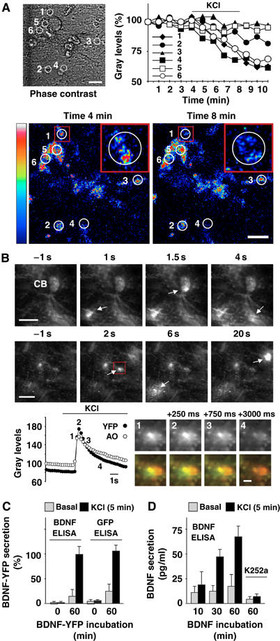
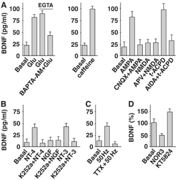
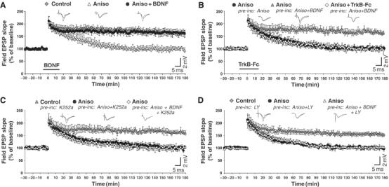
Regulation of brain-derived neurotrophic factor (BDNF) secretion plays a critical role in long-term potentiation (LTP). It is generally thought that the supply for this secretion is newly synthesized BDNF targeted to the synapse. Here we provide evidence that hippocampal neurons additionally recycle BDNF for activity-dependent secretion. Exogenously applied BDNF is internalized by cultured neurons and rapidly becomes available for activity-dependent secretion, which is controlled by the same mechanisms that regulate the secretion of newly synthesized BDNF. Moreover, BDNF recycling replaced the new synthesis pathway in mediating the maintenance of LTP in hippocampal slices: the late phase LTP, which is abolished by protein synthesis inhibition, was rescued in slices preincubated with BDNF. Thus, endocytosed BDNF is fed back to the activity-dependent releasable pool required for LTP maintenance.


Figures

Figure 1
Figure 1
TrkB-mediated BDNF endocytosis in cultured hippocampal neurons. (A) Time course of BDNF endocytosis in untreated neurons (0 min) or neurons exposed to BDNF (10 and 60 min) in the presence or absence of K252a pretreatment, obtained by immunocytochemistry and analyzed by confocal microscopy. Images are representative of 50 neurons analyzed in four independent experiments. Bar, 20 μm. (B) Regulation of plasma membrane expression of TrkB by BDNF. Plasma membrane proteins labeled with activated biotin were isolated by precipitation using agarose-conjugated streptavidin (IP Strept) from lysates of control neurons or neurons treated with BDNF for 60 min. Western blot analysis showed that TrkB, but not TrkB-t, is reduced by BDNF administration; this effect was prevented by pretreating neurons with K252a. Glycoconjugate proteins precipitated by WGA-agarose (IP WGA) showed total amount of TrkB and TrkB-t in whole-cell lysates. (C) Intracellular accumulation of exogenous BDNF measured by ELISA in the same lysates as in panel B. a.w. indicates the amount of BDNF immunoreactivity stripped from the cell surface by acid treatment. Data are expressed as percentage of the means±s.e.m. (n=6).
Figure 2
Figure 2
Internalization of BDNF–TrkB complexes in endocytic vesicles. (A) Magnetic beads coated with pan-Trk antibodies (pan-Trk bead) were used to trap Trk-containing vesicles from hippocampal neurons exposed to exogenous BDNF. Western blot analysis using anti-TrkB antibody revealed that purified vesicles express TrkB as a single immunoreactive protein of 135 kDa. (B) ELISA quantification of BDNF in pan-Trk bead and anti-syp bead purifications. Data are means±s.e.m. (n=4). (C) Quantification of vesicles isolated by pan-Trk beads. Vesicle number increased after BDNF exposure for 60 min. Representative image obtained by electron microscopy showing endocytic vesicles attached to a pan-Trk bead. (D) Quantification of vesicles isolated by anti-syp beads. Vesicle number did not change after BDNF exposure for 60 min. Representative image obtained by electron microscopy showing a vesicle with the typical size of a synaptic vesicle attached to an anti-syp bead. Bar, 100 nm. Data in panels C and D are means/100 beads±s.d.
Figure 3
Figure 3
Intracellular localization of endocytosed and newly synthesized BDNF. (A) Hippocampal neurons infected for 12 h with adenoviral vector transducing BDNF-myc in the presence or absence of TrkB-Fc were incubated with BDNF-YFP for 10 or 60 min. YFP fluorescence (green) and myc immunofluorescence (red) showed distinct intracellular localization in cell soma (CB) and neuronal processes (arrows). Images are representative of 25 neurons analyzed in five independent experiments. Colocalization between BDNF-YFP (B) or BDNF-myc (C) immunoreactivity (green) with that of EEA1, BiP and GM130 (red). Images are representative of 20 neurons analyzed in three independent experiments. Bar, 20 μm.
Figure 4
Figure 4
Activity-dependent secretion of endocytosed BDNF. (A) BDNF-YFP secretion was analyzed by time-lapse confocal imaging in circular areas 1–6 (phase contrast). Fluorescence intensity was expressed as percentage of gray levels and plotted with time. In resting conditions, fluorescence was unchanged, whereas KCl treatment induced a reduction in areas 1, 4 and 6. Confocal images document fluorescence intensity displayed in pseudo-color (scale palette on the left) before (min 4) and after (min 8) KCl treatment. Three-fold magnifications of area 1 (insets) showed BDNF-YFP fluorescence in a neuronal process. Note that fluorescence decreased within the white circle, and did not increase in the surrounding area delimited by the red square. This indicates that decreases in fluorescence intensity were due to BDNF-YFP secretion and not to its possible movements in the x–y axis. Data are representative of six independent experiments. Bars, 20 μm. (B) Exocytic fusion of vesicles analyzed by TIRF imaging in hippocampal neurons incubated with BDNF-YFP for 3 min. Sequential images document dequenching flashes of YFP fluorescence (arrows) in cell body (CB) and neuronal processes of a single neuron (upper sequence), or neuronal network (lower sequence). Times represent seconds before and after perfusion with KCl. Bars, 10 μm. Changes in fluorescence intensity (gray levels) of a single vesicle (red square) fusing to the plasma membrane. YFP and acridine-orange (AO) fluorescence was measured in a circular mask with an area of 120 pixels centered over the spots. Numbers (1–4) on the graph correspond to sequential images of YFP signal shown in gray on the right. The same sequential images showed overlap (yellow) of YFP (green) and acridine-orange (red) fluorescence. Times represent milliseconds from appearance (1), increase in brightness and diffusion (2), decrease (3) and disappearance (4) of the fluorescent signals. Bar, 2 μm. (C) ELISA quantification of BDNF-YFP in perfusates of control neurons or neurons previously treated with BDNF-YFP for 60 min. Cultured neurons did not show immunoreactivity for BDNF or GFP in sufficient amounts to be detected by two-site BDNF or GFP ELISAs, respectively. KCl treatment (5 min) increased BDNF-YFP immunoreactivity over the basal levels in BDMF-YFP-treated neurons. (D) ELISA quantification of BDNF in perfusates of neurons previously exposed to BDNF for 10, 30 or 60 min. KCl treatment (5 min) increased BDNF immunoreactivity over the basal levels and was prevented by K252a pretreatment. Data in panels C (n=5) and D (n=8) are means±s.e.m.
Figure 5
Figure 5
Pharmacology of endocytosed BDNF secretion. (A) BDNF secretion was evaluated by ELISA in perfusates of hippocampal neurons previously exposed to BDNF for 60 min. Glutamate (Glu) application elicited the secretion of endocytosed BDNF in both calcium-containing and calcium-free medium (EGTA). The effect was partially prevented by BAPTA-AM. BDNF secretion was also triggered by caffeine. AMPA- and t-ACPD-induced BDNF secretion was prevented by CNQX and AIDA, respectively. NMDA was not effective either in the presence or absence of APV. (B) BDNF secretion was induced by NT-4 and NT-3 but not NGF. This effect was prevented by K252a. (C) High-frequency stimulation (50 Hz) triggers BDNF secretion. This effect was prevented by TTX. (D) The NO donor NOR3 administered to BDNF-treated neurons elicited a decrease in BDNF secretion. Conversely, KT5823 triggered BDNF secretion. Data in panels A (n=12), B (n=9), C (n=12) and D (n=6) are means±s.e.m.
Figure 6
Figure 6
Endocytosed BDNF rescues LTP impaired by protein synthesis inhibition. (A) Field EPSPs evoked in CA1 area by Schaffer collaterals stimulation. TBS induced LTP that is maintained for 180 min (Control) (five slices, five rats). In slices perfused with anisomycin (Aniso) from 30 min before TBS to the end of the recording, LTP persisted for only 70–100 min (five slices, five rats). The effect of anisomycin was fully reversed by applying BDNF from 5 min before to 15 min after TBS (Aniso+BDNF) (six slices, five rats). (B) In slices incubated (pre-inc: Aniso) and perfused (Aniso) with anisomycin throughout the recording, LTP persisted for only 70–100 min (nine slices, seven rats). LTP was rescued in slices preincubated with BDNF (pre-inc: Aniso+BDNF) (12 slices, nine rats), an effect that was prevented by TrkB-Fc (Aniso+TrkB-Fc) (eight slices, six rats). (C) Pretreatment with K252a did not affect LTP maintenance (Control; pre-inc: K252a) (10 slices, seven rats). The presence of anisomycin in both preincubation and perfusion fluid reduced LTP duration to 70–100 min (Aniso; pre-inc: Aniso+K252a) (nine slices, six rats). The blocking action of anisomycin on LTP persisted in slices preincubated with BDNF and K252a (Aniso; pre-inc: Aniso+BDNF+K252a) (11 slices, eight rats). (D) Pretreatment with LY294002 (LY) (five slices, five rats) led to the same effects observed in the presence of K252a (panel C). Data in panels A–D are means±s.e.m. of field EPSP slopes plotted as percentages of the baseline. Representative field EPSP traces recorded before and 180 min after TBS are shown on the top of the panels.

References

    1. Aicardi G, Argilli E, Cappello S, Santi S, Riccio M, Thoenen H, Canossa M (2004) Induction of long-term potentiation and depression is reflected by corresponding changes in secretion of endogenous brain-derived neurotrophic factor. Proc Natl Acad Sci USA 101: 15788–15792 - PMC - PubMed
    1. Ashby MC, Ibaraki K, Hemley JM (2004) It's green outside: tracking cell surface proteins with pH-sensitive GFP. Trends Neurosci 27: 257–261 - PubMed
    1. Avery J, Ellis DJ, Lang T, Holroyd P, Riedel D, Henderson RM, Edwardson JM, Jahn R (2000) A cell-free system for regulated exocytosis in PC12 cells. J Cell Biol 148: 317–324 - PMC - PubMed
    1. Balkowiec A, Katz DM (2000) Activity-dependent release of endogenous brain-derived neurotrophic factor from primary sensory neurons detected by ELISA in situ. J Neurosci 20: 7417–7423 - PMC - PubMed
    1. Balkowiec A, Katz DM (2002) Cellular mechanisms regulating activity-dependent release of native brain-derived neurotrophic factor from hippocampal neurons. J Neurosci 22: 10399–10407 - PMC - PubMed

Publication types

MeSH terms