. 2006 Sep 20;25(18):4305-15.
doi: 10.1038/sj.emboj.7601277. Epub 2006 Sep 7.

New functions of XPC in the protection of human skin cells from oxidative damage

Affiliations

New functions of XPC in the protection of human skin cells from oxidative damage

Mariarosaria D'Errico et al. EMBO J. .

Abstract

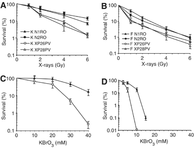
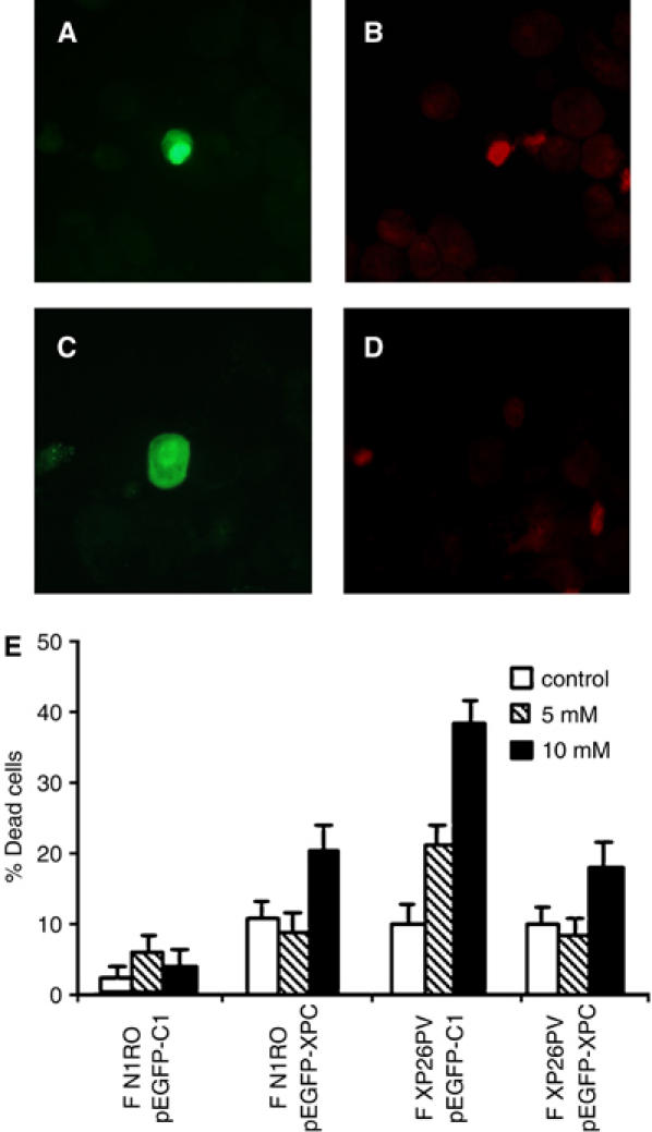
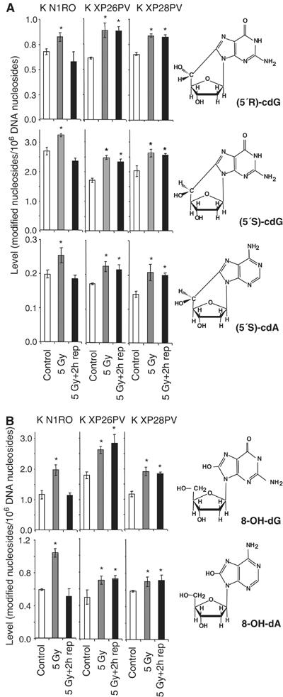
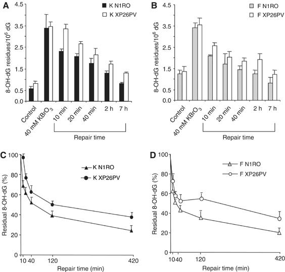
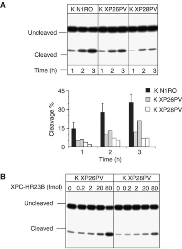
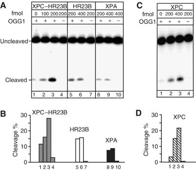
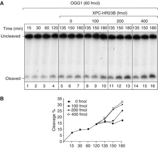
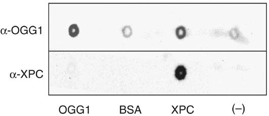
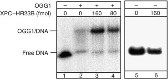
Xeroderma pigmentosum (XP) C is involved in the recognition of a variety of bulky DNA-distorting lesions in nucleotide excision repair. Here, we show that XPC plays an unexpected and multifaceted role in cell protection from oxidative DNA damage. XP-C primary keratinocytes and fibroblasts are hypersensitive to the killing effects of DNA-oxidizing agents and this effect is reverted by expression of wild-type XPC. Upon oxidant exposure, XP-C primary keratinocytes and fibroblasts accumulate 8,5'-cyclopurine 2'-deoxynucleosides in their DNA, indicating that XPC is involved in their removal. In the absence of XPC, a decrease in the repair rate of 8-hydroxyguanine (8-OH-Gua) is also observed. We demonstrate that XPC-HR23B complex acts as cofactor in base excision repair of 8-OH-Gua, by stimulating the activity of its specific DNA glycosylase OGG1. In vitro experiments suggest that the mechanism involved is a combination of increased loading and turnover of OGG1 by XPC-HR23B complex. The accumulation of endogenous oxidative DNA damage might contribute to increased skin cancer risk and account for internal cancers reported for XP-C patients.


Figures

Figure 1
Figure 1
XP-C keratinocytes and fibroblasts are hypersensitive to the killing effects of X-rays and KBrO3. Survival of primary keratinocytes (A, C) and fibroblasts (B, D) from two normal (N1RO and N2RO, closed symbols) and two XP-C (XP26PV and XP28PV, open symbols) donors after X-rays and KBrO3 treatment. (A–B) Survival of keratinocytes and fibroblasts after exposure to X-rays. (C–D) Survival of keratinocytes and fibroblasts after exposure to KBrO3. Survival was determined by colony formation assay. The reported values are the mean of at least two independent experiments, each performed in triplicate with standard errors always <10%.
Figure 2
Figure 2
The lack of XPC is responsible for the hypersensitivity to KBrO3 of human fibroblasts. Normal and XP-C fibroblasts were transiently transfected with an expression vector encoding for the EGFP-XPC fusion protein (pEGFP-XPC) or with the empty vector encoding for EGFP only (pEGFP-C1). At 24 h after transfection, cells were either untreated (control) or exposed to different KBrO3 doses (5 and 10 mM). At 48 h after transfection, cells were stained with PI to label in red dead cells, cytocentrifuged on slides and then analysed by fluorescence microscopy. (A–D) The photographs are representative fields of normal fibroblasts expressing the EGFP-XPC chimera (green fluorescence) and stained with PI to evaluate their viability following KBrO3 treatment (red fluorescence). Examples of cells expressing the ectopic protein and either dead (A–B) or alive (C–D). (E) The histograms report the percentages of dead cells following KBrO3 treatment of normal (F N1RO) and XP-C (F XP26PV) cells transfected with either the pEGFP-XPC or the pEGFP-C1 plasmid DNA. The reported values are the mean of at least three independent experiments and standard deviations are indicated.
Figure 3
Figure 3
XP-C keratinocytes accumulate cyclopurines and oxidized DNA bases induced by X-rays. Levels of oxidatively modified nucleosides in DNA of keratinocytes from normal (K N1RO) and XP-C (K XP26PV and K XP28PV) donors were measured by LC/MS or GC/MS. For each data point, DNA samples isolated from three independent experiments for cell strain were used. The chemical structures of the modified nucleosides are illustrated. (A) Induction by X-rays and repair of (5′S)-cdA, (5′R)-cdG and (5′S)-cdG. (B) Induction by X-rays and repair of 8-OH-dG and 8-OH-dA. DNA samples were isolated from untreated cells (control), cells exposed to X-rays (5 Gy), and exposed to X-rays and allowed to repair for 2 h (5 Gy+2 h rep). The data represent the mean of three independent experiments and standard deviations are reported. Statistical analysis was performed using one-way analysis of variance. The stars indicate statistically significant differences between control and 5 Gy, or control and 5 Gy+2 h rep with a P<0.05.
Figure 4
Figure 4
Repair of 8-OH-Gua is slower in XP-C keratinocytes and fibroblasts as compared with normal cells. 8-OH-dG levels were analysed in normal (N1RO) and XP-C (XP26PV) human fibroblasts and keratinocytes after exposure to 40 mM KBrO3 (1 h). Aliquots of cells were taken at the indicated times and the levels of 8-OH-Gua were measured by HPLC-ED. (A–B) 8-OH-dG levels in keratinocytes (A) and fibroblasts (B). (C–D) 8-OH-dG levels present at different repair times in keratinocytes (C) and fibroblasts (D) expressed as percentage of amounts at time 0.The data represent the mean of three independent experiments, and standard deviations (A–B) or standard errors (C–D) of the means are reported. Statistical analysis was performed using one- and two-way analyses of variance.
Figure 5
Figure 5
Cell extracts from XP-C keratinocytes are defective in 8-OH-Gua cleavage, but addition of purified XPC–HR23B restores normal cleavage activity. 30-mer duplex oligonucleotides (50 fmol) containing 8-OH-Gua were incubated with nuclear extracts (5 μg) of XP-C keratinocytes at 37°C for different periods of time as indicated. The 5′ end-labelled oligonucleotide was the 8-OH-Gua-containing strand. The products were separated by denaturing 20% PAGE. (A) Keratinocyte extracts from normal (K N1RO) and XP-C (K XP26PV and K XP28PV) donors. The relative percentages of 8-OH-Gua cleavage were obtained by electronic autoradiography of the gel (Instant Imager, Packard). Two or three independent extracts per cell strain were tested. (B) The 8-OH-Gua cleavage reaction by XP-C keratinocyte extracts was performed in the presence of varying concentrations of XPC–HR23B as indicated.
Figure 6
Figure 6
XPC–HR23B stimulates the activity of OGG1. DNA fragments (210 bp) containing a single 8-OH-Gua lesion were incubated with purified proteins, as indicated, at 37°C for 30 min. The 5′ end-labelled strand was the 8-OH-Gua-containing strand. The products were subjected to alkali treatment and separated by denaturing 8% PAGE. (A) The nicking assay was conducted in 10 μl (final volume) in the presence of 60 fmol of OGG1 and various concentrations of XPC–HR23B (lanes 2 and 3) or XPC-HR23B alone (lane 4); various concentrations of HR23B (lanes 5 and 6) or HR23B alone (lane 7); various concentrations of XPA (lanes 8 and 9) or XPA alone (lane 10), as indicated. (B) The amount of cleavage in each lane of (A) was evaluated by densitometric scanning of the autoradiography and expressed in arbitrary units following subtraction of the background. One representative experiment is shown. (C) The nicking assay was conducted in 10 μl (final volume) in the presence of 60 fmol of OGG1 and various concentrations of XPC (lanes 2 and 3) or XPC alone (lane 4). (D) The amount of cleavage in each lane of (C) was evaluated as in (B).
Figure 7
Figure 7
XPC–HR23B stimulates the turnover of OGG1 from its DNA substrate. 5′ end-labelled 210 bp fragments containing 8-OH-Gua were incubated with OGG1 (60 fmol) at 37°C for different periods of time, as indicated (lanes 1–4). After 120 min, different amounts of XPC–HR23B were added at the indicated concentrations and incubated further for 15, 30 or 60 min (lanes 5–16). The products were subjected to alkali treatment and separated by denaturing 8% PAGE. (A) Autoradiography of the gel. The incubation times indicated include the 120 min with OGG1 alone and the following periods in the presence of XPC–HR23B. (B) The amount of cleavage in each lane of (A) was evaluated by densitometric scanning of the autoradiography and expressed in arbitrary units following subtraction of the background. One representative experiment is shown.
Figure 8
Figure 8
Far Western analysis of OGG1 binding to XPC–HR23B. Purified proteins were spotted onto nitrocellulose and incubated with OGG1. OGG1 binding to proteins was tested by probing either with anti-XPC or anti-OGG1 polyclonal antibodies and visualization was performed by chemiluminescence. BSA and buffer alone were used as negative controls. Purified OGG1 was applied as positive control for antibody specificity.
Figure 9
Figure 9
The binding of OGG1 to 8-OH-Gua lesions is improved in the presence of XPC–HR23B. Electrophoretic mobility shift assay: mutant OGG1 (13 pmol), catalytically inactive, was incubated with a 250 bp fragment containing a single 8-OH-Gua residue. All the reactions mixtures were supplemented with BSA and undamaged plasmid DNA as described in Materials and methods. XPC–HR23B was added as indicated.

References

    1. Agar NS, Halliday GM, Barnetson RS, Ananthaswamy HN, Wheeler M, Jones AM (2004) The basal layer in human squamous tumors harbors more UVA than UVB fingerprint mutations: a role for UVA in human skin carcinogenesis. Proc Natl Acad Sci USA 101: 4954–4959 - PMC - PubMed
    1. Araki M, Masutani C, Takemura M, Uchida A, Sugasawa K, Kondoh J, Ohkuma Y, Hanaoka F (2001) Centrosome protein centrin 2/caltractin 1 is part of the xeroderma pigmentosum group C complex that initiates global genome nucleotide excision repair. J Biol Chem 276: 18665–18672 - PubMed
    1. Ballmaier D, Epe B (1995) Oxidative DNA damage induced by potassium bromate under cell-free conditions and in mammalian cells. Carcinogenesis 16: 335–342 - PubMed
    1. Birincioglu M, Jaruga P, Chowdhury G, Rodriguez H, Dizdaroglu M, Gates KS (2003) DNA base damage by the antitumor agent 3-amino-1,2,4-benzotriazine 1,4-dioxide (tirapazamine). J Am Chem Soc 125: 11607–11615 - PubMed
    1. Bjørås M, Luna L, Johnsen B, Hoff E, Haug T, Rognes T, Seeberg E (1997) Opposite base-dependent reactions of a human base excision repair enzyme on DNA containing 7,8-dihydro-8-oxoguanine and abasic sites. EMBO J 16: 6314–6322 - PMC - PubMed

Publication types

MeSH terms