. 2007 Jan 5;357(1):102-13.
doi: 10.1016/j.virol.2006.07.050. Epub 2006 Sep 7.

Adeno-associated virus interactions with B23/Nucleophosmin: identification of sub-nucleolar virion regions

Affiliations

Adeno-associated virus interactions with B23/Nucleophosmin: identification of sub-nucleolar virion regions

Joyce M Bevington et al. Virology. .

Abstract

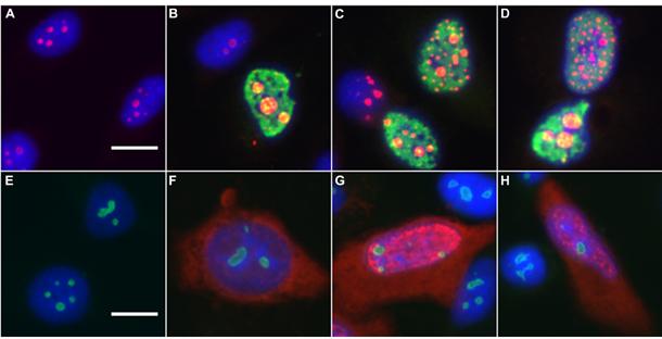
Adeno-associated virus (AAV) is a human parvovirus that normally requires a helper virus such as adenovirus (Ad) for replication. The four replication proteins (Rep78, 68, 52 and 40) encoded by AAV are pleiotropic effectors of virus integration, replication, transcription and virion assembly. Using Rep68 column chromatography and mass spectrometry, we have identified the nucleolar, B23/Nucleophosmin (NPM) protein as an Rep-interacting partner. Rep-NPM interactions were verified by co-immunofluorescence and chemical cross-linking studies. We have found that there is demonstrable, but limited co-localization between Rep and NPM in co-infected cells. In contrast, there was significant co-localization between NPM and AAV Cap proteins. In vitro experiments using purified MBPRep78 and NPM show that NPM stimulates MBPRep78 interactions with the AAV ITR as well as endonuclease activity. These studies suggest that NPM plays a role in AAV amplification affecting Rep function and virion assembly.


Figures

Figure 1
Figure 1
Co-localization of NPM and AAV proteins. HeLa cells were coinfected with Ad5 (10 m.o.i.) and AAV2 (250 m.o.i.) and fixed 20 to 30 hours post-infection. Fixed cells were stained for AAV Rep (panel A), Cap (panel D) and NPM (panels B and E). Merged images (panels C and F) show co-localization as yellow or white spots. Fixed cells were also stained for: Ad hexon protein (panels G and J), NPM protein (panel H), Ad hexon (panel J) and AAV Cap (panel K). These images are also merged (panels I and L). Nuclei were stained with DAPI and are depicted in blue in the merged images (C, F, I, and L). Scale bar is equivalent to 15 microns. Confocal microscopy was performed on AAV- and Ad- infected HeLa cells. Immunofluorescent staining of Cap (panels M and N) and Rep (panels O and P) are shown in green. NPM staining (panels M-P) is shown in red. Panels M and N and O and P are of the same field respectively and are separated by 1.5 microns. Yellow staining is the result of image merging and is indicative of co-localization.
Figure 2
Figure 2
Nucleolar remodeling during AAV and Ad coinfection. AAV and Ad coinfected HeLa cells (B-D), Ad infected HeLa cells (F-H) and uninfected cells (A, E) were fixed 20 p.i. NPM was stained with Alexa568 (A-D) or FITC (E-H). AAV Cap proteins were stained with Alexa488 (B-D) and Ad hexon was stained with Alexa568 (F-H). Alexa568 is a red fluorophore whereas FITC and Alexa488 are green fluorophores. Nuclei were countered stained with DAPI. Swollen nuclei and small punctate subnuclear structures are evident in panels B-D. Panels B-D and F-H are merged images. Scale bar is equivalent to 15 microns.
Figure 3
Figure 3
Coimmunoprecipitation of NPM and AAV proteins. Ad- or Ad- and AAV-coinfected Hela cells were harvested 22 h post infection, nuclei were isolated and chemically crosslinked with 2 mM DSP. Antibodies to Rep and NPM were used for immunoprecipitation, the crosslinked were reversed and the precipitates separated by SDS-PAGE. The separated proteins were analyzed by western blot using NPM, Rep or Cap antibodies. The extracts (Ext.) used were from Ad-infected (Ad) or AAV- and Ad-coinfected (Co) cells. The use of the DSP crosslinking agent (XL) is indicated by ‘+’ or ‘-’. Lanes 1 and 2 in each panel are crude extracts loaded on the gel without prior immunoprecipitation.
Figure 4
Figure 4
NPM stimulates MBPRep78 interaction with the AAV ITR. A 65-bp DNA fragment from the A-D component of the AAV ITR was radiolabeled and incubated with NPM and MBPRep78 (designated Rep78 in the figure). Protein-DNA complexes were separated by nondenaturing gel elctrophoresis and exposed to x-ray film. A. 3.4 nM MBPRep78 was used in lanes 4, 5, and 6, 14.3 nM GSTNPM was used in lanes 2 and 5, and 13.8 nM HisNPM was used in lanes 3 and 6. B. 8.9 nM MBPRep78 was used in lanes 3 to 8, and 4.7, 9.4, 18.7, 37.5, 74.9, and 93.5 nM GSTNPM was used in lanes 4, 5, 6, 7, 8, and 2, respectively. C. 3.4 nM MBPRep78 was used in lanes 3 to 8. 1.8, 3.5, 7, 14, 28, and 28 nM of BSA was used in lanes 4, 5, 6, 7, 8, and 2, respectively. Equimolar amounts of BSA:MBPRep78 and GSTNPM:MBPRep78 were used in the respective lanes (lanes 4 to 8) of Figure 4 B and C.
Figure 5
Figure 5
NPM stimulates MBPRep78 endonuclease activity. MBPRep78 (1.34 nM, designated Rep78) was incubated with radiolabeled ITR with ATP to induce endonuclease activity. The reactions were separated by denaturing polyacrylamide gel electrophoresis and exposed to x-ray film. A. Lanes 4, 5, 6, 7, and 2 contain to 0.8, 4, 20, 40, and 61 nM of HisNPM, respectively. B. Lanes 3, 4, 5, 6, and 1 contain 0.8, 4, 20, 40, and 60 nM of BSA, respectively.

References

    1. Adachi Y, Copeland TD, Hatanaka M, Oroszlan S. Nucleolar targeting signal of Rex protein of human T-cell leukemia virus type I specifically binds to nucleolar shuttle protein B-23. J Biol Chem. 1993;268(19):13930–4. - PubMed
    1. Basrur V, Yang F, Kushimoto T, Higashimoto Y, Yasumoto K, Valencia J, Muller J, Vieira WD, Watabe H, Shabanowitz J, Hearing VJ, Hunt DF, Appella E. Proteomic analysis of early melanosomes: identification of novel melanosomal proteins. J Proteome Res. 2003;2(1):69–79. - PubMed
    1. Bertwistle D, Sugimoto M, Sherr CJ. Physical and functional interactions of the Arf tumor suppressor protein with nucleophosmin/B23. Mol Cell Biol. 2004;24(3):985–96. - PMC - PubMed
    1. Borer RA, Lehner CF, Eppenberger HM, Nigg EA. Major nucleolar proteins shuttle between nucleus and cytoplasm. Cell. 1989;56(3):379–90. - PubMed
    1. Casper JM, Timpe JM, Dignam JD, Trempe JP. Identification of an adeno-associated virus Rep protein binding site in the adenovirus E2a promoter. J Virol. 2005;79(1):28–38. - PMC - PubMed

Publication types