. 2006 Sep 8;126(5):917-28.
doi: 10.1016/j.cell.2006.06.053.

Forespore engulfment mediated by a ratchet-like mechanism

Affiliations

Forespore engulfment mediated by a ratchet-like mechanism

Dan H Broder et al. Cell. .

Abstract

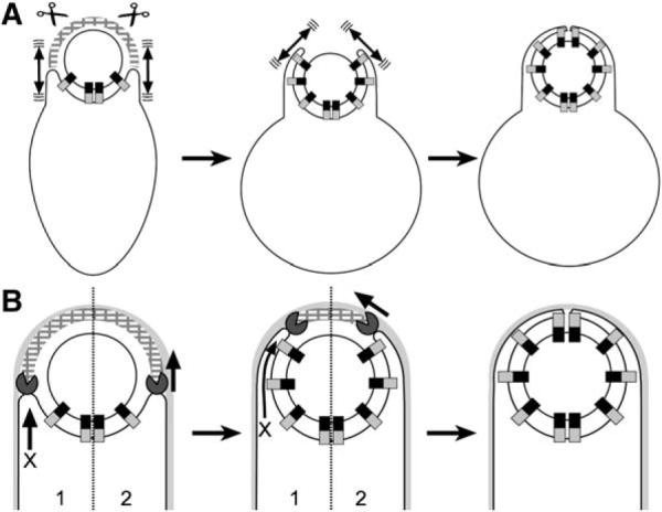
A key step in bacterial endospore formation is engulfment, during which one bacterial cell engulfs another in a phagocytosis-like process that normally requires SpoIID, SpoIIM, and SpoIIP (DMP). We here describe a second mechanism involving the zipper-like interaction between the forespore protein SpoIIQ and its mother cell ligand SpoIIIAH, which are essential for engulfment when DMP activity is reduced or SpoIIB is absent. They are also required for the rapid engulfment observed during the enzymatic removal of peptidoglycan, a process that does not require DMP. These results suggest the existence of two separate engulfment machineries that compensate for one another in intact cells, thereby rendering engulfment robust. Photobleaching analysis demonstrates that SpoIIQ assembles a stationary structure, suggesting that SpoIIQ and SpoIIIAH function as a ratchet that renders forward membrane movement irreversible. We suggest that ratchet-mediated engulfment minimizes the utilization of chemical energy during this dramatic cellular reorganization, which occurs during starvation.


Figures

Figure 1
Figure 1. The Process of Engulfment during B. subtilis Sporulation
(A) The smaller forespore (FS) and larger mother cell (MC) initially lie side by side. Engulfment commences with septal thinning (i), during which septal peptidoglycan (light gray) is degraded. The mother cell membrane then migrates around the forespore (steps ii–iii), until it meets and fuses to release the forespore into the mother cell cytoplasm (step iv). (B) Engulfment in intact cells requires three mother cell membrane proteins, SpoIID (Pac-Man), SpoIIM (dotted box), and SpoIIP (shaded lollipop), that localize to the septum and leading edge of the engulfing membrane. SpoIID is a peptidoglycan hydrolase, suggesting that engulfment might be mediated by the processive hydrolysis of the peptidoglycan adjacent to the forespore membrane, which could move the mother cell membrane around the forespore (in direction indicated by the arrow). Figure based on Abanes-De Mello et al. (2002). (C) The zipper-like interaction between the forespore membrane protein SpoIIQ (Q; gradient) and the mother cell membrane protein SpoIIIAH (AH; gray) localizes SpoIIIAH (Blaylock et al., 2004; Doan et al., 2005), which recruits additional mother cell proteins (Doan et al., 2005; Jiang et al., 2005). (D) A mechanical ratchet: the stationary pawl engages the ratchet teeth (or cogs), thereby preventing backward rotation of the wheel, allowing movement only in the direction indicated by the arrow. In a Brownian ratchet, random thermal energy is rectified, resulting in unidirectional rotation of the ratchet.
Figure 2
Figure 2. Time Lapse Fluorescence Microscopy Showing Protoplast Engulfment
Protoplasts were prepared from cells harvested 2.5 hr after the initiation of sporulation (t2.5) and stained with FM 4–64 (red). Images were collected at 45 s intervals. Time after first image is shown in bottom right corner of each frame (min:s). Arrows indicate sporangia that complete engulfment; arrowheads indicate sporangia with retracting membranes; yellow arrowheads indicate forespore of sporangia that separate; double yellow arrowheads indicate the mother cell of such sporangia. (A and C) FM 4–64-stained membranes (red) and mother cell produced SpoIIIJ-GFP (green; strain KP10068). (B and D) Surface plots showing the fluorescence intensity of FM 4–64-stained membranes (B) and mother cell GFP (D) from panels in (A) and (C) at the first (0 min) and last time points. Scale bar, 2 μm. (E) Alternate fates of mother cell-expressed SpoIIIJ-GFP. After protoplast engulfment, SpoIIIJ-GFP (green line) surrounds the forespore (upper pathway), while if engulfment does not occur, SpoIIIJ-GFP is restricted to the larger mother cell protoplast. (F–K) Time lapse microscopy showing protoplast engulfment in various mutants. Scale bar is 2 μm. (F) Wild-type (PY79), (G) spoIID298, spoIIMTn917, ΔspoIIPtet (KP4188), (H) ΔspoIIQspc (KP575), (I) spoIIIAG-HΩkan (KP896), (J) spoIIQ rbm13 (KP4124), (K) spoIIQ rbm9 (KP4138). (L) Quantification of protoplast engulfment, showing percent protoplasts that engulf (green), retract but remain attached (blue), or retract but separate (red). More than 50 protoplasts were scored for each strain.
Figure 3
Figure 3. Protoplast Engulfment Exhibits a Dosage-Dependent Requirement for SpoIIQ
(A) Immunoblot analysis of SpoIIQ in ribosome binding site mutants expressed in strains defective in SpoIIQ proteolysis (spoIVB)at t1, t2, t3, and t4. ΔspoIIQspec (KP4195), rbm2 (KP4203), rbm1 (KP4202), rbm12 (KP4201), rbm9 (KP4198), rbm14 (KP4199), rbm10 (KP4200), rbm11 (KP4196), rbm13 (KP4197), and wild-type (KP4194). (B) Quantification of protoplast engulfment (t2.5), spore titers (t24), SpoIIQ levels (t4), and the ability to support engulfment in the spoIIB mutant (t3.5) as described in Experimental Procedures.
Figure 4
Figure 4. Photobleaching Experiments Demonstrate that GFP-SpoIIQ Assembles a Static Structure
After photobleaching, images were collected, quantified, and plotted (see Experimental Procedures) to show the adjusted mean pixel intensity of the bleached (black squares) and unbleached (unfilled squares) regions and the theoretical pixel intensity value following equilibration between these regions (dashed line). Images of GFP-SpoIIQ (green) and FM 4–64-stained membranes (red) of photobleached cells are shown to the right of each graph. Images below each plot show GFP-SpoIIQ during the experiment. Bleached regions are indicated by a yellow circle. (A–D) FRAP of GFP-SpoIIQ (ΔspoIIQspc, gfp-spoIIQ) (KP845). (E–H) FRAP of GFP-SpoIIQ in the presence of SpoIIQ (gfp-spoIIQ) (KP866). (I) Frequency with which the various phenotypes were observed in individual cells subjected to FRAP. Recovery includes cells that reached ≥75% of the theoretical equilibration point; partial recovery reached 50%–75%; no recovery was less than 50%. (J) FRAP of MalF-GFP (malF-gfp) (KP834).
Figure 5
Figure 5. SpoIIQ and SpoIIIAH Contribute to Engulfment in Intact Cells
(A–L) Samples were harvested at t3 and a fusion assay was performed to assess the completion of engulfment. FM 4–64 is membrane impermeable (red); Mitotracker Green is permeable (green; MTG shown in overlay). Fused sporangia, indicated by arrows, have completed engulfment; the unfused sporangia, indicated by arrowheads, have not. Percent of sporangia fused at t3 (A–G) or percent fused relative to ΔspoIIQspc, amyEspoIIQΩcat, spoIIBerm (KP4369) at t3.5 (H–L) is shown (>200 sporangia scored for each strain). (A) Wild-type (PY79), (B) ΔspoIIQspc (KP575), (C) ΔspoIIIAH (MO1429), (D) ΔspoIIIAG-HΩkan (KP10359), (E) spoIID31 (KP38), (F) spoIID31, ΔspoIIQspc (KP4270), (G) spoIID31, ΔspoIIIAH (KP4296), (H) ΔspoIIQspc, amyEspoIIQ, spoIIBerm (KP4369), (I) ΔspoIIQspc, spoIIBerm (KP4276), (J) ΔspoIIQspc, amyEspoIIQ(rbm9), spoIIBerm (KP4373), (K) ΔspoIIQspc, amyEspoIIQ(rbm10), spoIIBerm (KP4377), (L) ΔspoIIQspc, amyEspoIIQ(rbm13), spoIIBerm (KP4370). (M) Cartoon of membrane fusion assay showing FM 4–64 (red) and Mitotracker Green (green).
Figure 6
Figure 6. Models for Engulfment in Protoplasts and Intact Cells
(A) Brownian ratchet model for protoplast engulfment. During protoplast formation, peptidoglycan hydrolases (scissors) remove the peptidoglycan (hatched arcs) that blocks the engulfing membrane, allowing the Q-AH ratchet to engage and prevent backward membrane movement. The mother cell membrane could move by Brownian motion (double-headed arrows). (B) Models for engulfment in intact cells. (1) Missing motor model. The DMP proteins (Pac-Man) might act like lysozyme, degrading the peptidoglycan that blocks engulfment. Since SpoIIQ and SpoIIIAH are not essential for engulfment in whole cells, this model requires additional unidentified force-generating proteins (X arrow). (2) Dual ratchet model. The membrane-anchored DMP proteins might act as a burnt-bridge Brownian ratchet, in which the enzymatic activity of SpoIID renders complex diffusion along the peptidoglycan unidirectional, thereby moving the engulfing membrane around the forespore. The Q-AH ratchet could contribute to the directionality and processivity of the DMP ratchet.

References

    1. Abanes-De Mello A, Sun YL, Aung S, Pogliano K. A cytoskeleton-like role for the bacterial cell wall during engulfment of the Bacillus subtilis forespore. Genes Dev. 2002;16:3253–3264. - PMC - PubMed
    1. Antal T, Krapivsky PL. “Burnt-bridge” mechanism of molecular motor motion. Phys. Rev. E Stat. Nonlin. Soft Matter Phys. 2005 in press. Published online October 6, 2005. 10.1103/PhysRevE.72.046104. - PubMed
    1. Bar-Nahum G, Epshtein V, Ruckenstein AE, Rafikov R, Mustaev A, Nudler E. A ratchet mechanism of transcription elongation and its control. Cell. 2005;120:183–193. - PubMed
    1. Blaylock B, Jiang X, Rubio A, Moran CP, Jr., Pogliano K. Zipper-like interaction between proteins in adjacent daughter cells mediates protein localization. Genes Dev. 2004;18:2916–2928. - PMC - PubMed
    1. Cutting S, Roels S, Losick R. Sporulation operon spoIVF and the characterization of mutations that uncouple mother-cell from forespore gene expression in Bacillus subtilis. J. Mol. Biol. 1991;221:1237–1256. - PubMed

Publication types

MeSH terms