. 2006 Sep 8;126(5):995-1004.
doi: 10.1016/j.cell.2006.07.025.

A rapid, reversible, and tunable method to regulate protein function in living cells using synthetic small molecules

Affiliations

A rapid, reversible, and tunable method to regulate protein function in living cells using synthetic small molecules

Laura A Banaszynski et al. Cell. .

Abstract

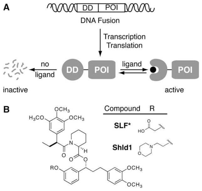
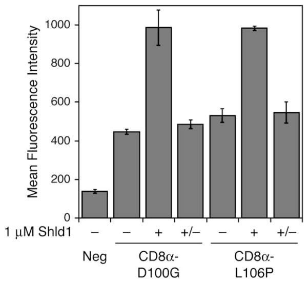
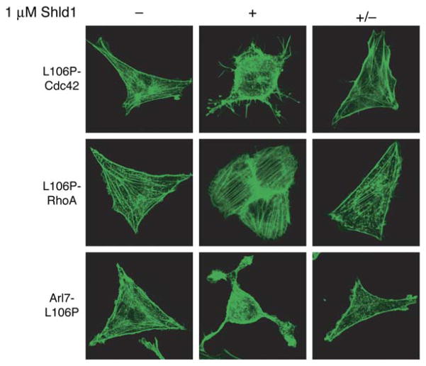
Rapid and reversible methods for perturbing the function of specific proteins are desirable tools for probing complex biological systems. We have developed a general technique to regulate the stability of specific proteins in mammalian cells using cell-permeable, synthetic molecules. We engineered mutants of the human FKBP12 protein that are rapidly and constitutively degraded when expressed in mammalian cells, and this instability is conferred to other proteins fused to these destabilizing domains. Addition of a synthetic ligand that binds to the destabilizing domains shields them from degradation, allowing fused proteins to perform their cellular functions. Genetic fusion of the destabilizing domain to a gene of interest ensures specificity, and the attendant small-molecule control confers speed, reversibility, and dose-dependence to this method. This general strategy for regulating protein stability should enable conditional perturbation of specific proteins with unprecedented control in a variety of experimental settings.


Figures

Figure 1
Figure 1. A General Method to Conditionally Control Protein Stability
(A) Genetic fusion of a destabilizing domain (DD) to a protein of interest (POI) results in degradation of the entire fusion. Addition of a ligand for the destabilizing domain protects the fusion from degradation. (B) Synthetic ligands for FKBP12 F36V.
Figure 2
Figure 2. Characterization of FKBP Mutants that Display Shld1-Dependent Stability
(A) Fluorescence of FKBP-YFP fusions expressed in NIH3T3 cells in the absence of Shld1 as determined by flow cytometry. (B) NIH3T3 cells stably expressing FKBP-YFP fusions were treated with 3-fold dilutions of Shld1 (1 μM to 0.1 nM) and monitored by flow cytometry. (C) NIH3T3 cells stably expressing FKBP-YFP fusions were either mock-treated (circles) or treated with 30 nM (squares), 100 nM (diamonds), 300 nM (crosses), or 1 μM (triangles) Shld1. Increases in fluorescence were monitored over time using flow cytometry. Mean fluorescence intensity (MFI) was normalized to 100% at 24 hr, 1 μM Shld1. (D) NIH3T3 cells stably expressing FKBP-YFP fusions were treated with 1 μM Shld1 for 24 hr, at which point the cells were washed with media to remove Shld1, and decreases in fluorescence were monitored using flow cytometry. Data for panels (A) through (D) are presented as the average MFI ± SEM relative to that of the maximum fluorescence intensity observed for the individual mutant. Experiments were performed in triplicate. (E) FKBP-YFP fusions were either mock-treated or treated with 1 μM Shld1 for 24 hr and immunoblotted with an anti-FKBP antibody. (F) NIH3T3 cells stably expressing F15S-YFP and L106P-YFP were treated with 1 μM Shld1 for 24 hr. Cells were then washed with media and treated with 10 μM MG132 in the presence or absence of 1 μM Shld1 for 4 hr. Immunoblotting was performed with an anti-YFP antibody. (G) HeLa cells were transfected with siRNA against lamin A/C and monitored over time. Time required for knockdown of lamin A/C is compared against time required for degradation of L106P-YFP upon removal of Shld1 from NIH3T3 cells stably expressing the fusion.
Figure 3
Figure 3. Fusion of an FKBP Destabilizing Domain to the N Terminus of YFP Results in Predictable and Reversible Small-Molecule Regulation of Intracellular Protein Levels
A population of NIH3T3 cells stably expressing L106P-YFP was treated with varying concentrations of Shld1 over the course of one week, and samples of the population were assayed by flow cytometry at the indicated time points. Data are presented as the average mean fluorescence intensity ± SEM relative to that of the maximum fluorescence intensity observed for L106P-YFP. Predicted fluorescence is based upon the dose response experiment shown in Figure 2B. The experiment was performed in triplicate.
Figure 4
Figure 4. FKBP Destabilizing Domains Confer Shld1-Dependent Stability to a Variety of Proteins
(A) FKBP mutants F15S and L106P were fused to the N termini of several different proteins and transduced into NIH3T3 cells. Cell populations stably expressing the fusions were then either mock-treated or treated with 1 mM Shld1, and cell lysates were immunoblotted with antibodies against the protein of interest. Endogenous proteins are shown as loading controls when detected, and Hsp90 serves this purpose in cases where they are not detected. (B) FKBP mutants D100G and L106P were fused to the C termini of several different proteins of interest and treated as above.
Figure 5
Figure 5. Destabilizing Domains Confer Shld1-Dependent Stability to a Transmembrane Protein
FKBP mutants D100G and L106P were fused to the C terminus of CD8α, and NIH3T3 cells stably expressing the fusions were split into three pools. The first population (−) was mock-treated and the second population (+) was treated with 1 mM Shld1 for 24 hr. The third population (+/−) was treated with 1 mM Shld1 for 24 hr, then washed with media and cultured for 24 hr in the absence of Shld1. Live cells were then probed with a FITC-conjugated anti-CD8a antibody and assayed by flow cytometry. Data are presented as the average mean fluorescence intensity ± SEM from an experiment performed in triplicate.
Figure 6
Figure 6. Stabilization of Specific Proteins with Shld1 Results in Predictable Changes in Cellular Morphologies
NIH3T3 cells stably expressing fusions of a constitutively active small GTPase to the L106P destabilizing domain were split into three pools. The first population (−) was mock-treated and the second population (+) was treated with 1 mM Shld1 for 24 hr. The third population (+/−) was treated with 1 mM Shld1 for 24 hr, then washed with media and cultured in the absence of Shld1 for 24 hr (RhoA Q63L) or 48 hr (Cdc42 Q61L, Arl7 Q72L). Cells were serum-starved for 12 hr, fixed, stained with Alexa Fluor 488-conjugated phalloidin, and visualized using confocal microscopy.

Comment in

Similar articles

Cited by

References

    1. Bachmair A, Finley D, Varshavsky A. In vivo half-life of a protein is a function of its amino-terminal residue. Science. 1986;234:179–186. - PubMed
    1. Bain J, McLauchlan H, Elliott M, Cohen P. The specificities of protein kinase inhibitors: an update. Biochem J. 2003;371:199–204. - PMC - PubMed
    1. Banaszynski LA, Wandless TJ. Conditional control of protein function. Chem Biol. 2006;13:11–21. - PubMed
    1. Bence NF, Sampat RM, Kopito RR. Impairment of the ubiquitin-proteasome system by protein aggregation. Science. 2001;292:1552–1555. - PubMed
    1. Bishop AC, Shah K, Liu Y, Witucki L, Kung CY, Shokat KM. Design of allele-specific inhibitors to probe protein kinase signaling. Curr Biol. 1998;8:257–266. - PubMed

Publication types